. 2013 Aug 15;89(2):31.
doi: 10.1095/biolreprod.113.110106. Print 2013 Aug.

Broad gap junction blocker carbenoxolone disrupts uterine preparation for embryo implantation in mice

Affiliations

Broad gap junction blocker carbenoxolone disrupts uterine preparation for embryo implantation in mice

Honglu Diao et al. Biol Reprod. .

Abstract

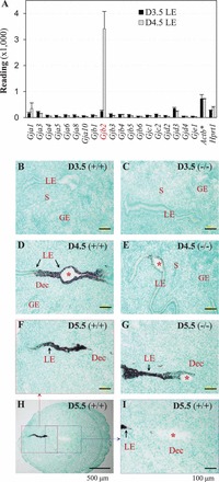
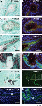
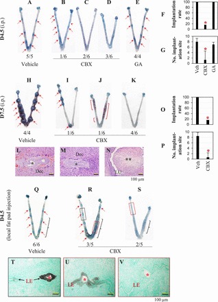
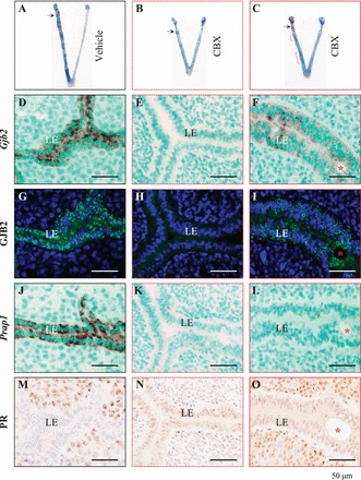
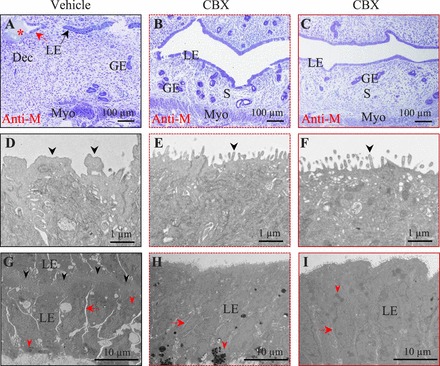
Gap junctions have an important role in cell-to-cell communication, a process obviously required for embryo implantation. Uterine luminal epithelium (LE) is the first contact for an implanting embryo and is critical for the establishment of uterine receptivity. Microarray analysis of the LE from peri-implantation mouse uterus showed low-level expression of 19 gap junction proteins in preimplantation LE and upregulation of gap junction protein, beta 2 (GJB2, connexin 26, Cx26) in postimplantation LE. Time course study using in situ hybridization and immunofluorescence revealed upregulation of GJB2 in the LE surrounding the implantation site before decidualization. Similar dynamic expression of GJB2 was observed in the LE of artificially decidualized mice but not pseudopregnant mice. To determine the potential function of uterine gap junctions in embryo implantation, carbenoxolone (CBX), a broad gap junction blocker, was injected i.p. (100 mg/kg) or via local uterine fat pad (10 mg/kg) into pregnant mice on Gestation Day 3 at 1800 h, a few hours before embryo attachment to the LE. These CBX treatments disrupted embryo implantation, suggesting local effects of CBX in the uterus. However, i.p. injection of glycyrrhizic acid (100 mg/kg), which shares similar structure and multiple properties with CBX but is ineffective in blocking gap junctions, did not affect embryo implantation. Carbenoxolone also inhibited oil-induced artificial decidualization, concomitant with suppressed molecular changes and ultrastructural transformations associated with uterine preparation for embryo implantation, underscoring the adverse effect of CBX on uterine preparation for embryo implantation. These data demonstrate that uterine gap junctions are important for embryo implantation.

Keywords: carbenoxolone; embryo implantation; gap junctions; uterine luminal epithelium.


Figures

FIG. 1
FIG. 1
Differential expression of Gjb2 (gap junction protein, beta 2, connexin 26) mRNA in the peri-implantation mouse uterine LE. A) Readings of gap junction proteins from microarray analysis of D3.5 and D4.5 LE. Actb, β-actin, a housekeeping gene; *, one tenth of the readings; Hprt1, hypoxanthine phosphoribosyltransferase 1, a housekeeping gene. n = 3. Error bars represent SD. BI) In situ hybridization of Gjb2 in D3.5 ∼ D5.5 WT (+/+) uterus and Lpar3-deficient (−/−) uterus with delayed implantation [19]. B) D3.5, +/+. C) D3.5, −/−. D) D4.5, +/+. E) D4.5, −/−. F) D5.5, +/+, enlarged view from the red box in H. G) D5.5, −/−. H) D5.5, +/+, low magnification (×4), the dotted red box and blue box shown in F and I, respectively. I) D5.5, +/+, enlarged view from the blue box in H. Bars = 100 μm (BG and I) and 500 μm (H). Red star, embryo; GE, glandular epithelium; S, stroma; Dec, decidual zone.
FIG. 2
FIG. 2
Upregulation of Gjb2 mRNA (via in situ hybridization, ISH) and GJB2 protein (via immunofluorescence, IF) in WT mouse uterine LE during early hours of embryo implantation (AJ) and GJB2 protein in the uterus with artificial decidualization (K and L). AJ) The sections for in situ hybridization and immunofluorescence at the same time point were from the same implantation sites. In situ hybridization (left panel): dark brown signal, Gjb2 mRNA. Immunofluorescence (right panel): green, GJB2 protein; blue, 4′6-diamidino-2-phenylindole dihydrochloride; red, integrin. A) Gestation Day 3 at 2300 h, ISH. B) On D3 at 2300 h, IF. C) On D4 at 0100 h, ISH. This section likely cut through the uterine luminal epithelial cells that cover the front view of the embryo, indicated by the dark brown signals detected in the location of the embryo but no GJB2 stain on the embryo in the section from the same implantation site in D, as well as no signal of Gjb2 or GJB2 on the embryos in A, B, and EH. D) On D4 at 0100 h, IF. E) On D4 at 0400 h, ISH. F) On D4 at 0400 h, IF. G) On D4 at 0600 h, ISH, enlarged view of the dotted red box in I. H) On D4 at 0600 h, IF, enlarged view of the dotted red box in J. I) On D4 at 0600 h, ISH, low magnification, the enlarged view of the dotted red box shown in G, the enlarged views of the dotted yellow box and white box shown in the two insets. J) On D4 at 0600 h, IF, low magnification, the enlarged view of the dotted red box shown in H, the enlarged views of the dotted yellow box and white box shown in the two insets. I and J) The insets showing gradient expression of Gjb2 mRNA and GJB2 protein away from the implantation site. K) GJB2 IF, D4 at 0600 h, intact uterine horn (Intact), contralateral to the uterine horn in L. L) GJB2 IF, D4 at 0600, oil-infused uterine horn to induce artificial decidualization (AD). Red star, embryo. Bar = 50 μm.
FIG. 3
FIG. 3
Effects of CBX on embryo implantation via i.p. injection (100 mg/kg, AP) and uterine fat pad injection (10 mg/kg, QV). AE) Uterine images from D4.5, i.p. injection. A) Vehicle treated. B) CBX treated, with six distinctive blue bands (red arrows) and two faint blue bands (red arrowheads), an indication of delayed embryo implantation. C) CBX treated, with no implantation and normal length. D) CBX treated, with no implantation and shorter appearance. E) Glycyrrhizic acid (GA) treated. F) Implantation rate on D4.5. G) Average number of implantation sites per mouse on D4.5. F and G) *P < 0.05 compared with vehicle (Veh)-treated and glycyrrhizic acid (GA)-treated groups. Error bars represent SD. n = 4–6. HK) Uterine images from D7.5, i.p. injection. H) Vehicle treated. I) CBX treated, with four smaller implantation sites compared with vehicle-treated control (H). J) CBX treated, with no implantation, shorter appearance, and bloody uterine segments (red brackets). K) CBX treated, with no implantation and shorter appearance. LN) Histology from D7.5 uteri. Hematoxylin-eosin stain. Bar = 100 μm. L) A section from an implantation site in H. M) A section from an implantation site in I. L and M) Dec, decidual zone; *, embryo. N) A section from a bloody segment in J. **, Tissue in the uterine lumen (LU) being reabsorbed. O) Implantation rate on D7.5. P) Average number of implantation sites per mouse on D7.5. O and P) *P < 0.05. Error bars represent SD. n = 4–6. QS) The D4.5 uterine images from uterine fat pad injection on the right side, with black brackets indicating the approximate locations of the injected uterine fat pad. Q) Vehicle injected. R) CBX injected, with implantation sites. S) CBX injected, with no implantation sites. TV) Detection of Gjb2 mRNA by in situ hybridization on longitudinal sections through an embryo in three areas indicated in R and S and processed on the same slide. *, Embryo. Bar = 100 μm. T) From the black dotted rectangle in R. U) From the red dotted rectangle in R. V) From the red dotted rectangle in S. In AE, HK, and Q and R, the number below each uterus indicates the number of uteri (uterus) with similar appearance in the same treatment group over the total number of mice in the same group; red arrows, implantation sites.
FIG. 4
FIG. 4
Effects of CBX on artificial decidualization and uterine gene expression detected on Pseudopregnant Day 4.5. Artificial decidualization (indicated by blue segments) was induced by oil infusion of the left uterine horn of pseudopregnant mice. The injection site for oil infusion is indicated by a black arrow on uterine images in AC. Fresh frozen longitudinal uterine sections (DO) were cut in the orientation of mesometrial and antimesometrial. Serial sections from a blue segment (indicating decidualization) of each uterus (except B) in AC are lined under the same column with the same outline (solid black from A, dotted red from B, solid red from C). A) Vehicle treated. B) CBX treated, representing the most suppressed decidualization upon CBX treatment, with shortened appearance. C) CBX treated, representing the least effect of CBX treatment, with shortened appearance; red bracket, suppressed decidualization. DF) In situ hybridization detection of Gjb2 mRNA in vehicle (D) and CBX-treated (E and F) uteri. GI) Immunofluorescence detection of GJB2 protein in vehicle (G) and CBX-treated (H and I) uteri. JL) In situ hybridization detection of proline-rich acidic protein 1 (PRAP1) mRNA in vehicle (J) and CBX-treated (K and L) uteri. MO) Immunohistochemistry detection of PR protein in vehicle (M) and CBX-treated (N and O) uteri. Red star, infused oil droplet. Bar = 50 μm.
FIG. 5
FIG. 5
Pseudopregnant Day 4.5 uterine structure of artificially decidualized uterus upon vehicle or CBX treatment. Uterine tissues were fixed for transmission electron microscopy. Solid black outlined images (A, D, and G) are from the vehicle-treated left uterine horn in Figure 4A. Dotted red line outlined images (B, E, and H) are from the CBX-treated left uterine horn in Figure 4B. Solid red line outlined images (C, F, and I) are from an area with blue dye reaction in the CBX-treated left uterine horn in Figure 4C. Longitudinal sections were cut in the orientation of mesometrial (top) and antimesometrial (Anti-M, bottom). AC) Semithin uterine sections. A thicker stromal compartment is seen in A. Uterine lumen is open in B and C. Stromal edema is obvious in C. Red star, oil droplet. Bar = 100 μm. DF) Ultrastructure of the apical surface of the uterine LE. Note the irregular projections (black arrowhead) on the apical surface of the vehicle-treated LE in D, which is from the area indicated by the red arrow in A. The apical surface of the CBX-treated LE is covered by thin, regular microvilli (black arrowhead) in E and F. Bar = 1 μm. GI) The LE structure. Bar = 10 μm. Red arrow, intercellular space between neighboring LE cells on the same side; red arrowhead, lipid droplet. G) The LE from the area indicated by the black arrow in A of a vehicle-treated uterus. The uterine lumen, indicated by the black arrowheads, is tightly closed by interdigitating cytoplasmic processes of the apical surface of the apposing LE cells. H) The LE from a CBX-treated uterus in B. I) The LE from a CBX-treated uterus in C. GE, glandular epithelium; S, stromal cell; Dec, decidual cell; Myo, myometrium.

Similar articles

Cited by

References

    1. Willecke K, Eiberger J, Degen J, Eckardt D, Romualdi A, Guldenagel M, Deutsch U, Sohl G. Structural and functional diversity of connexin genes in the mouse and human genome. Biol Chem 2002; 383: 725 737. - PubMed
    1. Maeda S, Tsukihara T. Structure of the gap junction channel and its implications for its biological functions. Cell Mol Life Sci 2011; 68: 1115 1129. - PMC - PubMed
    1. Söhl G, Willecke K. An update on connexin genes and their nomenclature in mouse and man. Cell Commun Adhes 2003; 10: 173 180. - PubMed
    1. Bosco D, Haefliger JA, Meda P. Connexins: key mediators of endocrine function. Physiol Rev 2011; 91: 1393 1445. - PubMed
    1. Juszczak GR, Swiergiel AH. Properties of gap junction blockers and their behavioural, cognitive and electrophysiological effects: animal and human studies. Prog Neuropsychopharmacol Biol Psychiatry 2009; 33: 181 198. - PubMed

Publication types

LinkOut - more resources