

Activation of the PKC pathway stimulates ovarian cancer cell proliferation, migration, and expression of MMP7 and MMP10
- PMID: 23843242
- PMCID: PMC4094197
- DOI: 10.1095/biolreprod.112.102327
Activation of the PKC pathway stimulates ovarian cancer cell proliferation, migration, and expression of MMP7 and MMP10
Abstract
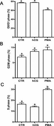
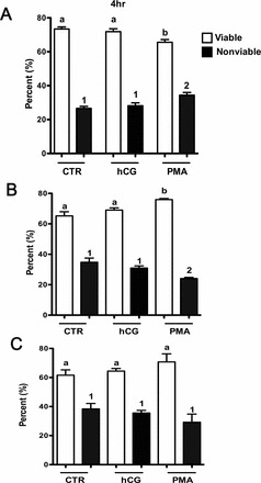
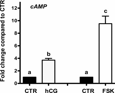
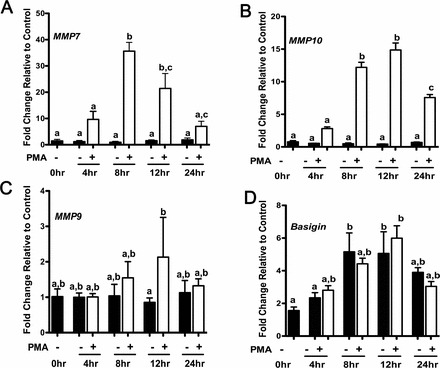
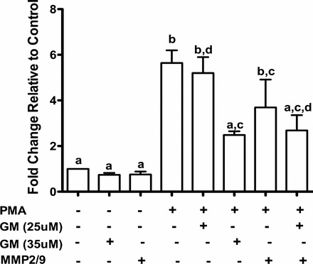
Postmenopausal women are at a higher risk of ovarian cancer due, in part, to increased levels of gonadotropins such as luteinizing hormone (LH). Gonadotropins and other stimuli are capable of activating two pathways, PKA and PKC, that are altered in ovarian cancer. To determine the role of LH on ovarian cancer, we explored the effects of human chorionic gonadotropin (hCG), an LH mimic, and an activator of the PKC pathway, phorbol-12-myristate 13-acetate (PMA), on ovarian cancer cell-cycle kinetics and apoptosis in Ovcar3 cells. PMA treatment increased cells in the S phase of the cell cycle and initially increased apoptosis after 4 h before diminishing apoptosis after 8 h. Treatment of ovarian cancer cells with hCG had no effect on these parameters. The PKC pathway is known to differentially regulate matrix metalloproteinase (MMP) expression. Results showed that ovarian cancer cells treated with PMA increased MMP7 and MMP10 mRNA levels after 8 h of treatment, and expression remained high after 12 h before decreasing at 24 h. The mRNA expression of extracellular matrix metalloproteinase inducer (BSG), an activator of MMPs, was unaffected by PMA. Due to the role that MMPs play in migration, we investigated the effect of PMA activation of MMPs on ovarian cancer cell migration. The use of the MMP inhibitor GM6001 blocked the increased migratory effects of PMA on ovarian cancer cells. Together, these studies show that activating the PKC pathway causes significant changes in cell cycle kinetics and selective expression of MMPs that are involved in enhancing ovarian cancer cell proliferation and migration.
Keywords: MMPs; cancer; gene expression; migration; ovary.
Figures
References
-
- Mandai M, Konishi I, Kuroda H, Fujii S. LH/hCG action and development of ovarian cancer—a short review on biological and clinical/epidemiological aspects. Mol Cell Endocrinol 2007; 269: 61 64. - PubMed
-
- Hillard PA. The 5-Minute Obstetrics & Gynecology Consult. Philadelphia: Lippincott Williams & Wilkins; 2008.
-
- Rossing MA, Daling JR, Weiss NS, Moore DE, Self SG. Ovarian tumors in a cohort of infertile women. N Engl J Med 1994; 331: 771 776. - PubMed
-
- Venn A, Watson L, Bruinsma F, Giles G, Healy D. Risk of cancer after use of fertility drugs with in-vitro fertilisation. Lancet 1999; 354: 1586 1590. - PubMed
-
- Venn A, Watson L, Lumley J, Giles G, King C, Healy D. Breast and ovarian cancer incidence after infertility and in vitro fertilisation. Lancet 1995; 346: 995 1000. - PubMed
Publication types
MeSH terms
Substances
Grants and funding
LinkOut - more resources
Full Text Sources
Other Literature Sources
Medical
Miscellaneous
