

Mild cognitive impairment in Parkinson's disease is associated with a distributed pattern of brain white matter damage
- PMID: 23843285
- PMCID: PMC6869219
- DOI: 10.1002/hbm.22302
Mild cognitive impairment in Parkinson's disease is associated with a distributed pattern of brain white matter damage
Abstract
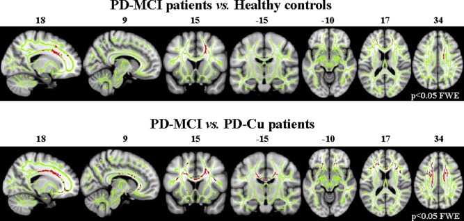
This study assesses the patterns of gray matter (GM) and white matter (WM) damage in patients with Parkinson's disease and mild cognitive impairment (PD-MCI) compared with healthy controls and cognitively unimpaired PD patients (PD-Cu). Three-dimensional T1-weighted and diffusion tensor (DT) magnetic resonance imaging (MRI) scans were obtained from 43 PD patients and 33 healthy controls. Cognition was assessed using a neuropsychological battery. Tract-based spatial statistics was applied to compare DT MRI indices between groups on a voxel-by-voxel basis. Voxel-based morphometry was performed to assess GM atrophy. Thirty PD patients were classified as MCI. Compared with healthy controls, PD-Cu and PD-MCI patients did not have GM atrophy. No region of WM damage was found in PD-Cu patients when compared with healthy controls. Relative to healthy controls and PD-Cu patients, PD-MCI patients showed a distributed pattern of WM abnormalities in the anterior and superior corona radiata, genu, and body of the corpus callosum, and anterior inferior fronto-occipital, uncinate, and superior longitudinal fasciculi, bilaterally. Subtle cognitive decline in PD is associated with abnormalities of frontal and interhemispheric WM connections, and not with GM atrophy. DT MRI might contribute to the identification of structural changes in PD-MCI patients prior to the development of dementia.
Keywords: MRI; Parkinson's disease; diffusion tensor MRI; mild cognitive impairment; white matter damage.
Copyright © 2013 Wiley Periodicals, Inc.
Figures
References
Publication types
MeSH terms
LinkOut - more resources
Full Text Sources
Other Literature Sources
Medical
