Review
. 2013 Aug;163C(3):144-56.
doi: 10.1002/ajmg.c.31369. Epub 2013 Jul 10.

Molecular mechanism of ventricular trabeculation/compaction and the pathogenesis of the left ventricular noncompaction cardiomyopathy (LVNC)

Affiliations
Review

Molecular mechanism of ventricular trabeculation/compaction and the pathogenesis of the left ventricular noncompaction cardiomyopathy (LVNC)

Wenjun Zhang et al. Am J Med Genet C Semin Med Genet. 2013 Aug.

Abstract

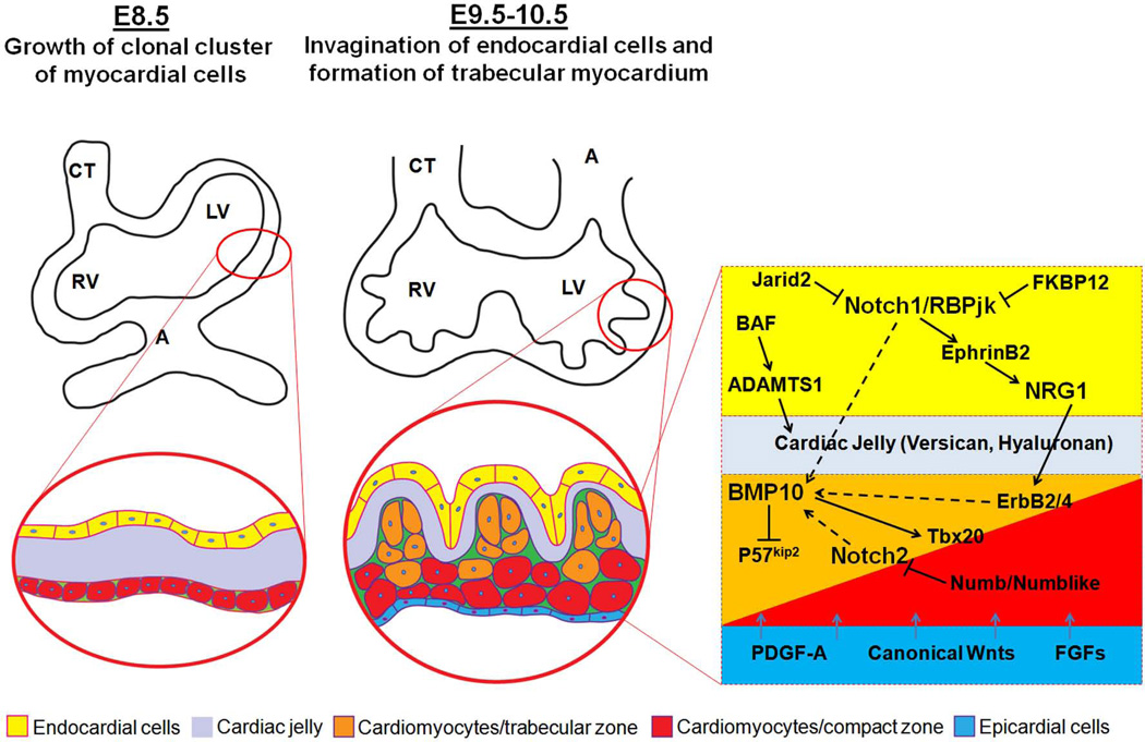
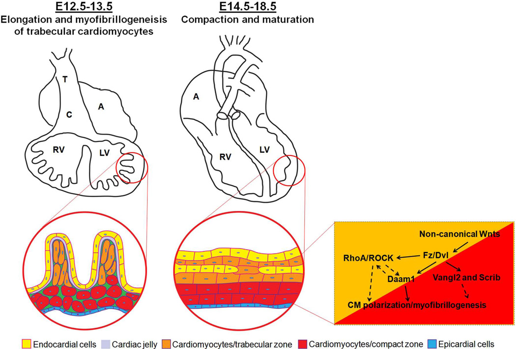
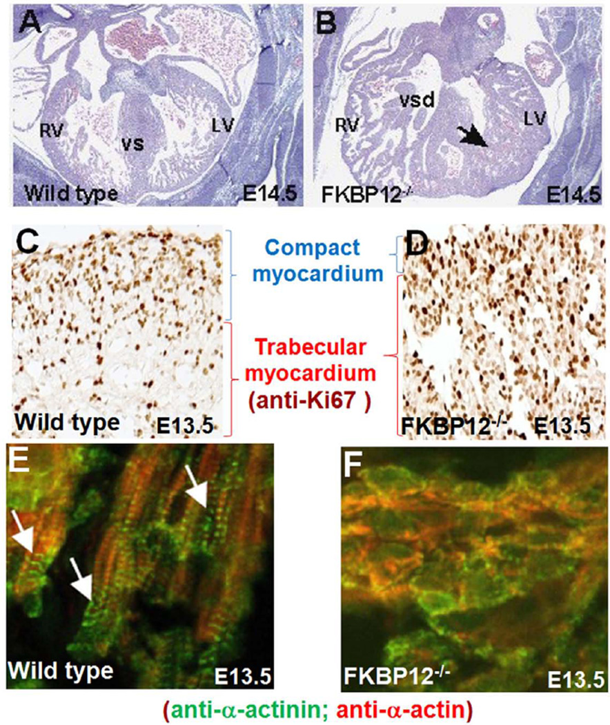
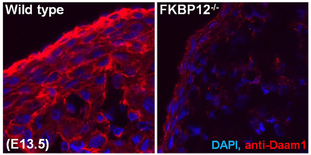
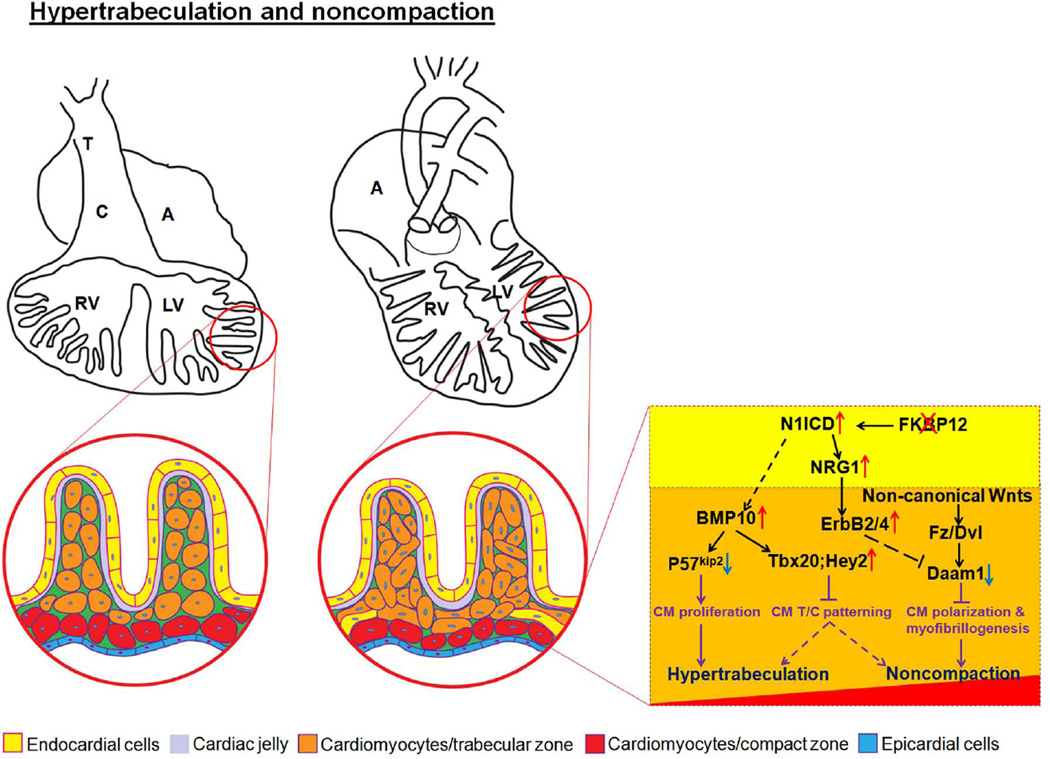
Ventricular trabeculation and compaction are two of the many essential steps for generating a functionally competent ventricular wall. A significant reduction in trabeculation is usually associated with ventricular compact zone deficiencies (hypoplastic wall), which commonly leads to embryonic heart failure and early embryonic lethality. In contrast, hypertrabeculation and lack of ventricular wall compaction (noncompaction) are closely related defects in cardiac embryogenesis associated with left ventricular noncompaction (LVNC), a genetically heterogenous disorder. Here we review recent findings through summarizing several genetically engineered mouse models that have defects in cardiac trabeculation and compaction.


Conflict of interest statement

The authors do not have conflict of interest to declare.

Figures

Figure 1
Figure 1
Early growth and development of ventricular wall. The interactive regulation of endocardial-, myocardial- and epicardial-derived growth factors and signaling networks is critical to the ventricular wall growth and trabeculation.
Figure 2
Figure 2
Ventricular wall trabeculation and compaction. Wnt/PCP signaling components are important in cardiomyocyte polarization and myofibrillogenesis and ventricular compaction.
Figure 3
Figure 3
Hypertrabeculation and noncompaction in FKBP12-deficient hearts. A and B, Cardiac histology of wild-type (A) and FKBP12-deficient heart (B) at E14.5. Black arrow denotes persistent trabecular myocardium in FKBP12 mutant heart. LV, left ventricle; RV, right ventricle; VS, ventricular septum; VSD, ventricular septal defect. C and D, Marked increase of cardiomyocyte proliferation in FKBP12 mutant heart. Immunohistochemical analysis of anti-Ki67 immune reactivity; the dark-brown nuclear signals are positive for Ki67, indicating proliferating cells. E and F, Disrupted cardiomyocyte polarization and myofibrillogenesis in FKBP12 mutant trabecular myocardium. Immunofluorescence staining using anti-α-actinin and anti-α-actin antibody, white arrows denote well organized sarcomeres in elongated normal trabecular cardiomyocytes.
Figure 4
Figure 4
Disrupted morphogenetic patterning of trabecular and compact myocardium. In situ hybridization analysis of Hey2 and Tbx20 expression; both Hey2 and Tbx20 expression are significantly higher in compact myocardium of wild-type hearts, but are expanded to trabecular myocardium in FKBP12 mutant hearts. Red arrows denote trabecular myocardium; blue arrows denote compact wall.
Figure 5
Figure 5
Daam1 down-regulated in FKBP12 mutant hearts (E14.5). Immunofluorescence staining using anti-Daam1 antibody.
Figure 6
Figure 6
Hypothetical model for the pathogenesis of ventricular hypertrabeculation and noncompaction. Two independent signaling pathways are associated with ventricular hypertrabeculation and noncompaction, in which abnormal regulation of cardiomyocyte proliferation (i.e., BMP10-mediated pathway) underlies the development of hypertrabeculation, while abnormal regulation of cardiomyocyte polarity and myofibrillogenesis (i.e., Daam1-mediated function) underlies the development of ventricular noncompaction.

References

    1. Anderson RH, Ho SY, Sanchez-Quintana D, Redmann K, Lunkenheimer PP. Heuristic problems in defining the three-dimensional arrangement of the ventricular myocytes. Anat Rec A Discov Mol Cell Evol Biol. 2006;288:579–586. - PubMed
    1. Anderson RH, Sanchez-Quintana D, Niederer P, Lunkenheimer PP. Structural-functional correlates of the 3-dimensional arrangement of the myocytes making up the ventricular walls. J Thorac Cardiovasc Surg. 2008;136:10–18. - PubMed
    1. Anderson RH, Sanchez-Quintana D, Redmann K, Lunkenheimer PP. How are the myocytes aggregated so as to make up the ventricular mass? Semin Thorac Cardiovasc Surg Pediatr Card Surg Annu. 2007:76–86. - PubMed
    1. Aras D, Tufekcioglu O, Ergun K, Ozeke O, Yildiz A, Topaloglu S, Deveci B, Sahin O, Kisacik HL, Korkmaz S. Clinical features of isolated ventricular noncompaction in adults long-term clinical course, echocardiographic properties, and predictors of left ventricular failure. J Card Fail. 2006;12:726–733. - PubMed
    1. Bao B, Zhang L, Hu H, Yin S, Liang Z. Deletion of a single-copy DAAM1 gene in congenital heart defect: a case report. BMC Med Genet. 2012;13:63. - PMC - PubMed

Publication types

MeSH terms

Substances

LinkOut - more resources