

Visualization of nigrosome 1 and its loss in PD: pathoanatomical correlation and in vivo 7 T MRI
- PMID: 23843466
- PMCID: PMC3775686
- DOI: 10.1212/WNL.0b013e31829e6fd2
Visualization of nigrosome 1 and its loss in PD: pathoanatomical correlation and in vivo 7 T MRI
Abstract
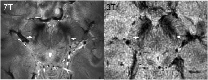
Objective: This study assessed whether high-resolution 7 T MRI allowed direct in vivo visualization of nigrosomes, substructures of the substantia nigra pars compacta (SNpc) undergoing the greatest and earliest dopaminergic cell loss in Parkinson disease (PD), and whether any disease-specific changes could be detected in patients with PD.
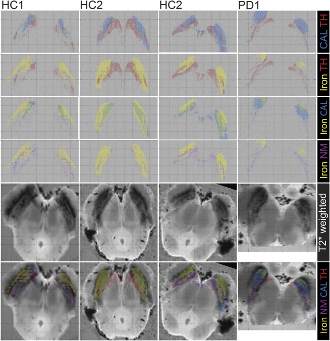
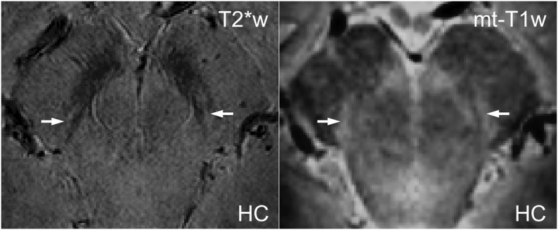
Methods: Postmortem (PM) midbrains, 2 from healthy controls (HCs) and 1 from a patient with PD, were scanned with high-resolution T2*-weighted MRI scans, sectioned, and stained for iron and neuromelanin (Perl), TH, and calbindin. To confirm the identification of nigrosomes in vivo on 7 T T2*-weighted scans, we assessed colocalization with neuromelanin-sensitive T1-weighted scans. We then assessed the ability to depict PD pathology on in vivo T2*-weighted scans by comparing data from 10 patients with PD and 8 age- and sex-matched HCs.
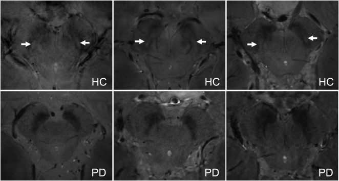
Results: A hyperintense, ovoid area within the dorsolateral border of the otherwise hypointense SNpc was identified in the HC brains on in vivo and PM T2*-weighted MRI. Location, size, shape, and staining characteristics conform to nigrosome 1. Blinded assessment by 2 neuroradiologists showed consistent bilateral absence of this nigrosome feature in all 10 patients with PD, and bilateral presence in 7/8 HC.
Conclusions: In vivo and PM MRI with histologic correlation demonstrates that high-resolution 7 T MRI can directly visualize nigrosome 1. The absence of nigrosome 1 in the SNpc on MRI scans might prove useful in developing a neuroimaging diagnostic test for PD.
Figures
Comment in
-
Visualization of nigrosome 1 and its loss in PD: pathoanatomical correlation and in vivo 7T MRI.Neurology. 2014 May 13;82(19):1752. doi: 10.1212/WNL.0000000000000398. Neurology. 2014. PMID: 24821935 No abstract available.
-
Author response.Neurology. 2014 May 13;82(19):1752. Neurology. 2014. PMID: 24936620 No abstract available.
References
-
- Lehéricy S, Sharman MA, Santos CLD, Paquin R, Gallea C. Magnetic resonance imaging of the substantia nigra in Parkinson's disease. Mov Disord 2012;27:822–830 - PubMed
-
- Auer DP. In vivo imaging markers of neurodegeneration of the substantia nigra. Exp Gerontol 2009;44:4–9 - PubMed
-
- Massey LA, Yousry TA. Anatomy of the substantia nigra and subthalamic nucleus on MR imaging. Neuroimaging Clin N Am 2010;20:7–27 - PubMed
-
- Haines DE. Fundamental Neuroscience for Basic and Clinical Applications. Philadelphia: Churchill Livingstone Elsevier; 2006
-
- Damier P, Hirsch EC, Agid Y, Graybiel AM. The substantia nigra of the human brain I: nigrosomes and the nigral matrix, a compartmental organization based on calbindin D 28K immunohistochemistry. Brain 1999;122:1421–1436 - PubMed
Publication types
MeSH terms
Substances
Grants and funding
LinkOut - more resources
Full Text Sources
Other Literature Sources
Medical
Miscellaneous