. 2013 Jul 10;33(28):11440-50.
doi: 10.1523/JNEUROSCI.1710-13.2013.

Rapid suppression of inhibitory synaptic transmission by retinoic acid

Affiliations

Rapid suppression of inhibitory synaptic transmission by retinoic acid

Federica Sarti et al. J Neurosci. .

Abstract

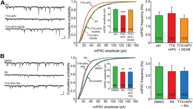
In brain, properly balanced synaptic excitation and inhibition is critically important for network stability and efficient information processing. Here, we show that retinoic acid (RA), a synaptic signaling molecule whose synthesis is activated by reduced neural activity, induces rapid internalization of synaptic GABAA receptors in mouse hippocampal neurons, leading to significant reduction of inhibitory synaptic transmission. Similar to its action at excitatory synapses, action of RA at inhibitory synapses requires protein translation and is mediated by a nontranscriptional function of the RA-receptor RARα. Different from RA action at excitatory synapses, however, RA at inhibitory synapses causes a loss instead of the gain of a synaptic protein (i.e., GABAARs). Moreover, the removal of GABAARs from the synapses and the reduction of synaptic inhibition do not require the execution of RA's action at excitatory synapses (i.e., downscaling of synaptic inhibition is intact when upscaling of synaptic excitation is blocked). Thus, the action of RA at inhibitory and excitatory synapses diverges significantly after the step of RARα-mediated protein synthesis, and the regulations of GABAAR and AMPAR trafficking are independent processes. When both excitatory and inhibitory synapses are examined together in the same neuron, the synaptic excitation/inhibition ratio is significantly enhanced by RA. Importantly, RA-mediated downscaling of synaptic inhibition is completely absent in Fmr1 knock-out neurons. Thus, RA acts as a central organizer for coordinated homeostatic plasticity in both excitatory and inhibitory synapses, and impairment of this overall process alters the excitatory/inhibitory balance of a circuit and likely represents a major feature of fragile X-syndrome.


Figures

Figure 1.
Figure 1.
RA mediates homeostatic downscaling of inhibitory synaptic transmission. A, Representative traces (left) and quantification of mIPSC amplitude (middle) and frequency (right) recorded from cultured rat hippocampal neurons treated with control vehicle, TTX + APV (24 h), or TTX + APV + DEAB (24 h). ***p < 0.001. B, Representative traces (left) and quantification (middle and right) of mIPSCs recorded from cultured neurons treated with DMSO, RA (1 μm, 0.5 h), or TTX + APV (24 h) followed by RA (0.5 h). **p < 0.01.
Figure 2.
Figure 2.
Downscaling of synaptic inhibition is independent of upscaling of synaptic excitation. A, Inhibitory synaptic scaling induced by TTX + CNQX (24 h). ***p < 0.001. B, Average mEPSC amplitude and frequency from cultured neurons overexpressing the C-terminal domain of GluR1 and treated with DMSO or 1 μm RA. ***p < 0.001. C, Average amplitude and frequency of mIPSCs from neurons overexpressing GluR1 C-terminal domain and treated with DMSO or 1 μm RA. ***p < 0.001.
Figure 3.
Figure 3.
Activity blockade or RA treatment led to a loss of total synaptic and surface GABAA receptors. A, Surface biotinylation assay for GABAAR β3 subunit in cultured hippocampal rat neurons treated with RA. **p < 0.01. B, Surface biotinylation assay for GABAAR β3 subunit in cultured neurons during activity blockade with TTX + APV. ***p < 0.0001. C, Immunolabeling of surface β2/3 subunit containing GABAARs (green) and VGAT (red) from 14 DIV rat hippocampal neurons treated with RA (1.5 h), TTX + APV (24 h), or TTX + CNQX (24 h). Scale bar, 10 μm. D, Quantification of synaptic GABAARβ2/3 punta (colocalized with VGAT). **p < 0.01. ***p < 0.001.
Figure 4.
Figure 4.
RA treatment enhances endocytosis of surface GABAARs. A, Representative images of endocytosis assay. Neurons were live-labeled with GABAAR β2/3 antibody, treated with DMSO or RA, and then fixed at different time points and stained for endocytosed (green) and remaining surface (red) GABAARs. Dendritic branches are labeled with MAP2 (blue). Scale bar, 10 μm. B, Quantification of remaining surface GABAAR (normalized to time 0) at different time points after DMSO or RA treatment (n/N = 25–34/3). *p < 0.05. C, Quantification of endocytosed GABAAR (normalized to time 0) at different time points after DMSO or RA treatment (n/N = 25–34/3). *p < 0.05. ***p < 0.001. D, mIPSC amplitude (top) and frequency (bottom) analysis from neurons overexpressing either wild-type dynamin-1 or the dominant-negative K44E, and treated with DMSO or RA. ***p < 0.0001. All graphs represent mean ± SEM. n/N indicates number of cells/number of independent experiments.
Figure 5.
Figure 5.
New protein synthesis and non-nuclear functions of RARα are required for downscaling of inhibitory synaptic transmission. A, Amplitude and frequency analysis of mIPSCs obtained from rat hippocampal neurons cotreated with transcription (actinomycin) or translation (anisomycin, cycloheximide) inhibitors and RA. **p < 0.01. B, Recording configuration for paired recordings of eIPSCs. Cultured hippocampal slices from RARα floxed mouse were infected with Cre-expressing lentivirus. Paired recordings of eIPSCs were obtained simultaneously from two neighboring CA1 pyramidal neurons: one infected (green), one uninfected (black). The stimulating electrodes were positioned in stratum radiatum as shown. C–F, Scatter plots of eIPSCs from individual pairs (gray circles) and group mean ± SEM (black squares) of simultaneously recorded neurons. Insets, Representative traces for infected neurons (green) and RARα floxed neurons (black) recorded after treatment with RA or DMSO. Neurons were infected with a lentiviral vector expressing the following: (C) Cre recombinase (to generate RARα KO neurons); (D) inactive recombinase ΔCre; (E) Cre and RARα LBD/F; and (F) Cre and RARα DBD. Calibration: 40 pA, 100 ms. n/N indicates number of cells/number of independent experiments.
Figure 6.
Figure 6.
RA induces downscaling of synaptic inhibition and increases excitation/inhibition ratio in acute hippocampal slices. A, Representative traces and amplitude analysis of mIPSC recordings obtained from CA1 pyramidal neurons of acute hippocampal slices incubated with DMSO or RA. ***p < 0.001. Calibration: 20 pA, 0.5 s. B, Frequency analysis of mIPSCs recorded in A. C, Example traces of 25 Hz five-pulse train evoked IPSCs and EPSCs recorded in the same wild-type CA1 pyramidal neurons treated with DMSO or RA. Calibration: 200 pA, 20 ms. D, Synaptic excitatory/inhibitory conductance ratio Ge/Gi (black line indicates mean; red or green shading indicates SEM) of the synaptic responses to train stimulation in wild-type neurons (n/N = 8/3). The peak of first pulse is cutoff at 1 for display purpose.
Figure 7.
Figure 7.
RA-mediated downscaling of synaptic inhibition is absent in Fmr1 KO neurons. A, Representative traces (top) and quantification (bottom) of mIPSCs recorded from WT or Fmr1 KO CA1 pyramidal neurons in cultured hippocampal slices treated with DMSO or TTX + CNQX. ***p < 0.001. B, Representative traces (top) and quantification (bottom) of mIPSCs recorded from WT or Fmr1 KO CA1 pyramidal neurons in cultured hippocampal slices treated with DMSO or RA. **p < 0.01. Calibration: 50 pA, 0.5 s. C, Example traces of 25 Hz five-pulse train evoked IPSCs and EPSCs recorded in the same Fmr1 KO CA1 pyramidal neurons treated with DMSO or RA. Calibration: 200 pA, 20 ms. D, Synaptic excitatory/inhibitory conductance ratio Ge/Gi (mean ± SEM) of the synaptic responses to train stimulation in Fmr1 KO neurons (n/N = 5/2).
Figure 8.
Figure 8.
RA alters neuronal excitability. A, Simulated EPSC/IPSC ratio in DMSO- or RA-treated wild-type neurons at resting membrane potential (−60 mV). EPSC/IPSC = (Vm − EEPSC)/(Vm − EIPSC) * (Ge/Gi), using Ge/Gi values obtained in Figure 6D. Gray area represents inhibition-dominating zone. For both treatments, synaptic excitation dominates at the beginning of each pulse because of large excitatory driving force (0 mV) and small inhibitory driving force (65 mV). B, Same simulation in wild-type neurons at membrane voltage just below action potential firing threshold (−50 mV). Because of a shift in excitatory and inhibitory driving force (50 and 15 mV, respectively), only synaptic responses in RA-treated neurons are consistently excitation-dominant. C, D, Simulation of EPSC/IPSC ratio in Fmr1 KO neurons at −60 mV and −50 mV. The effect of RA on boosting synaptic excitation is absent.

Similar articles

Cited by

References

    1. Aoto J, Nam CI, Poon MM, Ting P, Chen L. Synaptic signaling by all-trans retinoic acid in homeostatic synaptic plasticity. Neuron. 2008;60:308–320. doi: 10.1016/j.neuron.2008.08.012. - DOI - PMC - PubMed
    1. Brandon NJ, Delmas P, Kittler JT, McDonald BJ, Sieghart W, Brown DA, Smart TG, Moss SJ. GABAA receptor phosphorylation and functional modulation in cortical neurons by a protein kinase C-dependent pathway. J Biol Chem. 2000;275:38856–38862. doi: 10.1074/jbc.M004910200. - DOI - PubMed
    1. Brandon NJ, Jovanovic JN, Colledge M, Kittler JT, Brandon JM, Scott JD, Moss SJ. A-kinase anchoring protein 79/150 facilitates the phosphorylation of GABA(A) receptors by cAMP-dependent protein kinase via selective interaction with receptor beta subunits. Mol Cell Neurosci. 2003;22:87–97. doi: 10.1016/S1044-7431(02)00017-9. - DOI - PubMed
    1. Brandon N, Jovanovic J, Moss S. Multiple roles of protein kinases in the modulation of gamma-aminobutyric acid(A) receptor function and cell surface expression. Pharmacol Ther. 2002;94:113–122. doi: 10.1016/S0163-7258(02)00175-4. - DOI - PubMed
    1. Castillo PE, Chiu CQ, Carroll RC. Long-term plasticity at inhibitory synapses. Curr Opin Neurobiol. 2011;21:328–338. doi: 10.1016/j.conb.2011.01.006. - DOI - PMC - PubMed

Publication types

MeSH terms

LinkOut - more resources