

Proximodistal segregation of nonspatial information in CA3: preferential recruitment of a proximal CA3-distal CA1 network in nonspatial recognition memory
- PMID: 23843521
- PMCID: PMC6618684
- DOI: 10.1523/JNEUROSCI.4480-12.2013
Proximodistal segregation of nonspatial information in CA3: preferential recruitment of a proximal CA3-distal CA1 network in nonspatial recognition memory
Abstract
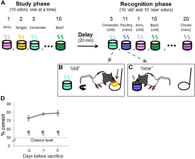
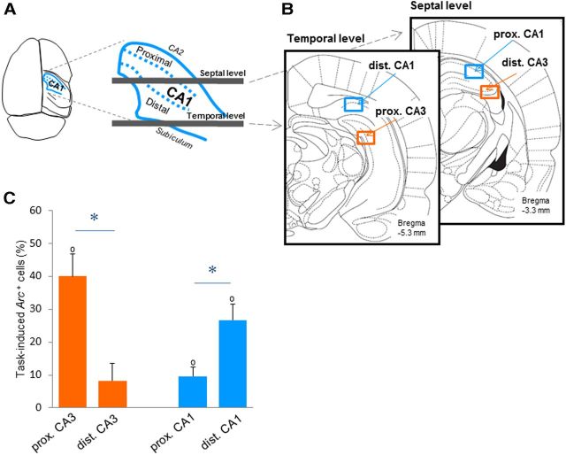
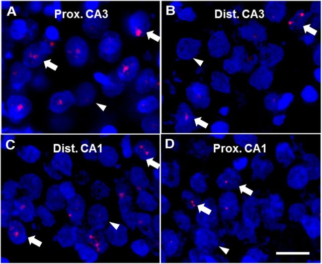
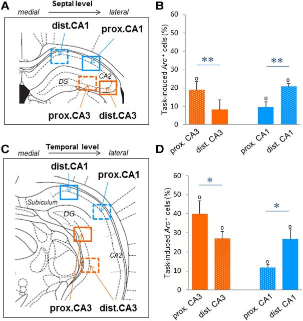
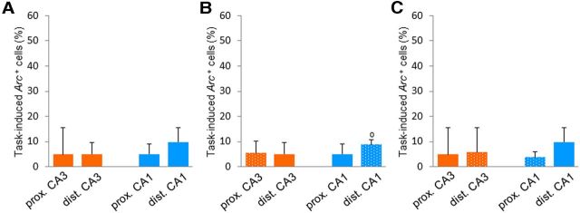
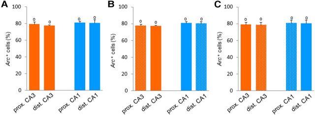
A prevailing view in memory research is that CA3 principally supports spatial processes. However, few studies have investigated the contribution of CA3 to nonspatial memory function. Interestingly, the proximal part of CA3 (close to the dentate gyrus) predominantly projects to distal CA1 (away from the dentate gyrus), which preferentially processes nonspatial information. Moreover, the cytoarchitecture and connectivity patterns in the proximal and distal parts of CA3 strongly differ, suggesting a functional segregation in this area. Here, we tested whether CA3 is recruited during nonspatial recognition memory, and whether nonspatial information is differentially represented along the proximodistal axis of CA3. Furthermore, we investigated whether the pattern of activation within CA3 would mirror that of CA1. We used a high-resolution imaging technique specifically designed to analyze brain activity in distant areas that is based on the detection of the expression of the immediate-early gene Arc, used as a marker of neuronal activation. We showed that proximal CA3 is strongly recruited during a nonspatial delayed nonmatching-to-sample recognition memory task in rats, while distal CA3 is not. In addition, distal CA1 was more activated than proximal CA1 in the same task. These findings suggest a functional segregation of CA3 that mirrors that of CA1, and potentially indicate the existence of a proximal CA3-distal CA1 hippocampal subnetwork that would preferentially process nonspatial information during recognition memory.
Figures
Similar articles
-
Spatial information is preferentially processed by the distal part of CA3: Implication for memory retrieval.Behav Brain Res. 2018 Nov 15;354:31-38. doi: 10.1016/j.bbr.2018.07.023. Epub 2018 Aug 8. Behav Brain Res. 2018. PMID: 30098839
-
Spatial information is preferentially processed by the distal part of CA3: implication for memory retrieval.Behav Brain Res. 2018 Jul 16;347:116-123. doi: 10.1016/j.bbr.2018.02.046. Epub 2018 Mar 5. Behav Brain Res. 2018. PMID: 29518437
-
Nonspatial sequence coding varies along the CA1 transverse axis.Behav Brain Res. 2018 Nov 15;354:39-47. doi: 10.1016/j.bbr.2017.10.015. Epub 2017 Oct 28. Behav Brain Res. 2018. PMID: 29107714 Free PMC article.
-
Mapping memory function in the medial temporal lobe with the immediate-early gene Arc.Behav Brain Res. 2013 Oct 1;254:22-33. doi: 10.1016/j.bbr.2013.04.048. Epub 2013 May 3. Behav Brain Res. 2013. PMID: 23648768 Review.
-
Functional differentiation in the transverse plane of the hippocampus: An update on activity segregation within the DG and CA3 subfields.Brain Res Bull. 2021 Jun;171:35-43. doi: 10.1016/j.brainresbull.2021.03.003. Epub 2021 Mar 13. Brain Res Bull. 2021. PMID: 33727088 Free PMC article. Review.
Cited by
-
Transversal functional connectivity and scene-specific processing in the human entorhinal-hippocampal circuitry.Elife. 2022 Oct 12;11:e76479. doi: 10.7554/eLife.76479. Elife. 2022. PMID: 36222669 Free PMC article.
-
Control of parallel hippocampal output pathways by amygdalar long-range inhibition.Elife. 2021 Nov 30;10:e74758. doi: 10.7554/eLife.74758. Elife. 2021. PMID: 34845987 Free PMC article.
-
Laminar activity in the hippocampus and entorhinal cortex related to novelty and episodic encoding.Nat Commun. 2014 Nov 26;5:5547. doi: 10.1038/ncomms6547. Nat Commun. 2014. PMID: 25424131 Free PMC article. Clinical Trial.
-
Integration of exteroceptive and interoceptive information within the hippocampus: a computational study.Front Syst Neurosci. 2015 Jun 5;9:87. doi: 10.3389/fnsys.2015.00087. eCollection 2015. Front Syst Neurosci. 2015. PMID: 26097448 Free PMC article.
-
Respiratory modulation of cognitive performance during the retrieval process.PLoS One. 2018 Sep 14;13(9):e0204021. doi: 10.1371/journal.pone.0204021. eCollection 2018. PLoS One. 2018. PMID: 30216372 Free PMC article.
References
-
- Andersen P, Bliss TV, Skrede KK. Lamellar organization of hippocampal pathways. Exp Brain Res. 1971;13:222–238. - PubMed
Publication types
MeSH terms
LinkOut - more resources
Full Text Sources
Other Literature Sources
Miscellaneous