. 2013 Jul 10;33(28):11703-14.
doi: 10.1523/JNEUROSCI.1631-13.2013.

Spinal μ and δ opioids inhibit both thermal and mechanical pain in rats

Affiliations

Spinal μ and δ opioids inhibit both thermal and mechanical pain in rats

Audrey Normandin et al. J Neurosci. .

Abstract

The expression and contribution of μ (MOPR) and δ opioid receptors (DOPR) in polymodal nociceptors have been recently challenged. Indeed, MOPR and DOPR were shown to be expressed in distinct subpopulation of nociceptors where they inhibit pain induced by noxious heat and mechanical stimuli, respectively. In the present study, we used electrophysiological measurements to assess the effect of spinal MOPR and DOPR activation on heat-induced and mechanically induced diffuse noxious inhibitory controls (DNICs). We recorded from wide dynamic range neurons in the spinal trigeminal nucleus of anesthetized rats. Trains of 105 electrical shocks were delivered to the excitatory cutaneous receptive field. DNICs were triggered either by immersion of the hindpaw in 49°C water or application of 300 g of mechanical pressure. To study the involvement of peptidergic primary afferents in the activation of DNIC by noxious heat and mechanical stimulations, substance P release was measured in the spinal cord by visualizing neurokinin type 1 receptor internalization. We found that the activation of spinal MOPR and DOPR similarly attenuates the DNIC and neurokinin type 1 receptor internalization induced either by heat or mechanical stimuli. Our results therefore reveal that the activation of spinal MOPR and DOPR relieves both heat-induced and mechanically induced pain with similar potency and suggest that these receptors are expressed on polymodal, substance P-expressing neurons.


Figures

Figure 1.
Figure 1.
Schematic representation of the electrophysiological experimental design. A, Sequences of 105 percutaneous electrical stimulations (0.66 Hz at 3 times the C-fiber threshold intensity) were applied to the excitatory receptive field. The DNICs were triggered by alternately immersing one hindpaw into a 49°C water bath (B) or applying a 300 g mechanical pressure (C) on the other hindpaw between the 36th and 60th electrical stimuli (corresponding to 37.5 s). DAMGO (5 μg in 10 μl) or Dlt II (8 μg in 10 μl) was injected to the lumbar spinal cord via an intrathecal catheter. B, C, Histograms showing the C-fiber-evoked responses of trigeminal WDR neurons to 105 successive electrical stimulations. B, Effect of the noxious heat stimulus on the C-fiber-evoked response of a trigeminal WDR neuron. C, Effect of the noxious mechanical stimulus on the C-fiber-evoked response of a trigeminal WDR neuron.
Figure 2.
Figure 2.
Intrathecal DAMGO and Dlt II both inhibit the DNIC induced by a noxious heat stimulus. A–C, E–G, Histograms showing representative C-fiber-evoked responses of two trigeminal WDR neurons to 105 successive electrical stimulations recorded before (A,E) and 10 min after intrathecal (i.t.) administration of DAMGO 5 μg (B) or 20 min after Dlt II 8 μg (F). Between the 36th and 60th stimulation, one hindpaw was immersed into a 49°C water bath. The DNICs triggered by heat noxious stimulation of the hindpaw are reduced after both DAMGO (B) and Dlt II (F) injection. The intravenous (i.v.) administration of naloxone (C) and naltrindole (G) reversed the effect of DAMGO and Dlt II, respectively. D, H, Graphic representation showing the mean (n = 8 for D, and n = 9 for H) percentage of inhibition of C-fiber-evoked action potentials before and 10–20 min and 30–40 min after the intrathecal injection of the opioids. The data are individually normalized to those before administration of the opioids. The selective MOPR and DOPR agonists significantly reduced the percentage of inhibition of C-fiber-evoked action potentials either 10–20 or 30–40 min after their administration. Naloxone significantly reversed those opioidergic-induced effects, and naltrindole significantly reversed the DOPR-mediated effect 10–20 min after Dlt II administration. **p < 0.01 (one-way ANOVA for repeated measures with Bonferroni's post hoc test). ***p < 0.001 (one-way ANOVA for repeated measures with Bonferroni's post hoc test). Error bars indicate the SEM.
Figure 3.
Figure 3.
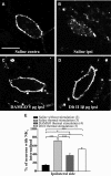
Intrathecal DAMGO and Dlt II reduce heat-induced NK1 receptor internalization. Internalization of NK1 receptors was induced by immersing the right hindpaw of male Sprague Dawley rats in a 49°C water bath for 38 s, and lamina I NK1 receptor-immunoreactive neurons were observed by immunofluorescence. The noxious heat stimulation was applied 10 min after intrathecal injection of saline (A,B), DAMGO 5 μg (C), or Dlt II 10 μg (D). Confocal images of neurons on the contralateral (A) and ipsilateral (B–D) sides of the spinal cord are shown. On the contralateral side of the saline-injected animals, immunolabeling of NK1 receptors appeared to be at the cell surface (A). However, on the ipsilateral side of the same animals, the noxious heat stimulation induced a significant increase in NK1 receptor internalization, as evidenced by the intensely labeled intracellular vesicle-like structures (B). When DAMGO (C) or Dlt II (D) was injected, a significant reduction in NK1 receptor internalization was observed on the ipsilateral side compared with the same side in saline-injected rats. The animals that had received a saline injection but no noxious stimulation had low basal proportions of neurons with internalized NK1 receptors. This result is illustrated in the graphic representation showing the percentage of neurons with NK1 receptor internalization induced by heat stimulation for ipsilateral side of the lumbar spinal cord (E). *p < 0.05 (one-way ANOVA with Bonferroni's post hoc test). ***p < 0.001 (one-way ANOVA with Bonferroni's post hoc test). The numbers in parentheses represent the number of animals per group. Error bars indicate the SEM. Scale bar: A, 30 μm.
Figure 4.
Figure 4.
Intrathecal DAMGO and Dlt II both inhibit the DNIC triggered by a noxious mechanical stimulus. A–C, E–G, Histograms showing representative C-fiber-evoked responses of two trigeminal WDR neurons to 105 successive electrical stimulations recorded before (A,E) and 30 min after intrathecal (i.t.) administration of DAMGO 5 μg (B) or 10 min after Dlt II 8 μg (F). Between the 36th and 60th stimulation, a 300 g mechanical pressure was applied on one hindpaw of the animal with calibrated forceps. The DNICs triggered by mechanical noxious stimulation of the hindpaw are reduced after both DAMGO (B) and Dlt II (F) injection. The intravenous (i.v.) administration of naloxone (C) and naltrindole (G) reversed the effect of DAMGO and Dlt II, respectively. D, H, Graphic representation showing the mean (n = 8 for D, and n = 9 for H) percentage of inhibition of C-fiber-evoked action potentials before and 10–20 min and 30–40 min after the intrathecal injection of the opioids. The data are individually normalized to those before administration of the drugs. The selective-MOPR agonist significantly reduced the percentage of inhibition of C-fiber-evoked action potentials either 10–20 or 30–40 min after its administration. Naloxone significantly reversed those opioidergic-induced effects (D). The selective-DOPR agonist significantly reduced the percentage of inhibition of C-fiber-evoked action potentials only 10–20 min after its administration and naltrindole significantly reversed this DOPR-mediated effect (D). *p < 0.05 (one-way ANOVA for repeated measures with Bonferroni's post hoc test). **p < 0.01 (one-way ANOVA for repeated measures with Bonferroni's post hoc test). ***p < 0.001 (one-way ANOVA for repeated measures with Bonferroni's post hoc test). Error bars indicate the SEM.
Figure 5.
Figure 5.
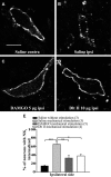
Intrathecal DAMGO and Dlt II reduce mechanical stimulus-induced NK1 receptor internalization. Internalization of NK1 receptors was induced by applying a 300 g mechanical pressure on the right hindpaw with calibrated forceps for 38 s. Lamina I NK1 receptor-immunoreactive neurons were observed by immunofluorescence. The noxious mechanical stimulation was applied 10 min after intrathecal injection of saline (A,B), DAMGO 5 μg (C), or Dlt II 10 μg (D). Confocal images showed that application of the mechanical pressure did not affect the cell-surface localization of NK1 receptors in the contralateral side of the lumbar spinal cord of saline-injected rats (A). In contrast, application of the same stimulus induced a strong internalization of NK1 receptors in the ipsilateral side for the same animal, as shown by the intracellular localization of the immunolabeling (B). The injection of DAMGO or Dlt II both significantly inhibited mechanically induced NK1 receptor internalization (E). For comparison purposes, data from the saline-injected group without noxious stimulation presented in Fig. 3E are reported in panel E. *p < 0.05 (one-way ANOVA with Bonferroni's post hoc test). **p < 0.01 (one-way ANOVA with Bonferroni's post hoc test). ***p < 0.001 (one-way ANOVA with Bonferroni's post hoc test). Values in parentheses represent the number of animals per group. Error bars indicate the SEM. Scale bar: A, 30 μm.
Figure 6.
Figure 6.
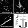
Heat-induced and mechanically induced NK1 receptor internalization in mice. In mice, internalization of NK1 receptors was induced by immersion of the right hindpaw for 38 s in a 49°C water bath or by applying (with calibrated forceps) a 200 g mechanical pressure on the right hindpaw, also for 38 s. Animals were perfused without receiving any noxious stimulation (A), 10 min after receiving the noxious thermal (B) or the noxious mechanical (C) stimulation. Lamina I NK1 receptor-immunoreactive neurons were observed by immunofluorescence. Confocal images showed that nonstimulated animals present a cell-surface localization of NK1 receptor labeling (A). Application of the thermal stimulus (B) or the mechanical pressure (C) both induces strong internalization of NK1 receptors in the ipsilateral side of the lumbar segment of the spinal cord (D). **p < 0.01 (one-way ANOVA with Bonferroni's post hoc test). ***p < 0.001 (one-way ANOVA with Bonferroni's post hoc test). Values in parentheses represent the number of animals per group. Error bars indicate the SEM. Scale bar: A, 30 μm.

Similar articles

Cited by

References

    1. Abbadie C, Trafton J, Liu H, Mantyh PW, Basbaum AI. Inflammation increases the distribution of dorsal horn neurons that internalize the neurokinin-1 receptor in response to noxious and non-noxious stimulation. J Neurosci. 1997;17:8049–8060. - PMC - PubMed
    1. Abrahamsen B, Zhao J, Asante CO, Cendan CM, Marsh S, Martinez-Barbera JP, Nassar MA, Dickenson AH, Wood JN. The cell and molecular basis of mechanical, cold, and inflammatory pain. Science. 2008;321:702–705. doi: 10.1126/science.1156916. - DOI - PubMed
    1. Allen BJ, Rogers SD, Ghilardi JR, Menning PM, Kuskowski MA, Basbaum AI, Simone DA, Mantyh PW. Noxious cutaneous thermal stimuli induce a graded release of endogenous substance P in the spinal cord: imaging peptide action in vivo. J Neurosci. 1997;17:5921–5927. - PMC - PubMed
    1. Arnér S, Meyerson BA. Lack of analgesic effect of opioids on neuropathic and idiopathic forms of pain. Pain. 1988;33:11–23. doi: 10.1016/0304-3959(88)90198-4. - DOI - PubMed
    1. Back SK, Lee J, Hong SK, Na HS. Loss of spinal μ-opioid receptor is associated with mechanical allodynia in a rat model of peripheral neuropathy. Pain. 2006;123:117–126. doi: 10.1016/j.pain.2006.02.017. - DOI - PubMed

Publication types

Substances