. 2013 Sep 15;126(Pt 18):4173-86.
doi: 10.1242/jcs.126102. Epub 2013 Jul 10.

14-3-3 protein targets misfolded chaperone-associated proteins to aggresomes

Affiliations

14-3-3 protein targets misfolded chaperone-associated proteins to aggresomes

Zhe Xu et al. J Cell Sci. .

Abstract

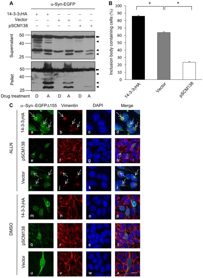
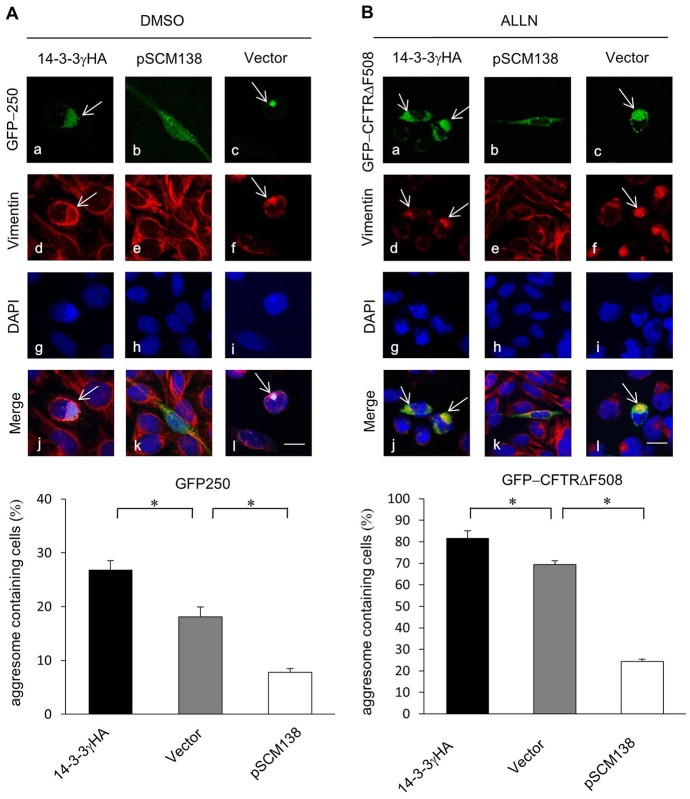
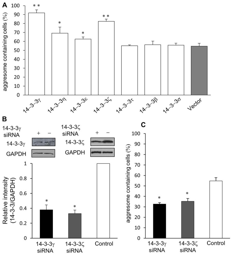
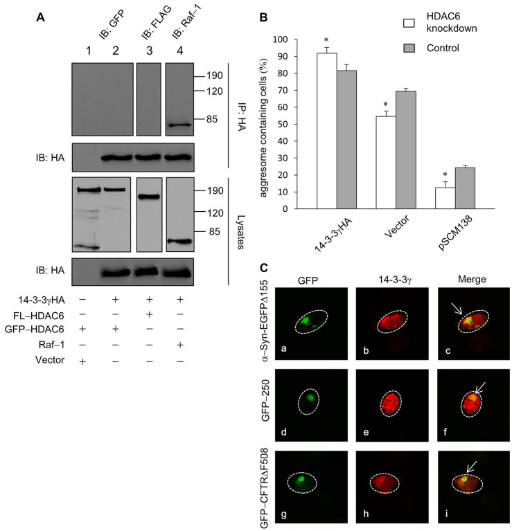
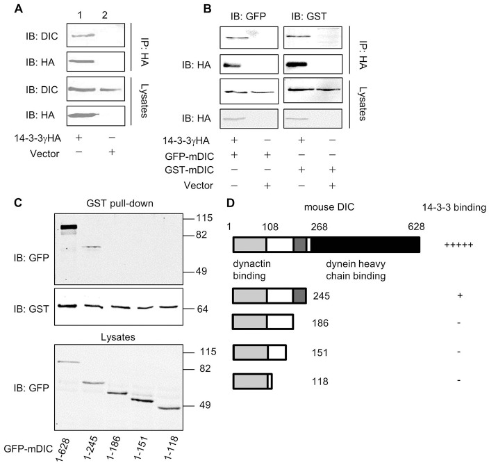
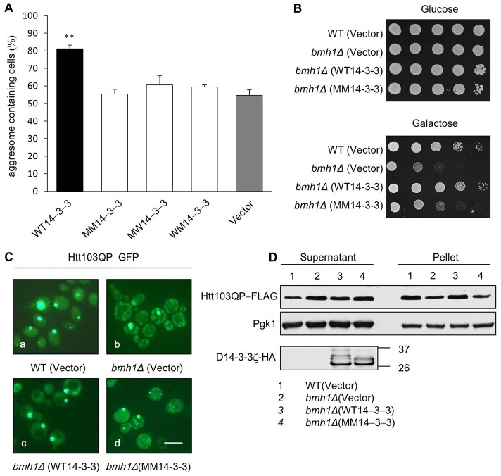
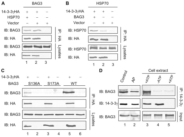
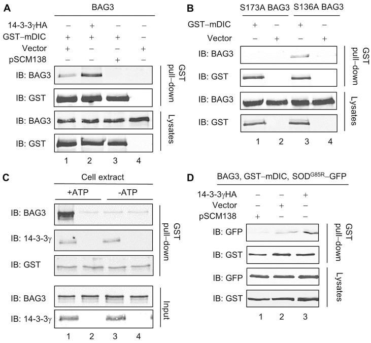
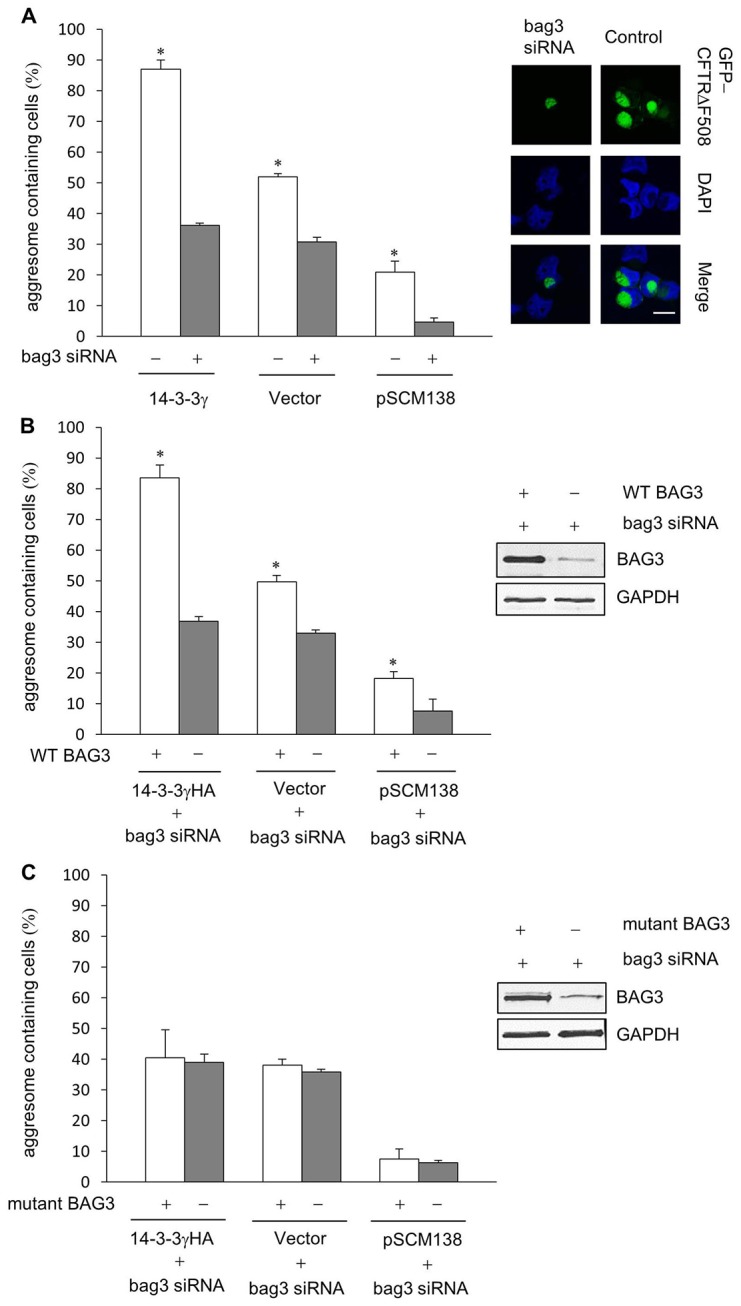
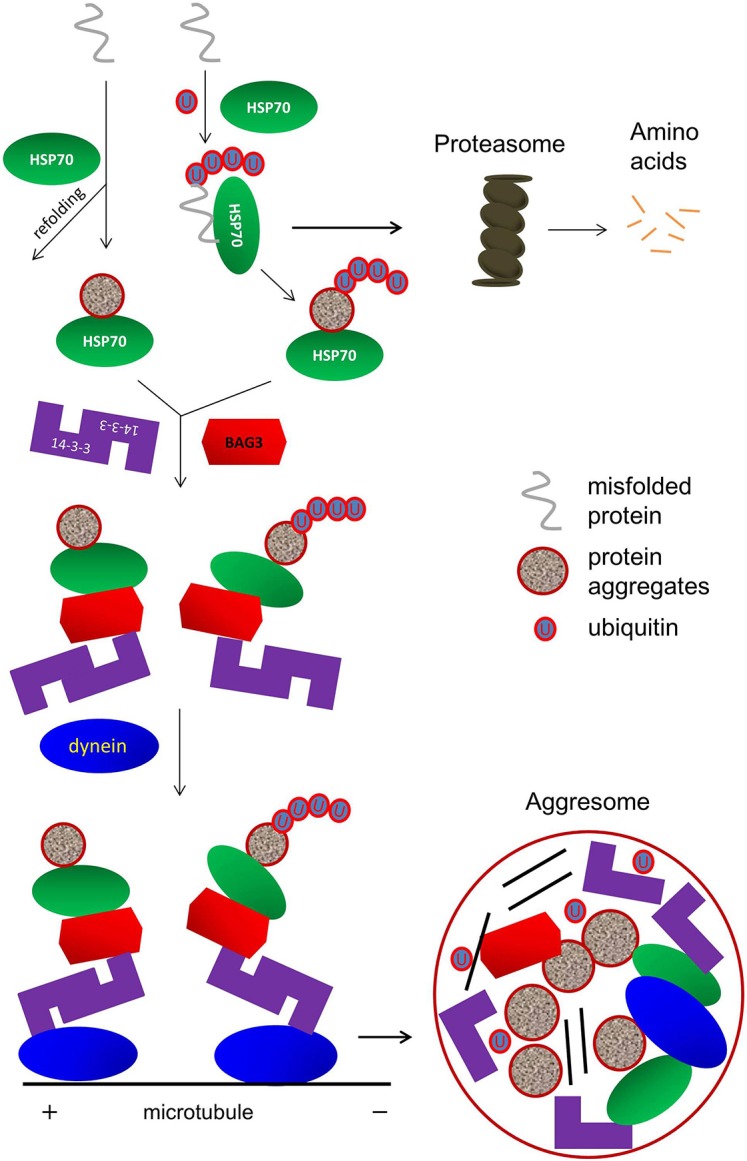
The aggresome is a key cytoplasmic organelle for sequestration and clearance of toxic protein aggregates. Although loading misfolded proteins cargos to dynein motors has been recognized as an important step in the aggresome formation process, the molecular machinery that mediates the association of cargos with the dynein motor is poorly understood. Here, we report a new aggresome-targeting pathway that involves isoforms of 14-3-3, a family of conserved regulatory proteins. 14-3-3 interacts with both the dynein-intermediate chain (DIC) and an Hsp70 co-chaperone Bcl-2-associated athanogene 3 (BAG3), thereby recruiting chaperone-associated protein cargos to dynein motors for their transport to aggresomes. This molecular cascade entails functional dimerization of 14-3-3, which we show to be crucial for the formation of aggresomes in both yeast and mammalian cells. These results suggest that 14-3-3 functions as a molecular adaptor to promote aggresomal targeting of misfolded protein aggregates and may link such complexes to inclusion bodies observed in various neurodegenerative diseases.

Keywords: 14-3-3; Adaptor; Aggresome; BAG3; Dynein.


Figures

Fig. 1.
Fig. 1.
14-3-3 promotes α-Syn aggresome formation. (A) α-Syn-EGFP was transfected into tsA201 cells together with 14-3-3γHA, empty vector or pSCM138. Followed by a treatment of either ALLN (proteasome inhibitor, 50 µM) (A) or DMSO (D) as control for 24 h, whole-cell lysates were separated into Triton X-100 soluble (supernatant) and insoluble (pellet) fractions, and samples from both fractions were analyzed by western blotting using antibody against α-Syn. Arrows and arrowheads denote full-length and truncated α-Syn-EGFP, respectively. (B) The percentage of cells bearing large α-Syn-containing inclusion bodies are shown in the bar graph (*P<0.05, n = 5). (C) Representative confocal images of CHO cells into which α-Syn-EGFPΔ155 was co-transfected with 14-3-3γHA, pSCM138 or a control vector. After treatment with either DMSO or ALLN for 24 h, cells were immunostained using specific antibodies against α-Syn (panels a, e, i, m, q and u; green) or vimentin (panels b, f, j, n, r and v; red), and nuclei were visualized by DAPI (panels c, g, k, o, s and w; blue). Superimposed images (panels d, h, l, p, t and x; merge) show colocalization of α-Syn-EGFPΔ155 and vimentin in aggresomes (indicated by arrows). Scale bar: 10 µm.
Fig. 2.
Fig. 2.
14-3-3 promotes aggresome formation of GFP-250 and GFP-CFTRΔF508. (A,B) Representative confocal images of CHO cells in which 14-3-3γHA, pSCM138 or a control vector was co-transfected with GFP-250 (A, panels a–l) or GFP-CFTRΔF508 (B, panels a–l). Cells transfected with GFP-CFTRΔF508 were treated with ALLN (50 µM) for 24 h. All cells were stained using anti-vimentin antibody (A, panels d–f; B, panels d–f; red) and DAPI (A, panels g–i; B, panels g–i; blue). Superimposed (merged) images show colocalization of vimentin and aggresomes (visualized through GFP) as indicated by arrows (A, panels j–l; B, panels j–l). Scale bars: 10 µm. Percentages of cells bearing GFP-250 and CFTRΔF508 aggresomes are shown in the lower bar graphs (*P<0.05, n = 5). Note that the green signals in pSCM138-co-transfected cells (A, panels b and k; B panels b and k) include GFP-250 or GFP-CFTRΔF508, as well as spectrally overlapping YFP-difopein that is minimally present in the aggregates.
Fig. 3.
Fig. 3.
14-3-3 exhibits isoform specificity in promoting aggresome formation. (A) Quantification of GFP-CFTRΔF508-induced aggresome formation in HDAC6-knockdown A549 cells co-transfected with one of the seven 14-3-3 isoforms (γ, η, ε, ζ, τ, β and σ). (B) tsA201 cells were transfected with siRNA oligonucleotids specific for either 14-3-3γ or 14-3-3ζ, followed by western blot analysis using antibodies as indicated. Quantification of levels of endogenous 14-3-3γ and 14-3-3ζ was done by comparing the amount of endogenous 14-3-3 proteins normalized with levels of GAPDH in control cells or those transfected with siRNAs targeting 14-3-3. (C) Percentages of cells bearing CFTRΔF508 induced aggresomes in control tsA201 cells and in those transfected with 14-3-3γ or ζ (*P<0.05, **P<0.001, n = 5).
Fig. 4.
Fig. 4.
14-3-3 is independent of HDAC6 and localizes to aggresomes. (A) Western blot analyses of immunoprecipitates and cell lysates from tsA201 cells in which 14-3-3γHA was co-transfected with a control vector (lane 1), GFP-HDAC6 (lane 2), FL-HDAC6 (lane 3) or Raf-1 (lane 4). 14-3-3 co-immunoprecipitates with a known 14-3-3 binding partner, Raf-1, but not with the HDAC6 protein. (B) Quantification of GFP-CFTRΔF508 aggresome formation in HDAC6-knockdown and control cells in % (*P<0.05, **P<0.001, n = 5). (C) Representative confocal images of tsA201 cells in which 14-3-3γHA was co-transfected with α-Syn-EGFPΔ155 (a, b and c), GFP-250 (d, e and f) or GFP-CFTRΔF508 (g, h and i). Cells were immunostained with an antibody against 14-3-3γ (b, e and h; red). Superimposed images (merge, c, f and i) show colocalization of 14-3-3γHA with several aggresomes as indicated by arrows. Dashed lines indicate the borders of respective cells.
Fig. 5.
Fig. 5.
14-3-3 interacts with dynein-intermediate chain. (A,B) 14-3-3 co-immunoprecipitates with both endogenous dynein-intermediate chain (DIC) (A) and exogenously transfected GFP-mDIC or GST-mDIC (B) in tsA201 cells. Exogenously expressed 14-3-3γHA was immunoprecipitated with an anti-HA antibody. (C) Deletion analyses reveal the region in DIC that is crucial for 14-3-3 binding. GFP-tagged full-length or C-terminal-truncated mDIC-deletion mutants were expressed in tsA201 cells. Their interactions with 14-3-3 were assessed in GST pull-down assays using GST-14-3-3, and in western blots (top panel). (D) Diagram that summarizes the effect of progressive truncation of the DIC C-terminus on its binding to 14-3-3.
Fig. 6.
Fig. 6.
Dimerization of 14-3-3 is required for aggresome formation. (A) Quantification of GFP-CFTRΔF508 induced aggresome formation in A549 HDAC6-knockdown cells that had been co-transfected with WT14-3-3, dimerization-deficient mutant MM14-3-3, MW14-3-3, WM14-3-3 or a control vector (**P<0.001, n = 5). (B) Expression of WT14-3-3 but not the dimerization-deficient mutant 14-3-3, suppresses the Htt103QP-induced growth defects in bmh1Δ cells. WT and bmh1Δ yeast cells with integrated PGALFLAG-Htt103QP-GFP plasmids were transformed with vector WT14-3-3 or MM14-3-3 plasmids. The saturated transformants were 10-fold diluted and then spotted on either glucose or galactose plates to examine the growth after 3-day incubation at 30 °C. (C) Representative fluorescence images of WT and bmh1Δ yeast cells with PGALFLAG-Htt103QP-GFP plasmids that were incubated in galactose medium. Transformation of WT14-3-3 (c), but not MM14-3-3 (d), restores aggresome formation in bmh1Δ cells. Aggresomes are shown as large green dots. Scale bar: 3 µm. (D) The yeast cells were incubated in galactose medium for 12 h, and separated into Triton X-100-soluble (supernatant) and -insoluble (pellet) fractions. Samples from both fractions were analyzed by western blotting using anti-FLAG (for Htt103QP) or anti-HA (for expressed WT and MM D14-3-3ζ–HA) antibody, respectively. Pgk1 was used as a loading control.
Fig. 7.
Fig. 7.
14-3-3 interacts with chaperone proteins Hsp70 and BAG3. Western blot analyses of 14-3-3 immunoprecipitates and cell lysates from tsA201 cells expressing various combinations of proteins as indicated. (A) BAG3 and 14-3-3γHA co-immunoprecipitate from co-transfected tsA201 cells (lane 2), and the extent of BAG3-14-3-3 interaction is not altered by exogenously expressed Hsp70 (lane 1). (B) Hsp70 co-immunoprecipitates with 14-3-3γHA in co-transfected cells (lane 2), but its binding to 14-3-3 is increased when co-transfected with BAG3 (lane 1). (C) Residues S173 and S136 in BAG3 are necessary for 14-3-3 binding. Compared with WT (lane 5), the S136A BAG3 has reduced binding to 14-3-3 (lane 1), whereas the S173A mutation abolishes the interaction completely (lane 3). (D) Interaction between 14-3-3 and BAG3 is regulated by phosphorylation. (Left panels) co-immunoprecipitation of 14-3-3 and BAG3 is reduced by alkaline phosphatase (AP) treatment of cell lysates. (Right panels) In vitro phosphorylation of recombinant BAG3 with crude tsA201 cell extract significantly enhances its binding to 14-3-3γ, as assessed by co-immunoprecipitation and western blotting. For controls, either ATP (lane 2) or recombinant 14-3-3γ (lane 3) was omitted.
Fig. 8.
Fig. 8.
14-3-3 is crucial for associations of BAG3 and cargos with dynein. Western blot analyses of GST pull-down assays and cell lysates from tsA201 cells expressing various combinations of proteins as indicated. (A) The BAG3–DIC association is enhanced by exogenously expressed 14-3-3γHA (lane 2), but eliminated by co-transfection of pSCM138 (lane 3). (B) Compared with WT BAG3, the S136A BAG3 mutant has a reduced binding to DIC, and the S173A BAG3 mutant does not co-precipitate with GST-mDIC. (C) Binding of recombinant GST-mDIC and BAG3 is enhanced by in vitro phosphorylation of BAG3 with tsA201 cell extract (lane 1). This effect is not observed when 14-3-3 (lane 2) or ATP (lanes 3, 4) is absent. (D) The association of SODG85R-GFP with GST-mDIC is enhanced by exogenously expressed 14-3-3γHA (lane 3), and virtually abolished when pSCM138 (lane 1) is co-transfected.
Fig. 9.
Fig. 9.
The interaction between 14-3-3 and BAG3 is required for aggresome formation. (A) Aggresome formation promoted by 14-3-3 is impaired in cells treated with siRNA targeting Bag3. The bar graph shows GFP-CFTRΔF508 aggresome formation in control cells (white bars) or BAG3-knockdown cells (gray bars) co-transfected with 14-3-3γHA, empty vector or pSCM138. Representative confocal images of aggresomes in control cells and cells treated with BAG3 siRNA are shown on the right. Scale bar: 10 µm. (B) Percentage of cells containing aggresomes in BAG3-knockdown cells that had BAG3 reintroduced through transfection of BAG3-siRNA-resistant wild-type BAG3 (WT BAG3). (C) Percentage of cells that contain aggresomes in cells that had been transfected with BAG3-siRNA (to knock down endogenous Bag3) and a 14-3-3-binding-deficient Bag3 mutant (S136A/S173A BAG3) that is resistant to BAG3 siRNA (mutant BAG3) (*P<0.05, n = 5). Insets in B and C show the levels of exogenously expressed BAG3-siRNA-resistant wild-type BAG3 (WT BAG3) and BAG3-siRNA-resistant 14-3-3-binding-deficient BAG3 (mutant BAG3), respectively, in cells treated with BAG3 siRNA.
Fig. 10.
Fig. 10.
Model for 14-3-3-mediated aggresome-targeting pathway. Several steps of this process are described in the Discussion.

Similar articles

Cited by

References

    1. Aitken A. (2002). Functional specificity in 14-3-3 isoform interactions through dimer formation and phosphorylation. Chromosome location of mammalian isoforms and variants. Plant Mol. Biol. 50, 993–1010 10.1023/A:1021261931561 - DOI - PubMed
    1. Boston P. F., Jackson P., Thompson R. J. (1982). Human 14-3-3 protein: radioimmunoassay, tissue distribution, and cerebrospinal fluid levels in patients with neurological disorders. J. Neurochem. 38, 1475–1482 10.1111/j.1471-4159.1982.tb07928.x - DOI - PubMed
    1. Chen H. K., Fernandez-Funez P., Acevedo S. F., Lam Y. C., Kaytor M. D., Fernandez M. H., Aitken A., Skoulakis E. M., Orr H. T., Botas J. et al.(2003). Interaction of Akt-phosphorylated ataxin-1 with 14-3-3 mediates neurodegeneration in spinocerebellar ataxia type 1. Cell 113, 457–468 10.1016/S0092-8674(03)00349-0 - DOI - PubMed
    1. Chin L. S., Olzmann J. A., Li L. (2010). Parkin-mediated ubiquitin signalling in aggresome formation and autophagy. Biochem. Soc. Trans. 38, 144–149 10.1042/BST0380144 - DOI - PMC - PubMed
    1. Datta S. R., Katsov A., Hu L., Petros A., Fesik S. W., Yaffe M. B., Greenberg M. E. (2000). 14-3-3 proteins and survival kinases cooperate to inactivate BAD by BH3 domain phosphorylation. Mol. Cell 6, 41–51 - PubMed

Publication types