. 2013 Sep 15;126(Pt 18):4136-46.
doi: 10.1242/jcs.123000. Epub 2013 Jul 10.

The ire-1 ER stress-response pathway is required for normal secretory-protein metabolism in C. elegans

Affiliations

The ire-1 ER stress-response pathway is required for normal secretory-protein metabolism in C. elegans

Modi Safra et al. J Cell Sci. .

Abstract

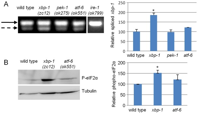
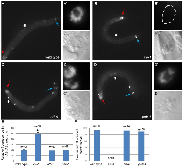
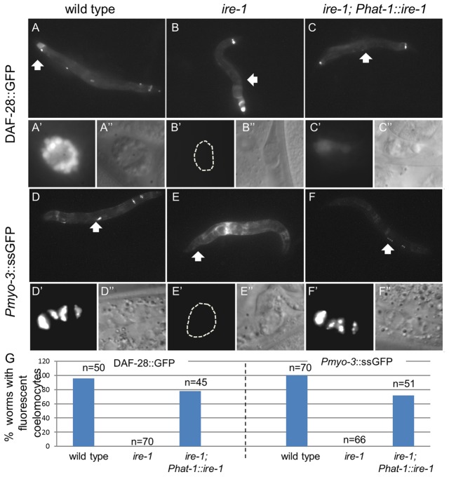
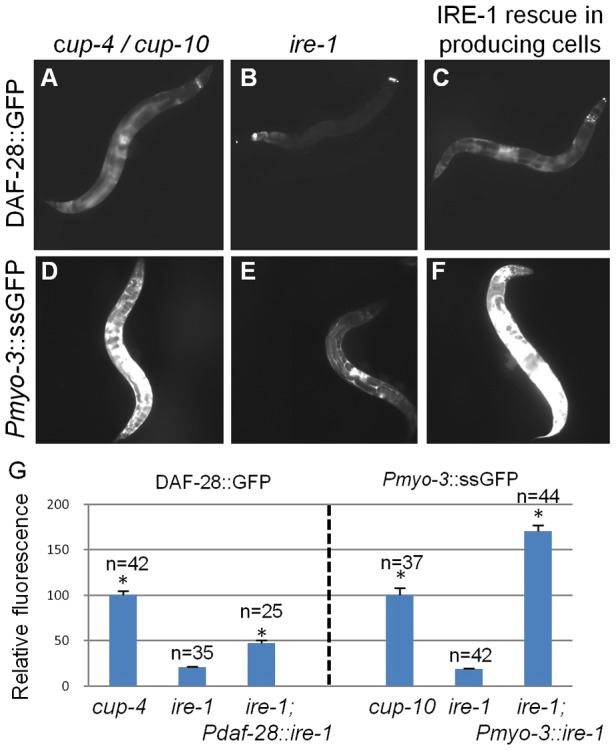
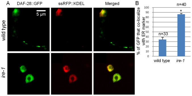
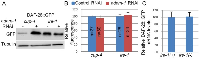
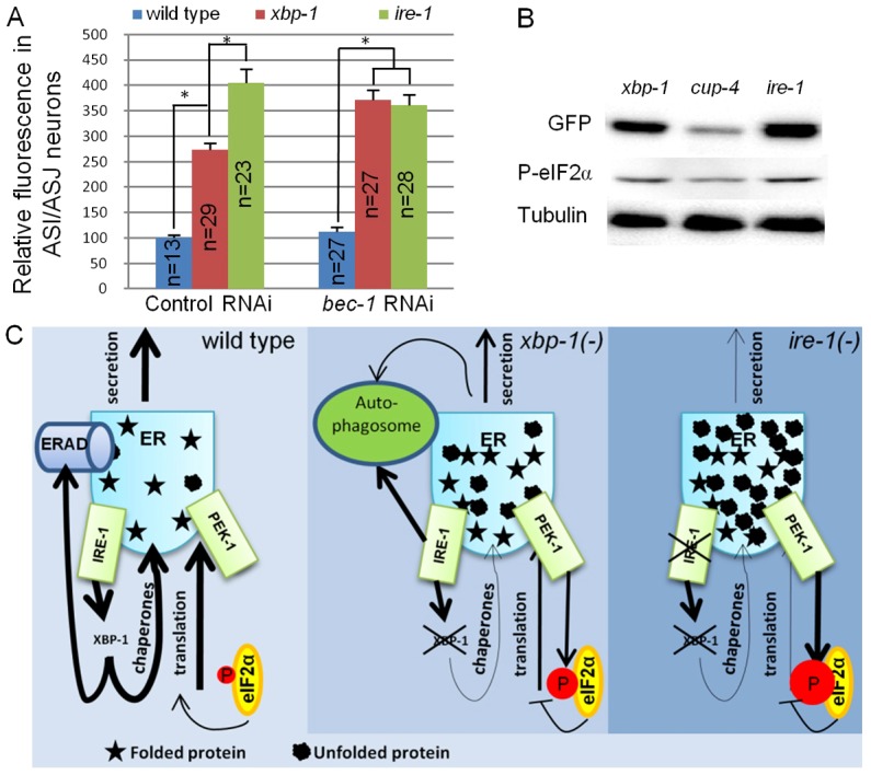
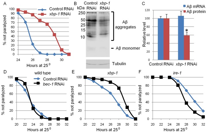
The unfolded protein response (UPR) allows cells to cope with endoplasmic reticulum (ER) stress by adjusting the capacity of the ER to the load of ER-associated tasks. The UPR is important for maintaining ER homeostasis under extreme ER stress. UPR genes are important under normal growth conditions as well, but what they are required for under these conditions is less clear. Using C. elegans, we show that the ire-1/xbp-1 arm of the UPR plays a crucial role in maintaining ER plasticity and function also in the absence of external ER stress. We find that during unstressed growth conditions, loss of ire-1 or xbp-1 compromises basic ER functions required for the metabolism of secreted proteins, including translation, folding and secretion. Notably, by compromising ER-associated degradation (ERAD) and phagocytosis, loss of ire-1 hinders the clearance of misfolded proteins from the ER as well as the clearance of proteins that were secreted into the pseudocoleom. Whereas the basal activity of the UPR is beneficial under normal conditions, it accelerates the pathology caused by toxic Aβ protein in a C. elegans model of Alzheimer's disease. Taken together, our findings indicate that UPR genes are critical for maintaining secretory protein metabolism under normal growth conditions.

Keywords: Alzheimer's disease; Caenorhabditis elegans; Coelomocytes; ER stress; ERAD; UPR.


Figures

Fig. 1.
Fig. 1.
xbp-1 deficiency activates the ire-1 and pek-1 arms of the UPR. (A) Representative steady-state RT-PCR products of unspliced xbp-1 mRNA (solid arrow) and spliced xbp-1 mRNA (dashed arrow) of day-1 animals of the indicated genotypes. The xbp-1 segment was amplified by a single set of PCR primers encompassing the putative intron region. Note that no spliced xbp-1 mRNA was detected in ire-1 mutants and that a basal level of spliced xbp-1 was detected in wild-type animals. The bar graph shows the normalized mean ratio of spliced/unspliced xbp-1 transcripts in three independent biological experiments. (B) Representative western blot of phosphorylated eIF2α and tubulin of day-1 wild-type, xbp-1 and atf-6 animals. Bar graph presents the normalized mean ratio of phosphorylated eIF2α levels relative to tubulin in three independent experiments. *P<0.05 of the indicated genotype relative to wild type (Student's t-test).
Fig. 2.
Fig. 2.
ire-1 deficiency alters DAF-28::GFP localization. (A–D) Representative fluorescence micrographs (original magnification, 100×) of day-3 wild-type, ire-1(ok799), atf-6(ok551) and pek-1(ok275) adults harboring an integrated DAF-28::GFP transgene. Blue arrows point to the ASI/ASJ neurons and red arrows indicate the hind gut. Wide white arrows point to coelomocytes, which are shown at high magnification in (A′–D′; fluorescence images) and (A′′–D′′; Nomarski images) (E) Quantification of fluorescence in ASI/ASJ neurons of the different strains, normalized to wild-type fluorescence levels. *P<0.0001 (Student's t-test). Similar results were obtained in two additional independent experiments. (F) Percentage of worms of the different genetic backgrounds in which fluorescent coelomocytes were detected. n, the number of animals analyzed. See supplementary material Fig. S2 for confocal images of DAF-28::GFP within the producing cells of wild-type animals and of ire-1, atf-6 or pek-1 mutants.
Fig. 3.
Fig. 3.
ire-1 is required cell-autonomously for coelomocyte function. Representative fluorescence micrographs (original magnification, 100×) of day-3 adults harboring an integrated DAF-28::GFP transgene (A–C) and of day-1 adults harboring an integrated Pmyo-3::ssGFP transgene (D–F). In ire-1(ok799) mutants no GFP-labeled coelomocytes were detected (B,E). Rescue of ire-1 in the coelomocytes, achieved by the expression of Phat-1::ire-1 (see supplementary material Fig. S5 for rescue transgene expression) restored GFP fluorescence in coelomocytes (C,F). Wide white arrows point at coelomocytes shown at higher magnification in A′–F′′ (original magnification, 630×); fluorescence (A′–F′) and Nomarski (A″–F″) photographs are presented. (G) Percentage of animals in which fluorescent coelomocytes were detected in the different genetic backgrounds. n, the number of animals analyzed.
Fig. 4.
Fig. 4.
ire-1 deficiency reduces accumulation of secreted proteins in the body cavity of coelomocyte-defective animals. Representative fluorescence micrographs (100×) of (A–C) day-3 adults harboring an integrated DAF-28::GFP transgene, and (D–F) day-1 adults harboring a Pmyo-3::ssGFP transgene. Coelomocyte-defective cup-4(ok837) (A) and cup-10(ar479) (D) mutants accumulate secreted proteins in their body cavities. Coelomocyte-defective ire-1(ok799) mutants accumulate significantly less GFP in their body cavities (B,E). Expressing ire-1 specifically in the producing cells of ire-1(ok799) mutants using the daf-28 or the myo-3 promoters (see supplementary material Fig. S5 for rescue transgene expression) restored accumulation of secreted proteins in the body cavity of ire-1 mutants (C,F). (G) Bar graph showing the relative mean fluorescence measured in the whole body of the animals of the indicated genotypes. n, the number of animals analyzed. Similar results were obtained in two additional independent experiments. *P<0.0001compared with ire-1 mutants (Student's t-test).
Fig. 5.
Fig. 5.
ire-1 deficiency traps DAF-28::GFP in the ER. (A) Representative fluorescence confocal micrographs (original magnification, 630×) of a 1.5-µm-thick section of ASI/ASJ neurons in day-3 wild type and ire-1 mutants co-expressing RFP (fused to a secretion signal and a KDEL-ER retention signal) and DAF-28::GFP. In ire-1 mutants most of the DAF-28::GFP colocalizes with the ER, whereas in wild-type animals the DAF-28::GFP is detected in the ER as well as in the axons. (B) Colocalization of DAF-28::GFP and the ER marker was quantified. n, the number of cells analyzed. *P<0.0001 (Student's t-test).
Fig. 6.
Fig. 6.
Misfolded DAF-28::GFP accumulates in ire-1 mutants. (A) Representative western blot of GFP and tubulin of coelomocyte-defective day-2 cup-4 and ire-1(ok799) mutants harboring an integrated DAF-28::GFP transgene. Animals were treated with control RNAi or edem-1 RNAi. edem-1 inactivation increased DAF-28::GFP levels in cup-4 mutants, but not in ire-1 mutants, whose basal DAF-28::GFP levels were high even before edem-1 inactivation. (B) Bar graph showing the relative mean whole-body fluorescence of DAF-28::GFP in day-2 cup-4 or ire-1 mutants treated with control RNAi (blue) or edem-1 RNAi (red). Fluorescence was calculated relative to control RNAi treatment of each strain. edem-1 inactivation did not increase DAF-28::GFP fluorescence. n, the number of animals analyzed. Similar results were obtained in two additional independent experiments. (C) Bar graph showing the average relative mRNA levels of daf-28::gfp in ire-1(+); cup-4(−) and ire-1(−) mutants, measured by qRT-PCR in three independent experiments. daf-28::gfp mRNA levels were not increased in ire-1 mutants (P>0.45, Student's t-test).
Fig. 7.
Fig. 7.
DAF-28::GFP accumulation in ASI/ASJ neurons is enhanced in ire-1 mutants and is bec-1 dependent. (A) Bar graph showing the relative mean fluorescence of DAF-28::GFP measured within the ASI/ASJ DAF-28::GFP-producing cells in wild type, xbp-1(tm2457) and ire-1(ok799) mutants treated with control or bec-1 RNAi. n, the number of animals analyzed. Similar results were obtained in two additional independent experiments. *P<0.0001 (Student's t-test). (B) Representative western blot of DAF-28::GFP, phosphorylated eIF2α and tubulin of day-1 xbp-1, cup-4 and ire-1 mutants expressing the daf-28::gfp transgene. (C) Model for disruption of ER homeostasis in ire-1 and xbp-1 mutants. Normally, xbp-1 transcribes target genes that promote ER homeostasis by preventing the accumulation of misfolded proteins in the ER. In xbp-1-deficient animals, misfolded proteins are not cleared by ERAD. These accumulate and activate IRE-1. ire-1 cannot activate xbp-1. Instead, it induces autophagosomes that clear some of the misfolded proteins from the ER. In ire-1-deficient animals, misfolded proteins accumulate in the ER but the ERAD and ire-1-induced autophagosomes do not clear them. High levels of misfolded proteins burden the ER, activate the remaining UPR arms and interfere with protein secretion.
Fig. 8.
Fig. 8.
xbp-1 inactivation reduces Aβ levels and delays paralysis. Eggs from animals expressing inducible Aβ in their muscles were grown at the permissive temperature and treated with control RNAi, xbp-1 RNAi or bec-1 RNAi for 48 hours. Animals were then shifted to the non-permissive temperature and Aβ expression was induced. (A) Paralyzed animals were scored each hour between 24 to 31 hours after temperature shift. xbp-1 RNAi significantly delayed paralysis of Aβ-expressing animals (control RNAi: mean = 25.9 hours; n = 124; xbp-1 RNAi: mean = 28.7 hours, n = 134; P<0.0001) (B) Representative western blots of Aβ (top panel) and tubulin (bottom panel) of Aβ-expressing animals treated with control RNAi and xbp-1 RNAi. Most of the Aβ protein was detected in high-molecular-mass aggregates. A very weak Aβ band was in a monomeric form. (C) Bar graph showing the average relative protein and mRNA levels of Aβ in control and xbp-1-RNAi-treated animals measured by qRT-PCR (blue bars) and western blot (red bars). Measurements were normalized to actin mRNA and tubulin protein levels, respectively. Values are the average of three independent experiments. *P>0.05, Student's t-test. (D–F) Paralyzed animals were scored every 2 hours between 20 and 32 hours after temperature shift. (D) bec-1 RNAi did not delay paralysis of wild-type Aβ-expressing animals (control RNAi: mean = 24.1 hours; n = 600; bec-1 RNAi: mean = 24.2 hours, n = 317; P = 0.27). (E) bec-1 RNAi significantly delayed paralysis of Aβ-expressing xbp-1 mutants (control RNAi: mean = 27.4 hours, n = 577; bec-1 RNAi: mean = 29.6 hours, n = 488; P<0.0001). (F) bec-1 RNAi did not delay paralysis of Aβ-expressing ire-1 mutants (control RNAi: mean = 30.4 hours, n = 380; bec-1 RNAi: mean = 28.8 hours, n = 250; P<0.0001).

Similar articles

Cited by

References

    1. Bicknell A. A., Babour A., Federovitch C. M., Niwa M. (2007). A novel role in cytokinesis reveals a housekeeping function for the unfolded protein response. J. Cell Biol. 177, 1017–1027 10.1083/jcb.200702101 - DOI - PMC - PubMed
    1. Brenner S. (1974). The genetics of Caenorhabditis elegans. Genetics 77, 71–94 - PMC - PubMed
    1. Calfon M., Zeng H., Urano F., Till J. H., Hubbard S. R., Harding H. P., Clark S. G., Ron D. (2002). IRE1 couples endoplasmic reticulum load to secretory capacity by processing the XBP-1 mRNA. Nature 415, 92–96 10.1038/415092a - DOI - PubMed
    1. Fares H., Greenwald I. (2001). Genetic analysis of endocytosis in Caenorhabditis elegans: coelomocyte uptake defective mutants. Genetics 159, 133–145 - PMC - PubMed
    1. Fonseca S. G., Urano F., Weir G. C., Gromada J., Burcin M. (2012). Wolfram syndrome 1 and adenylyl cyclase 8 interact at the plasma membrane to regulate insulin production and secretion. Nat. Cell Biol. 14, 1105–1112 10.1038/ncb2578 - DOI - PMC - PubMed

Publication types

MeSH terms

Substances