Review
. 2013 Jul 15;126(Pt 14):2979-89.
doi: 10.1242/jcs.079475. Epub 2013 Jul 10.

Signaling inputs to invadopodia and podosomes

Affiliations
Review

Signaling inputs to invadopodia and podosomes

Daisuke Hoshino et al. J Cell Sci. .

Abstract

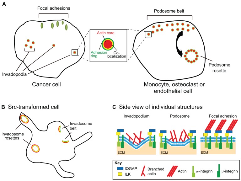
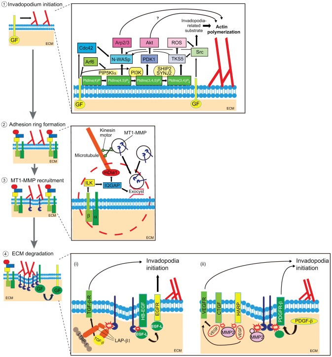
Remodeling of extracellular matrix (ECM) is a fundamental cell property that allows cells to alter their microenvironment and move through tissues. Invadopodia and podosomes are subcellular actin-rich structures that are specialized for matrix degradation and are formed by cancer and normal cells, respectively. Although initial studies focused on defining the core machinery of these two structures, recent studies have identified inputs from both growth factor and adhesion signaling as crucial for invasive activity. This Commentary will outline the current knowledge on the upstream signaling inputs to invadopodia and podosomes and their role in governing distinct stages of these invasive structures. We discuss invadopodia and podosomes as adhesion structures and highlight new data showing that invadopodia-associated adhesion rings promote the maturation of already-formed invadopodia. We present a model in which growth factor stimulation leads to phosphoinositide 3-kinase (PI3K) activity and formation of invadopodia, whereas adhesion signaling promotes exocytosis of proteinases at invadopodia.

Keywords: Actin cytoskeleton; Adhesion; Growth factor; Invadopodia; PI3K; Podosome; phosphoinositide 3-kinase.


Figures

Fig. 1.
Fig. 1.
Structural features of invadopodia, podosomes and focal adhesions. (A) Comparison of invasive adhesion structures in cancer and normal cells. Focal adhesions (green ovals in figure) are streak-like structures consisting of >150 molecular components and >700 direct interactions (Zaidel-Bar and Geiger, 2010). In general, they serve as mechanical and signaling connections between the actin cytoskeleton and ECM-bound integrins. This occurs through multiple interactions between scaffolding and signaling proteins, integrins and the actin cytoskeleton. Invadopodia and podosomes (shown as circular structures in the figure) are punctate branched actin-rich structures defined, in part, by the presence of a substantial amount of actin regulatory proteins, such as the Arp2/3 complex, cortactin and N-WASp, along with TKS5, active Src kinase, tyrosine phosphorylated proteins and the proteinase MT1-MMP (Murphy and Courtneidge, 2011; Weaver, 2006). Recent proteomic studies have respectively identified ∼200 putative podosome components and ∼60 putative invadopodia components (Attanasio et al., 2011; Cervero et al., 2012). Although invadopodia-producing cells generally only form punctate actin structures, podosome puncta can assemble into higher-order structures, such as podosome rosettes and podosome belts (the double layer of podosomes at cell periphery). Podosome belts can go on to form the sealing zone in osteoclasts. Both podosomes and invadopodia contain adhesion proteins and are surrounded by adhesion rings that that typically contain vinculin and paxillin (green rings in figure). Actin filaments radiate from the inner puncta and are likely to connect to the outer adhesion ring (yellow overlap) (Luxenburg et al., 2007). Adhesion proteins have also been localized to actin puncta (Alexander et al., 2008; Bowden et al., 1999; Mueller et al., 1999). (B) Src-transformed cells also form invadopodia- and podosome-like structures termed invadosomes. Invadosomes can organize into a larger rosette that consists of giant actin rings with colocalized adhesion molecules (Brábek et al., 2004). By light microscopy, invadosome rosettes probably best resemble osteoclast podosome belts which form at the periphery (Destaing et al., 2003) or sealing zones which are highly specialized structures formed by fusion of individual podosomes (Luxenburg et al., 2007); however, it is unclear at this point how closely rosette formation by Src-transformed cells resembles the osteoclast sealing ring formation. Owing to their robust formation of invadosomes, Src-transformed cells are extremely useful as a model for studying the basic core machinery that is common to invadopodia and podosomes. However, it is likely that the presence of constitutively active Src inherently bypasses and/or alters some upstream signals that would otherwise come from growth factors and/or adhesions. (C) Side view of invasive adhesion structures. Adhesion components include integrins, IQGAP and ILK, along with typical markers such as vinculin.
Fig. 2.
Fig. 2.
A model of invadopodia and podosome stages. (1) Invadopodia/podosome initiation. Growth factors (GF) signaling via GF receptors (yellow bars) leads to invadopodia initiation and actin polymerization (branched actin filaments shown in red). The inset on the right illustrates a proposed scheme of synergistic molecular interactions that lead to invadopodia/podosome initiation. Signals from activated lipid and protein kinases and small GTPases converge on N-WASp which activates the Arp2/3 complex to promote actin assembly. (2, 3) Adhesion ring formation and vesicle capture. The next step is the formation of the adhesion ring (indicated by molecules in the red dashed circle, especially integrins and ILK, as shown in the enlarged scheme to the right), which helps to capture vesicles (black circles) that contain proteinases, such as MT1-MMP (dark blue). The inset illustrates key molecules involved in vesicle capture at invadopodia/podosomes, including the formin mDia1, ILK, IQGAP and the exocyst complex (Branch et al., 2012; Liu et al., 2009; Lizárraga et al., 2009; Sakurai-Yageta et al., 2008). (4) ECM degradation and signaling feedback. In addition to ECM degradation, proteinase activity at invadopodia and podosomes might provide signal feedback to affect either the lifetime of existing invadosomes or formation of new invadosomes. (i) An enlarged model of potential signal feedback from proteinase activity. MT1-MMP can release growth factors (indicated by the red flash) by multiple mechanisms, including cleavage of the TGFβ-binding protein, latency-associated protein (red, LAP-β1 complex) to release active TGF-β1 (Mu et al., 2002). In addition, MT1-MMP also cleaves HB-EGF, which then releases an EGF-like domain (EGF-L) that could activate EGFR and trigger invadosome initiation (Díaz et al., 2013; Hayes et al., 2012; Koshikawa et al., 2010). (ii) MT1-MMP also cleaves other proteinases such as MMP2 (Sato et al., 1994). MMP2 is reported to disrupt vascular endothelial growth factor (VEGF)–heparin affin regulatory peptide (HARP) and VEGF–connective tissue growth factor (CTGF) angiogenic inhibitory complexes, which might release VEGF (Dean et al., 2007) and then trigger invadosome initiation (Lucas et al., 2010). Finally, MT1-MMP activation of PDGFR signaling (Lehti et al., 2005) is another potential mechanism that could promote invadosome formation (Eckert et al., 2011).

Similar articles

Cited by

References

    1. Abram C. L., Seals D. F., Pass I., Salinsky D., Maurer L., Roth T. M., Courtneidge S. A. (2003). The adaptor protein fish associates with members of the ADAMs family and localizes to podosomes of Src-transformed cells. J. Biol. Chem. 278, 16844–16851 10.1074/jbc.M300267200 - DOI - PubMed
    1. Alexander N. R., Branch K. M., Parekh A., Clark E. S., Iwueke I. C., Guelcher S. A., Weaver A. M. (2008). Extracellular matrix rigidity promotes invadopodia activity. Curr. Biol. 18, 1295–1299 10.1016/j.cub.2008.07.090 - DOI - PMC - PubMed
    1. Artym V. V., Zhang Y., Seillier-Moiseiwitsch F., Yamada K. M., Mueller S. C. (2006). Dynamic interactions of cortactin and membrane type 1 matrix metalloproteinase at invadopodia: defining the stages of invadopodia formation and function. Cancer Res. 66, 3034–3043 10.1158/0008-5472.CAN-05-2177 - DOI - PubMed
    1. Artym V. V., Yamada K. M., Mueller S. C. (2009). ECM degradation assays for analyzing local cell invasion. Methods Mol. Biol. 522, 211–219 10.1007/978-1-59745-413-1_15 - DOI - PubMed
    1. Artym V. V., Matsumoto K., Mueller S. C., Yamada K. M. (2011). Dynamic membrane remodeling at invadopodia differentiates invadopodia from podosomes. Eur. J. Cell Biol. 90, 172–180 10.1016/j.ejcb.2010.06.006 - DOI - PMC - PubMed

Publication types

Substances

LinkOut - more resources