Review
. 2013 Jul 15;126(Pt 14):2965-78.
doi: 10.1242/jcs.093609. Epub 2013 Jul 10.

Interactions between sarco-endoplasmic reticulum and mitochondria in cardiac and skeletal muscle - pivotal roles in Ca²⁺ and reactive oxygen species signaling

Affiliations
Review

Interactions between sarco-endoplasmic reticulum and mitochondria in cardiac and skeletal muscle - pivotal roles in Ca²⁺ and reactive oxygen species signaling

Verónica Eisner et al. J Cell Sci. .

Abstract

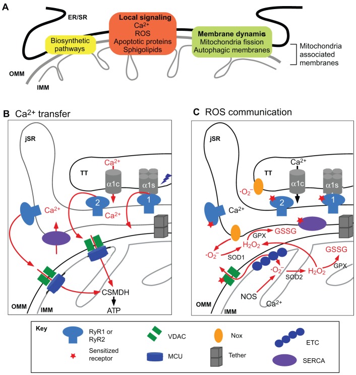
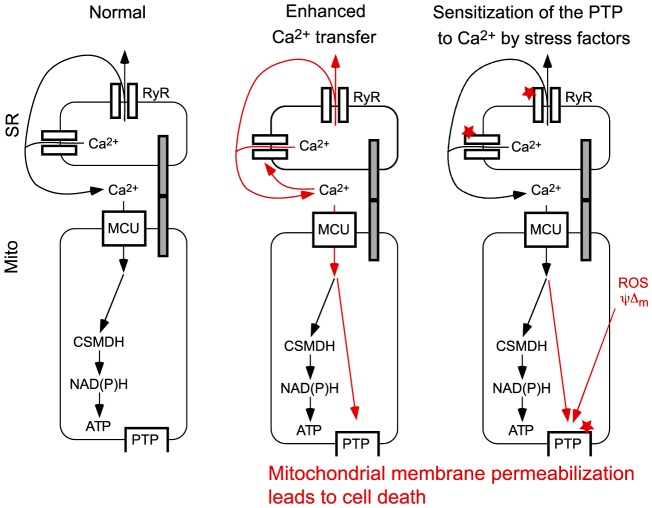
Mitochondria are strategically and dynamically positioned in the cell to spatially coordinate ATP production with energy needs and to allow the local exchange of material with other organelles. Interactions of mitochondria with the sarco-endoplasmic reticulum (SR/ER) have been receiving much attention owing to emerging evidence on the role these sites have in cell signaling, dynamics and biosynthetic pathways. One of the most important physiological and pathophysiological paradigms for SR/ER-mitochondria interactions is in cardiac and skeletal muscle. The contractile activity of these tissues has to be matched by mitochondrial ATP generation that is achieved, at least in part, by propagation of Ca(2+) signals from SR to mitochondria. However, the muscle has a highly ordered structure, providing only limited opportunity for mitochondrial dynamics and interorganellar interactions. This Commentary focuses on the latest advances in the structure, function and disease relevance of the communication between SR/ER and mitochondria in muscle. In particular, we discuss the recent demonstration of SR/ER-mitochondria tethers that are formed by multiple proteins, and local Ca(2+) transfer between SR/ER and mitochondria.

Keywords: Ca2+ signalling; MAM; Mitochondria; Mitochondria-associated membranes; ROS; Ryanodine receptor; Sarcoplasmic reticulum; Uniporter; reactive oxygen species.


Figures

Fig. 1.
Fig. 1.
The ‘social life’ of mitochondria. (A) Illustration of the interactions of SR/ER with mitochondria. Fluorescent proteins targeted to ER (ER–GFP) and mitochondria (MitoRFP) were expressed in RLB-2H3 cells and viewed using confocal microscopy followed by three-dimensional reconstruction. The physical tethering between ER and mitochondria (blue rectangle in scheme) facilitates local transfer of Ca2+ (green arrow) and membrane constituents. (B) Intermitochondrial coupling mediated by soluble molecules. Imaging of the mitochondrial membrane potential loss in H9c2 myotubes as reported by a potentiometric probe (TMRE) illustrates the spatial organization of mitochondrial apoptosis as a regenerative wave. The propagation of membrane permeabilization among individual mitochondria is mediated by factors that are released from the first responding mitochondria during permeabilization and promote permeabilization of the neighboring mitochondria. (C) Intermitochondrial content exchange mediated by fusion. Time course of a typical mitochondrial transient fusion is shown by photoactivatable fluorescent protein technology. The donor mitochondrion containing the photoactivated Kindling protein (MitoKP) aligns with the acceptor (MitoGFP), followed by content mixing. Within seconds the pair has reseparated at the apparent site of fusion and moved apart. (D) Mitochondrial transport along microtubules. In the image mitochondria (MitoDSRed) are aligned with microtubules (TubulinGFP). The images shown in A and C are adapted with permission from Spät et al., 2008 with permission from Elsevier, and Liu et al., 2009, respectively. Images shown in B and D were acquired as described in Pacher and Hajnóczky, 2001 and Yi et al., 2004.
Fig. 2.
Fig. 2.
SR–mitochondrial physical coupling in the muscle. (A) Close proximity of SR and mitochondria in a Flexor Digitorum Brevis (FDB) SM fiber is illustrated by labeling with an SR-specific dye, BODIPY–Ryanodine (left) and the potentiometric dye TMRE (middle). The overlay image on the right shows that SR (green) and mitochondria (red) run in parallel, both in longitudinal and transversal orientation. The yellow areas indicate the close proximity between SR and mitochondria. (B) Visualization of the sites of close SR/ER–mitochondrial associations in a live cardiac-muscle-derived cell (H9c2) cell by drug-inducible synthetic interorganellar linkers. The scheme on the left illustrates the rapamycin (Rapa)-inducible bridge-forming modules. Specifically, the OMM- and SR/ER-targeting sequences were coupled with the two components of the FKBP (FK506 binding protein 12)-FRB (FRB domain of mTOR) heterodimerization system, respectively. Addition of rapamycin causes heterodimerization between adjacent FKBP and FRB domains to rapidly connect the SR/ER- and OMM-targeted anchors. Induction of the bridge formation is initially confined to the areas where the SR/ER and OMM were naturally close. On the right, confocal images show the broad SR/ER and mitochondrial distribution of the respective fluorophores before and enrichment (colocalization of the CFP and RFP tags) at the sites of the SR/ER–mitochondrial interface after 3 minutes of 100 nM rapamycin treatment (bottom row). Note the rapamycin-induced colocalization of CFP and mRFP appears in white on the overlay images. For technical details see Csordás et al., 2010. The scheme is adapted with permission from Csordás et al., 2010 with permission from Elsevier. (C) TEM image of a membrane complex formed by TT and jSR (the diad) in close association with a mitochondrion (M) in cardiac muscle. In the magnified schematics of the jSR-mitochondrial interface (right), two forms of the tethering mechanisms are depicted. ‘Professional’ tethers refer to those proteins that are known to provide only structural support for the interface. MFN2 is a candidate to support SR-to-mitochondria juxtaposition. Signaling complexes provide both structural support and communication between SR/ER and mitochondria, and include the σ1 receptor, mitostatin, RAB32, phosphofurin acidic cluster sorting protein 2 (PACS2), ryanodine receptor (RyR) and voltage-dependent anion channels (VDACs). TT, transversal tubule; jSR, junctional SR; OMM, outer mitochondrial membrane; IMM, inner mitochondrial membrane.
Fig. 3.
Fig. 3.
Specific functions preferentially localized at the SR–mitochondrial interface. (A) A range of cellular activities, including several biosynthetic and signaling pathways and events involving membrane dynamics take place at the ER/SR–mitochondrial interface. (B) Local Ca2+ transport between RyR and mitochondria (red arrows). Upon action potential stimulation, the voltage dependent Ca2+ channel Cav1.2 activates RyR. In the heart, the Cav1.2-specific subunit α1c permeates Ca2+ from the extracellular medium to activate the RyR2. In skeletal muscle, Cav1.2 α1s interacts physically with RyR1, and the activation of Cav1.2 α1s directly promotes RyR1-mediated release of Ca2+ from the jSR to the cytoplasm. A high-Ca2+ microdomain is generated at the mouth of the RyRs, and it is sensed by the mitochondrial Ca2+ uniporter. Mitochondrial Ca2+ uptake activates matrix Ca2+-dependent dehydrogenases (CSMDH) and ATP synthesis. (C) Local interactions between ROS producers and ROS-sensitive proteins at the SR–mitochondria interface. The mitochondria electron transport chain generates superoxide anion (· O2) at the level of complex I and III (ETC, electron transport chain), which is converted into H2O2 by superoxide dismutase 2 (SOD2) in the mitochondrial matrix, or SOD1 in the cytosol or intermembrane space. H2O2 serves as substrate for glutathione peroxidase (GPX) that mediates the conversion of glutathione (GSH) to glutathione disulfide (GSSG; oxidized glutathione). GSSG targets proteins that contain reactive cysteines, such as RyR or the SR calcium transport ATPase (SERCA). SR- and TT-localized NADPH oxidases (NOXs) also contribute to · O2 generation. For further details see Csordás and Hajnóczky, 2009.
Fig. 4.
Fig. 4.
Misregulation of SR/ER–mitochondrial communication as a potential source of muscle injury. (A) Under normal conditions, activation of RyR-mediated Ca2+ release is propagated to the mitochondria to activate oxidative metabolism. (B) Augmented Ca2+ release through increased RyR activity leads to a mitochondrial Ca2+ overload and the activation of the permeability transition pore (PTP). (C) Stress factors (e.g. ROS) sensitize (indicated by the red stars) either RyR-mediated Ca2+ release, or activation of the PTP by Ca2+. Prolonged PTP activation initiates mitochondrial membrane permeabilization, leading to dissipation of the mitochondrial membrane potential and release of apoptotic factors from the intermembrane space to the cytoplasm, which then can initiate cell death. MCU, mitochondrial Ca2+ uniporter.

Similar articles

Cited by

References

    1. Andersson D. C., Betzenhauser M. J., Reiken S., Meli A. C., Umanskaya A., Xie W., Shiomi T., Zalk R., Lacampagne A., Marks A. R. (2011). Ryanodine receptor oxidation causes intracellular calcium leak and muscle weakness in aging. Cell Metab. 14, 196–207 10.1016/j.cmet.2011.05.014 - DOI - PMC - PubMed
    1. Andrienko T. N., Picht E., Bers D. M. (2009). Mitochondrial free calcium regulation during sarcoplasmic reticulum calcium release in rat cardiac myocytes. J. Mol. Cell. Cardiol. 46, 1027–1036 10.1016/j.yjmcc.2009.03.015 - DOI - PMC - PubMed
    1. Anelli T., Bergamelli L., Margittai E., Rimessi A., Fagioli C., Malgaroli A., Pinton P., Ripamonti M., Rizzuto R., Sitia R. (2012). Ero1α regulates Ca2+ fluxes at the endoplasmic reticulum-mitochondria interface (MAM). Antioxid. Redox Signal. 16, 1077–1087 10.1089/ars.2011.4004 - DOI - PubMed
    1. Anesti V., Scorrano L. (2006). The relationship between mitochondrial shape and function and the cytoskeleton. Biochim. Biophys. Acta 1757, 692–699 10.1016/j.bbabio.2006.04.013 - DOI - PubMed
    1. Aon M. A., Cortassa S., Marbán E., O'Rourke B. (2003). Synchronized whole cell oscillations in mitochondrial metabolism triggered by a local release of reactive oxygen species in cardiac myocytes. J. Biol. Chem. 278, 44735–44744 10.1074/jbc.M302673200 - DOI - PubMed

Publication types

MeSH terms

Substances