. 2013 Sep 15;126(Pt 18):4195-207.
doi: 10.1242/jcs.127779. Epub 2013 Jul 10.

Phosphorylation of serine 4,642 in the C-terminus of plectin by MNK2 and PKA modulates its interaction with intermediate filaments

Affiliations

Phosphorylation of serine 4,642 in the C-terminus of plectin by MNK2 and PKA modulates its interaction with intermediate filaments

Jamal-Eddine Bouameur et al. J Cell Sci. .

Abstract

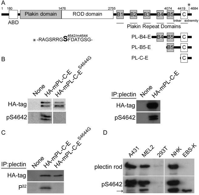
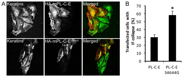
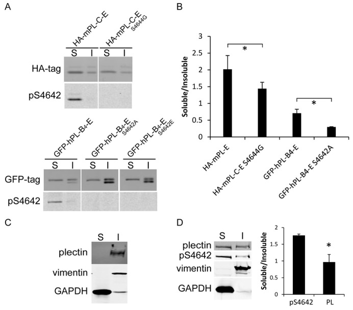
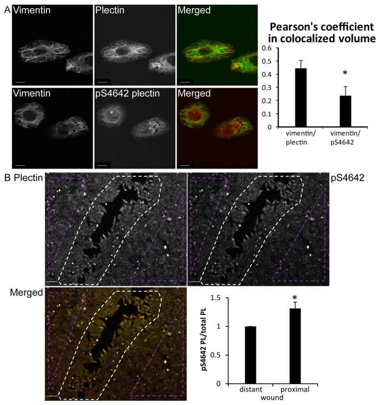
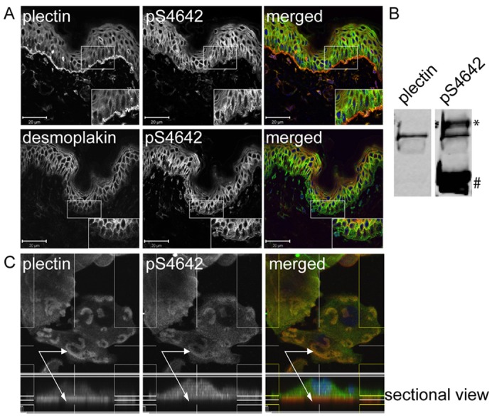
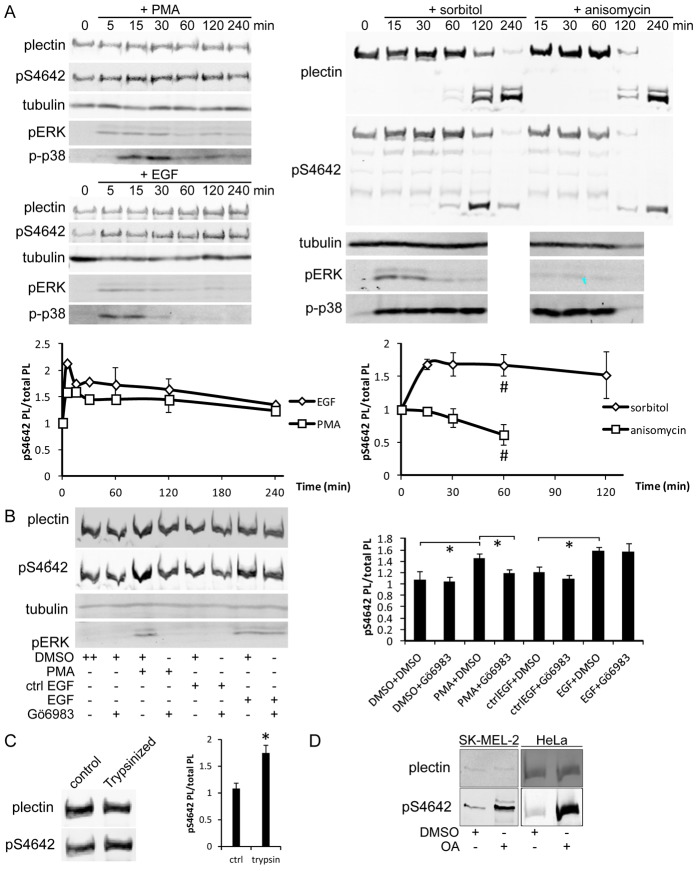
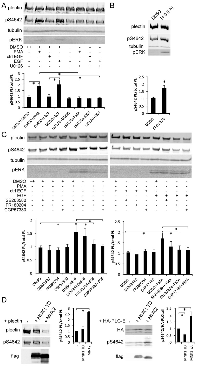
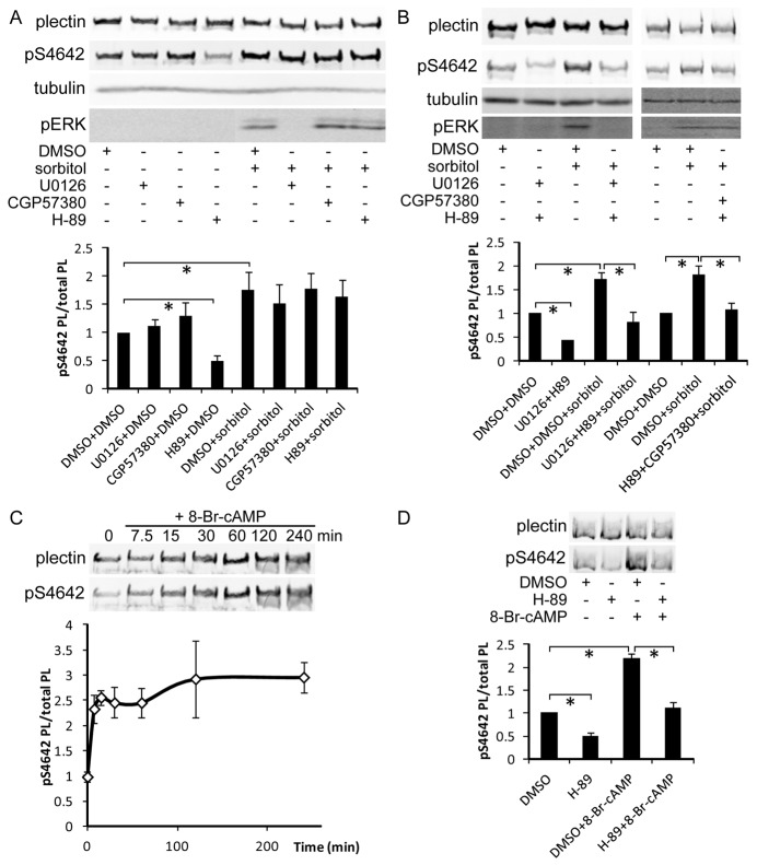
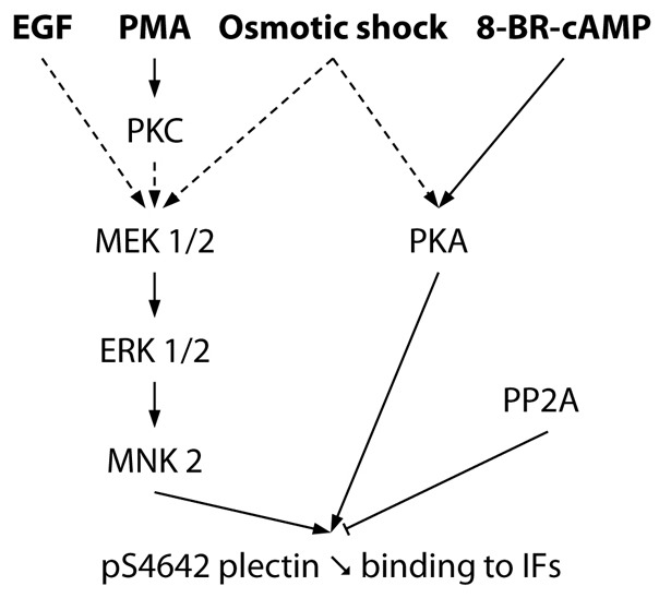
Plectin is a versatile cytolinker of the plakin family conferring cell resilience to mechanical stress in stratified epithelia and muscles. It acts as a critical organizer of the cytoskeletal system by tethering various intermediate filament (IF) networks through its C-terminal IF-binding domain (IFBD). Mutations affecting the IFBD cause devastating human diseases. Here, we show that serine 4642, which is located in the extreme C-terminus of plectin, is phosphorylated in different cell lines. Phosphorylation of S4642 decreased the ability of plectin IFBD to associate with various IFs, as assessed by immunofluorescence microscopy and cell fractionation studies, as well as in yeast two-hybrid assays. Plectin phosphorylated at S4642 was reduced at sites of IF network anchorage along cell-substrate contacts in both skin and cultured keratinocytes. Treatment of SK-MEL-2 and HeLa cells with okadaic acid increased plectin S4642 phosphorylation, suggesting that protein phosphatase 2A dephosphorylates this residue. Moreover, plectin S4642 phosphorylation was enhanced after cell treatment with EGF, phorbol ester, sorbitol and 8-bromo-cyclic AMP, as well as during wound healing and protease-mediated cell detachment. Using selective protein kinase inhibitors, we identified two different kinases that modulate the phosphorylation of plectin S4642 in HeLa cells: MNK2, which is downstream of the ERK1/2-dependent MAPK cascade, and PKA. Our study indicates that phosphorylation of S4642 has an important regulatory role in the interaction of plectin with IFs and identifies a novel link between MNK2 and the cytoskeleton.

Keywords: Cytoskeleton; Intermediate filaments; Plakin; Plectin; Protein phosphorylation.


Figures

Fig. 1.
Fig. 1.
Phosphorylation of plectin S4642 in cultured cells. (A) Schematic representation of human plectin isoform 1. The different domains, with the corresponding amino acid positions, are depicted (ABD: actin-binding domain). The C-terminal region consists of six plakin repeat domains (PRDs) (boxes B1 to B5 and C) separated by connecting segments, with an important role of the linker, bridging B5 to C, for the binding of plectin to IFs. The serine residue, equivalent to desmoplakin S2849, is located within the C-terminal extremity at position 4642 [residues 4644 in mouse (m) plectin isoform 1]. (B) Extracts and plectin immunoprecipitates from PtK2 cells, transfected with plasmids encoding HA–mPL-C-E or HA–mPL-C-ES4644G (see A), were analyzed by western blotting (WB, ECL) with anti-HA and anti-pS4642 antibodies as indicated. (C) Transfected PtK2 cells were 32P-radiolabeled in vivo prior to immunoprecipitation of recombinant proteins from cell extracts with the anti-plectin antibodies GP21. Immunoprecipitates were analyzed by WB with anti-HA antibodies and autoradiography. (D) Anti-pS4642 and anti-plectin rod antibodies recognized a protein with the same electrophoretic mobility on WB (ECL) prepared with extracts from A431, SK-MEL-2 and NHK cells (HEK 293T cells express very low levels of plectin). The rod-less isoform(s) of plectin whose migration is indicated by the arrow, was also detected by the anti-pS4642 antibodies [MD-EBS keratinocytes only express rod-less plectin (Koster et al., 2004) whereas desmoplakin migrates below plectin rod-less isoform(s)].
Fig. 2.
Fig. 2.
Frequency of IF collapse is higher in transfected PtK2 cells expressing HA–mPL-C-ES4644G than HA–mPL-C-E. (A) Immunofluorescence microscopy images of transfected PtK2 cells, expressing HA–mPL-C-E or HA–mPL-C-ES4644G, with anti-HA antibodies (red) and anti-panKeratin antibodies (green). (B) Quantification of bundling and collapse of the IF network (arrows) in transfected cells (means ± s.d.). Scale bars: 10 µm. More than 100 transfected cells were counted per slide. *P<0.01.
Fig. 3.
Fig. 3.
pS4642/4 plectin-IFBD proteins are predominantly found in the TX-100-soluble fraction from transfected HeLa cells. (A) HeLa cells, transfected with the indicated plectin constructs (see Fig. 1A) fused to HA or GFP tag, were fractionated in TX-100 buffer. Soluble (S) and insoluble (I) fractions were analyzed by WB with anti-tag and anti-pS4642 plectin antibodies (the cause for the presence of a doublet in some fractions is unknown). (B) WB quantitative analysis. *P<0.05 (n = 3). (C) WB analysis of the solubility of endogenous plectin, vimentin and GAPDH in HeLa cell extracts lyzed in TX-100 buffer. (D) Cells lyzed in TX-100-2 buffer. Graphs show means ± s.d. *P<0.05 (n = 3).
Fig. 4.
Fig. 4.
pS4642 plectin has an altered cytoplasmic distribution and increases during wound healing in SK-MEL-2 cells. (A) Confocal immunofluorescence microscopy images of SK-MEL-2 cells, immunostained with antibodies against plectin or pS4642 and vimentin as indicated. Degree of colocalization was determined by measuring the Pearson's coefficient using the Imaris software. Scale bar: 10 µm. More than 100 cells counted per slide. *P<0.01. (B) Confluent SK-MEL-2 cells were scratched, fixed 20 hours later and immunostained with anti-plectin and pS4642 antibodies. Scale bar: 100 µm. The ratio pS4642 plectin to plectin was determined in wound-proximal (white dotted area) and scratch-distant cells (violet dotted areas). Graphs show means ± s.d. *P<0.05 (n = 3).
Fig. 5.
Fig. 5.
Plectin is less phosphorylated on S4642 at sites of cell-substrate contact in human epidermis and PAJEB-β4 keratinocytes. (A) Acetone-fixed cryosections of human skin were co-immunostained for plectin and pS4642 or for desmoplakin and pS4642. High-power views of the boxed areas are depicted in the insets. In the basal layer of the epidermis, there was no correlation between plectin and pS4642 signals whereas the immunostained patterns of desmoplakin and pS4642 were similar showing that plectin is weakly phosphorylated in hemidesmosomes. Scale bars: 20 µm. (B) Epidermal cell extracts were immunoblotted with anti-plectin and anti-pS4642 antibodies, indicating plectin S4642 phosphorylation in the skin (*, immunoreactive protein migrating above plectin; #, p2849 desmoplakin). (C) PAJEB-β4 keratinocytes, cultured in high Ca2+ for 24 hours, were fixed with formaldehyde and co-immunostained for plectin (red) and pS4642 (green) as indicated. Z-stack sectional view of plectin (red) and pS4642 (green) showing the dichotomy between the two signals in hemidesmosome-like structures (arrows indicate cell-substrate contact/hemidesmosome-like structures with a prevalence of unphosphorylated plectin in the lower part).
Fig. 6.
Fig. 6.
Cell treatment with PMA, EGF, sorbitol, trypsin or OA increases plectin S4642 phosphorylation. (A) Starved HeLa cells were treated with 100 nM PMA, 100 ng/ml EGF, 400 mM sorbitol or 1 µg/ml anisomycin for the indicated times and the levels of plectin, pS4642, tubulin, pERK1/2 and p-p38 in whole cell extracts were assessed by WB. Ratios of plectin pS4642/total plectin in response to these treatments are depicted in the graphs (# indicate the onset of plectin proteolysis). (B) Starved HeLa cells were pretreated with 100 nM Gö6983 or DMSO (control) for 1 hour and then treated or not for 15 minutes with 100 nM PMA or 100 ng/ml EGF as indicated. WB analysis was carried out as in A. *P<0.05 (n≥3). (C) HeLa cells were detached by trypsinization and levels of plectin and pS4642 analyzed as in A. *P <0.05 (n = 3). (D) SK-MEL-2 and HeLa cells were treated with 100 nM OA for 3 hours and analyzed as in A. Graphs show means ± s.d.
Fig. 7.
Fig. 7.
Inhibitors of the ERK1/2 MAPK pathway and MNKs block EGF-induced and PMA-induced S4642 phosphorylation. (A) Starved HeLa cells were pretreated for 1 hour with 10 µM U0126 (MEK1/2 and 5 inhibitor) or DMSO (control) and then treated or not with PMA or EGF (100 nM or 100 ng/ml respectively for 15 minutes). WB analysis was performed as in Fig. 6. *P<0.05 (n≥3). (B) Starved HeLa cells were treated with 10 µM BI-D1870 (RSKs inhibitor) for 1 hour and pERK and pS4642 levels analyzed by WB. *P<0.05 (n = 3). (C) Starved HeLa cells were pretreated for 1 hour with 10 µM FR 180204 (ERK1/2 inhibitor), 40 µM CGP 57380 (MNK inhibitor), 10 µM SB 203580 (p38 inhibitor) or DMSO (control) and then treated or not with PMA or EGF (100 nM or 100 ng/ml respectively for 15 minutes), WB analysis as in A. *P<0.05 (n≥3). (D) WB analysis of whole extracts from HEK 293T cells co-transfected with plasmids encoding either HA–plectin or HA–plectin-C-E and pcDNA3, pcDNA3–FLAG–MNK2, or pcDNA3–FLAG–MNK1-T344D. *P<0.05 (n = 3). For unclear reasons, the amount of HA–plectin in cells co-transfected with pcDNA3–FLAG–MNK2 was systematically lower than with the empty vector or pcDNA3–FLAG–MNK1-T344D. All graphs show means ± s.d.
Fig. 8.
Fig. 8.
PKA stimulation increases plectin S4642 phosphorylation. (A) Starved HeLa cells were pretreated for 1 hour with DMSO (control) or the following PK inhibitors: 10 µM U0126 (MEK 1/2, 5), 40 µM CGP 57380 (MNKs) or 10 µM H-89 (PKA) prior to hyperosmotic shock induction with 400 mM sorbitol for 30 minutes. Levels of plectin, pS4642, tubulin and pERK1/2 were analyzed by WB. H-89 decreased pS4642 levels in unstimulated cells but none of the inhibitors blocked the sorbitol-induced increase in pS4642 levels. *P<0.05 (n≥3). (B) Starved HeLa cells were pretreated for 1 hour with combinations of PK inhibitors as indicated and then treated and analyzed as in A. Sorbitol-induced pS4642 increase was blocked by combining H-89 with U0126 or with CGP 57380. *P<0.05 (n≥3). (C) Starved HeLa cells were incubated with 0.5 mM 8-Br-cAMP for the indicated time periods and pS4642/total plectin ratios were measured by WB. *P<0.05 (n = 3). (D) Starved HeLa cells were pretreated for 1 hour with 10 µM H-89 or DMSO (control) prior to 30 minutes of stimulation with 0.5 mM 8-Br-cAMP and processed as in C. Graphs show means ± s.d. *P<0.05 (n = 3).
Fig. 9.
Fig. 9.
Signaling pathways regulating the phosphorylation of plectin S4642 in HeLa cells. EGF, PMA and hyperosmotic shock (sorbitol) stimulate the phosphorylation of plectin S4642 via the ERK1/2 MAPK pathway (dotted arrows indicate indirect activations). EGF effect is independent of PKC. Sorbitol-induced hyperosmotic stress also stimulates plectin S4642 phosphorylation in a PKA-dependent manner. Phosphorylation of plectin S4642 decreases its binding to IFs. Protein Phosphatase 2A (PP2A) directly or indirectly mediates pS4642 dephosphorylation.

Similar articles

Cited by

References

    1. Adler J., Parmryd I. (2010). Quantifying colocalization by correlation: the Pearson correlation coefficient is superior to the Mander's overlap coefficient. Cytometry 77A, 733–742 10.1002/cyto.a.20896 - DOI - PubMed
    1. Andrä K., Lassmann H., Bittner R., Shorny S., Fässler R., Propst F., Wiche G. (1997). Targeted inactivation of plectin reveals essential function in maintaining the integrity of skin, muscle, and heart cytoarchitecture. Genes Dev. 11, 3143–3156 10.1101/gad.11.23.3143 - DOI - PMC - PubMed
    1. Andrä K., Kornacker I., Jörgl A., Zörer M., Spazierer D., Fuchs P., Fischer I., Wiche G. (2003). Plectin-isoform-specific rescue of hemidesmosomal defects in plectin (-/-) keratinocytes. J. Invest. Dermatol. 120, 189–197 10.1046/j.1523-1747.2003.12027.x - DOI - PubMed
    1. Bagowski C. P., Besser J., Frey C. R., Ferrell J. E., Jr (2003). The JNK cascade as a biochemical switch in mammalian cells: ultrasensitive and all-or-none responses. Curr. Biol. 13, 315–320 - PubMed
    1. Bass-Zubek A. E., Hobbs R. P., Amargo E. V., Garcia N. J., Hsieh S. N., Chen X., Wahl J. K., III, Denning M. F., Green K. J. (2008). Plakophilin 2: a critical scaffold for PKC alpha that regulates intercellular junction assembly. J. Cell Biol. 181, 605–613 10.1083/jcb.200712133 - DOI - PMC - PubMed

Publication types

MeSH terms