. 2013 Sep;87(18):10148-62.
doi: 10.1128/JVI.01002-13. Epub 2013 Jul 10.

Contribution of myocyte enhancer factor 2 family transcription factors to BZLF1 expression in Epstein-Barr virus reactivation from latency

Affiliations

Contribution of myocyte enhancer factor 2 family transcription factors to BZLF1 expression in Epstein-Barr virus reactivation from latency

Takayuki Murata et al. J Virol. 2013 Sep.

Abstract

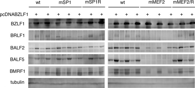
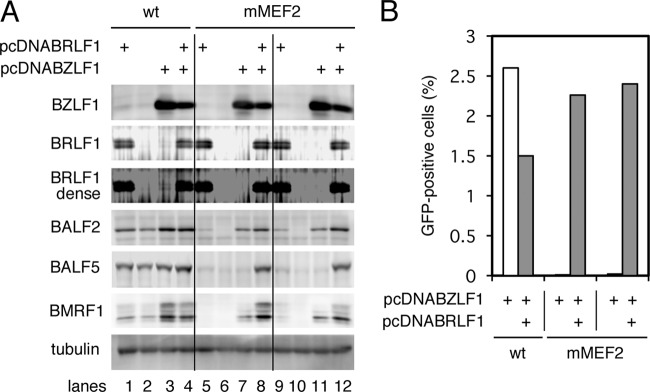
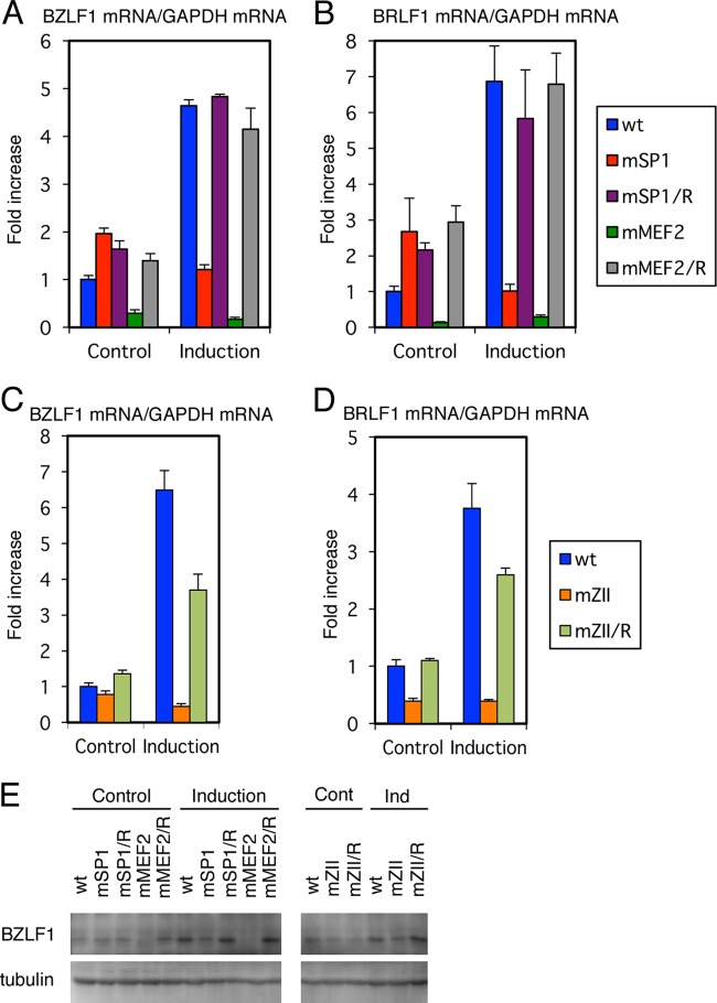
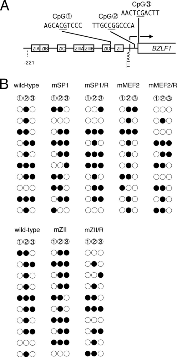
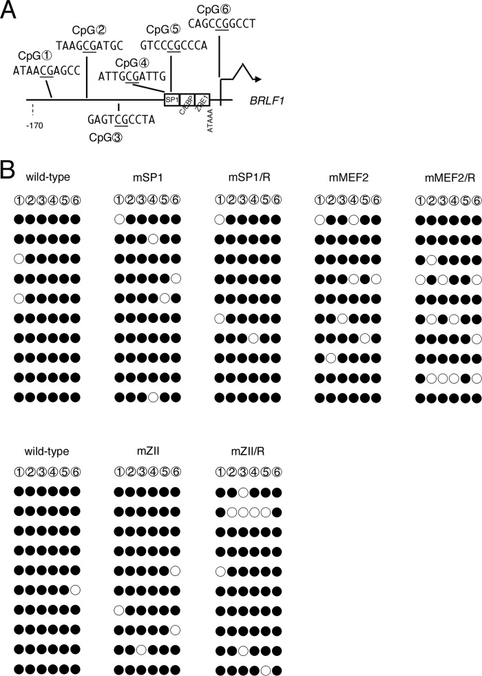
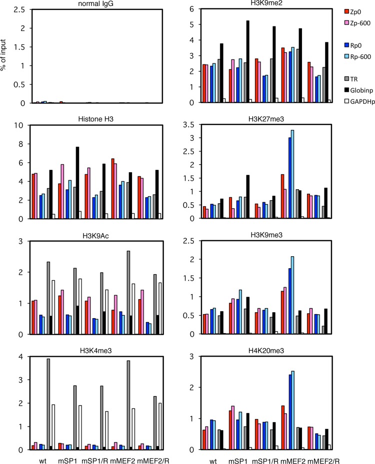
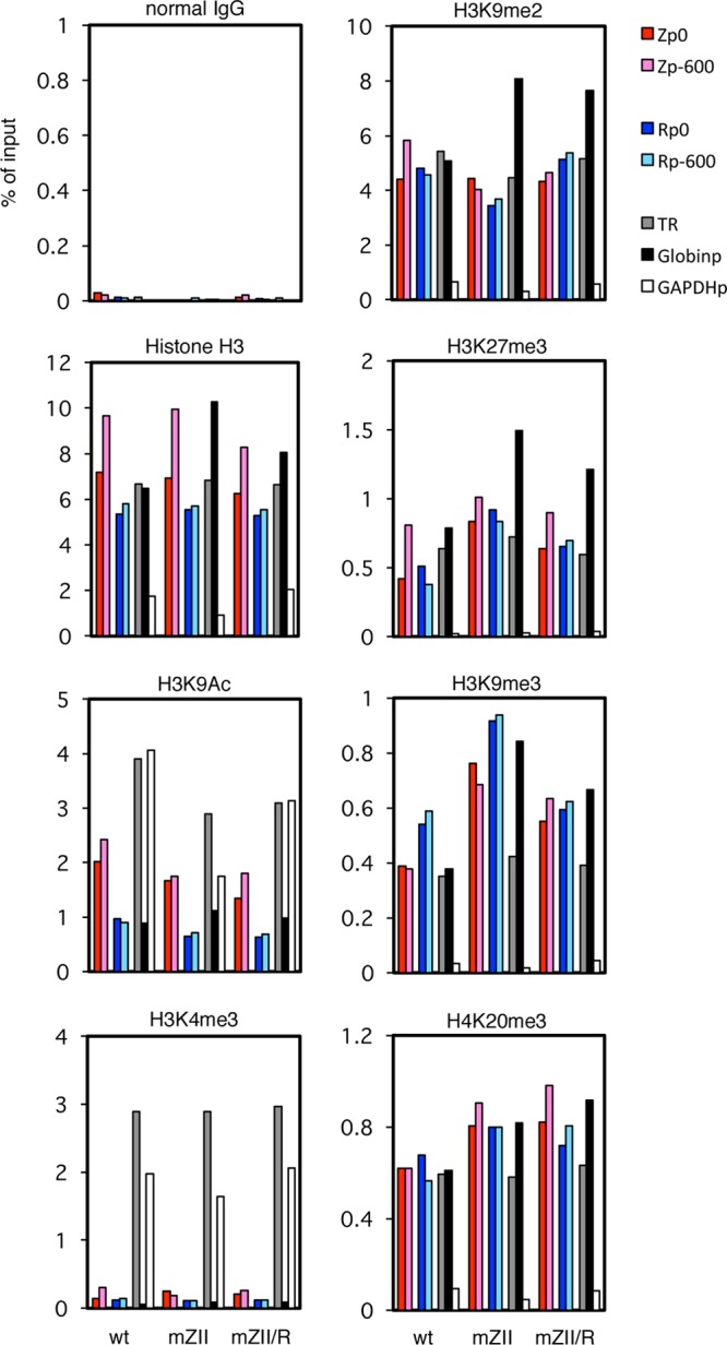
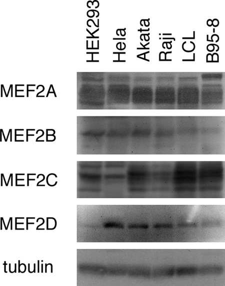
Reactivation of Epstein-Barr virus (EBV) from latency is dependent on expression of the viral transactivator BZLF1 protein, whose promoter (Zp) normally exhibits only low basal activity but is activated in response to chemical or biological inducers. Using a reporter assay system, we screened for factors that can activate Zp and isolated genes, including those encoding MEF2B, KLF4, and some cellular b-Zip family transcription factors. After confirming their importance and functional binding sites in reporter assays, we prepared recombinant EBV-BAC, in which the binding sites were mutated. Interestingly, the MEF2 mutant virus produced very low levels of BRLF1, another transactivator of EBV, in addition to BZLF1 in HEK293 cells. The virus failed to induce a subset of early genes, such as that encoding BALF5, upon lytic induction, and accordingly, could not replicate to produce progeny viruses in HEK293 cells, but this restriction could be completely lifted by exogenous supply of BRLF1, together with BZLF1. In B cells, induction of BZLF1 by chemical inducers was inhibited by point mutations in the ZII or the three SP1/KLF binding sites of EBV-BAC Zp, while leaky BZLF1 expression was less affected. Mutation of MEF2 sites severely impaired both spontaneous and induced expression of not only BZLF1, but also BRLF1 in comparison to wild-type or revertant virus cases. We also observed that MEF2 mutant EBV featured relatively high repressive histone methylation, such as H3K27me3, but CpG DNA methylation levels were comparable around Zp and the BRLF1 promoter (Rp). These findings shed light on BZLF1 expression and EBV reactivation from latency.


Figures

Fig 1
Fig 1
Alignment of wild-type and mutated sequences of the EBV minimal BZLF1 promoter. cis-regulatory element sequences are shown in capitals. Mutated nucleotides are boxed and shown in capitals.
Fig 2
Fig 2
Library screening identified factors involved in BZLF1 promoter activation. (A) HEK293T cells were transfected with 10 ng of the reporter plasmid pZp-luc, 1 ng of pCMV-RL, and 100 ng of expression plasmids for the indicated genes. (B) As in panel A, HEK293T cells were transfected with 10 ng of the reporter plasmid pZp-luc (wt) or its promoterless parental vector, pGL4.10 (empty); 1 ng of pCMV-RL; and 100 ng of expression plasmids for the indicated genes. Luciferase assays were carried out as described in Materials and Methods. Luciferase activity is shown as fold activation over that with the control vector. Each bar represents the mean and standard deviation (SD) of three independent transfections.
Fig 3
Fig 3
Effects of b-Zip, SP1/KLF, and MEF2 family transcription factors on the BZLF1 promoter. (A) b-Zip family transcription factor activation of the BZLF1 promoter. HEK293T cells were transfected with 10 ng of the reporter plasmid pZp-luc or its derivatives, 1 ng of pCMV-RL, and 100 ng of expression plasmids for the indicated genes. Luciferase assays were carried out as described in Materials and Methods. Luciferase activity is shown as fold activation over that with the control vector (Cont). Each bar represents the mean and SD of three independent transfections. Derivatives of pZp-luc (wt), pZpmZII-luc (mZII), or pZpmZIII-luc (mZIII) contained point mutations in the binding motifs for cellular b-Zip transcription factors or for the viral b-Zip transcription factor BZLF1. The pZpmZII+ZIII-luc vector (mZII,III) contained both mutations. (B) KLF4, SP1, and MEF2B activation of the BZLF1 promoter. HEK293T cells were transfected with 10 ng of the reporter plasmid pZp-luc (wt), 100 ng of pGL4.70(hRluc), and 100 ng of expression plasmid for KLF4, SP1, or MEF2B. pZp-luc reporters with point mutations in SP1-binding sites (mSP1) and MEF2 (mMEF2) binding sites were also used. Luciferase assays were carried out as described in Materials and Methods. Luciferase activity is shown as fold activation over that with control vector and pZp-luc (wt). Each bar represents the mean and SD of three independent transfections. Diagrams below the graphs show constructs of the reporter plasmid pZp-luc and its derivatives.
Fig 4
Fig 4
Construction of recombinant EBV featuring point mutations in SP1 binding sites (A) and MEF2 binding sites (B) of the BZLF1 gene promoter. Shown is a schematic arrangement of the recombination of the EBV genome using tandemly arranged neomycin resistance and streptomycin sensitivity genes (NeoSt+). The BZLF1 promoter region was first replaced with the NeoSt+ cassette, which was then replaced with point-mutated sequences (asterisks) to construct EBV-BAC mSP1 or mMEF2. The mutated sequence was replaced again with the NeoSt+ marker cassette and swapped with the wild-type promoter sequence to prepare the revertant clone, EBV-BAC mSP1/R or mMEF2/R. The recombinant EBV genomes were digested with BamHI or EcoRI and separated in an agarose gel. PCR products produced by the indicated primers were electrophoresed to show successful recombinations.
Fig 5
Fig 5
Expression of viral proteins from recombinant viruses in HEK293 cells. The recombinant EBV-BAC DNAs shown in Fig. 5 were introduced into HEK293 cells, followed by hygromycin selection. The resultant cell clones were tested for viral protein expression. Two or three clones of each recombinant virus were transfected with the BZLF1 expression vector (pcDNABZLF1) or its empty control vector. After 24 h, cell proteins were harvested and immunoblotting was performed using anti-BZLF1, -BRLF1, -BALF2, -BALF5, -BMRF1, and -tubulin antibodies.
Fig 6
Fig 6
Suppression of early gene expression with MEF2 binding site mutation of the BZLF1 promoter could be reversed with exogenous BRLF1. (A) HEK293 cells latently infected with the wt or the mMEF2 mutant of recombinant EBV-BAC DNA were transfected with the BZLF1 expression vector (pcDNABZLF1), the BRLF1 expression vector (pcDNABRLF1), and/or its empty vector. After 24 h, cell proteins were harvested and immunoblotting was performed using anti-BZLF1, -BRLF1, -BALF2, -BALF5, -BMRF1, and -tubulin antibodies. (B) HEK293 cells latently infected with the wt or the mMEF2 mutant of recombinant EBV-BAC DNA were transfected with BZLF1 expression vector (pcDNABZLF1) (white bars) or BZLF1 expression vector plus BRLF1 expression vector (pcDNABRLF1) (gray bars). The total amount of transfected DNA was adjusted with the empty vector, and 72 h after transfection, the culture supernatants were collected and cocultured with Akata(−) cells for 48 h, and then GFP-positive cells were counted by fluorescence-activated cell sorter (FACS).
Fig 7
Fig 7
Expression of viral IE genes in B cells. (A to E) LCLs latently infected with recombinant EBV-BAC prepared from HEK293 EBV-BAC cells were treated (Induction) or not (Control) with TPA (200 ng/ml), A23187 (0.1 μM), and sodium butyrate (10 mM). After 24 h, cell RNAs were harvested and subjected to real-time RT-PCR using specific primers for BZLF1, BRLF1, and GAPDH. Relative BZLF1mRNA (A and C) and BRLF1 mRNA (B and D) levels are shown after normalization to GAPDH mRNA levels. Each bar represents the mean and SD of three independent treatments. (E) LCLs were treated likewise, and cell proteins were subjected to Western blotting using anti-BZLF1 and -tubulin antibodies.
Fig 8
Fig 8
CpG DNA methylation of the BZLF1 promoter in mutant LCLs. (A) Schematic illustration of the minimal BZLF1 promoter. The distribution of three CpG dinucleotides analyzed in panel B is shown with circled numbers. cis-element motifs and the BZLF1 coding region are also depicted. (B) CpG DNA methylation of the BZLF1 promoter in B cells. DNA from LCLs latently infected with wild-type or mutant recombinant EBV-BAC was subjected to bisulfite modification, followed by sequencing. Filled circles, methylated; open circles, unmethylated.
Fig 9
Fig 9
CpG DNA methylation of the BRLF1 promoter in mutant LCLs. (A) Schematic illustration of the BRLF1 promoter. The distribution of six CpG dinucleotides analyzed in panel B is shown with circled numbers. cis-element motifs and BRLF1 coding region are also depicted. (B) CpG DNA methylation of the BRLF1 promoter in B cells. DNA from LCLs latently infected with wild-type or mutant recombinant EBV-BAC was subjected to bisulfite modification, followed by sequencing. Filled circles, methylated; open circles, unmethylated.
Fig 10
Fig 10
Histone modification of BZLF1/BRLF1 promoters in mSP1 and mMEF2 mutant LCLs. LCLs latently infected with recombinant EBV-BAC were cross-linked and subjected to ChIP experiments as described in Materials and Methods using normal IgG and anti-histone H3, -H3K9Ac, -H3K4me3, -H3K9me2, -H3K27me3, -H3K9me3, and -H4K20me3 antibodies, followed by DNA extraction and real-time PCR to detect DNA fragments using the indicated primers. The Zp/Rp numbers indicate the sequence positions relative to the transcription start site. As controls, the TR region of EBV, the β-globin promoter (Globinp), and the GAPDH promoter (GAPDHp) were also quantified.
Fig 11
Fig 11
Histone modification of BZLF1/BRLF1 promoters in mZII mutant LCLs. LCLs latently infected with recombinant EBV-BAC were cross-linked and subjected to ChIP experiments as described in Materials and Methods using normal IgG and anti-histone H3, -H3K9Ac, -H3K4me3, -H3K9me2, -H3K27me3, -H3K9me3, and -H4K20me3 antibodies, followed by DNA extraction and real-time PCR to detect DNA fragments using the indicated primers. The Zp/Rp numbers indicate the sequence positions relative to the transcription start site. As controls, the TR region of EBV, the β-globin promoter (Globinp), and the GAPDH promoter (GAPDHp) were also quantified.
Fig 12
Fig 12
Expression profile of MEF2 family transcription factors. Cell proteins from HEK293, HeLa, Akata, Raji, and B95-8 cells and LCLs were subjected to Western blotting using anti-MEF2A, -MEF2B, -MEF2C, -MEF2D, and -tubulin antibodies.

Similar articles

Cited by

References

    1. Tsurumi T, Fujita M, Kudoh A. 2005. Latent and lytic Epstein-Barr virus replication strategies. Rev. Med. Virol. 15:3–15 - PubMed
    1. Amon W, Farrell PJ. 2005. Reactivation of Epstein-Barr virus from latency. Rev. Med. Virol. 15:149–156 - PubMed
    1. Speck SH, Chatila T, Flemington E. 1997. Reactivation of Epstein-Barr virus: regulation and function of the BZLF1 gene. Trends Microbiol. 5:399–405 - PubMed
    1. Miller G, El-Guindy A, Countryman J, Ye J, Gradoville L. 2007. Lytic cycle switches of oncogenic human gammaherpesviruses. Adv. Cancer Res. 97:81–109 - PubMed
    1. Sinclair AJ. 2003. bZIP proteins of human gammaherpesviruses. J. Gen. Virol. 84:1941–1949 - PubMed

Publication types

LinkOut - more resources