

Whipple's Disease: Our Own Experience and Review of the Literature
- PMID: 23843784
- PMCID: PMC3703430
- DOI: 10.1155/2013/478349
Whipple's Disease: Our Own Experience and Review of the Literature
Abstract
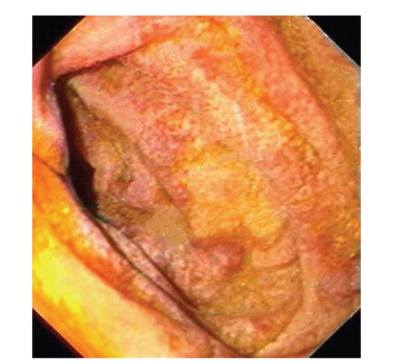
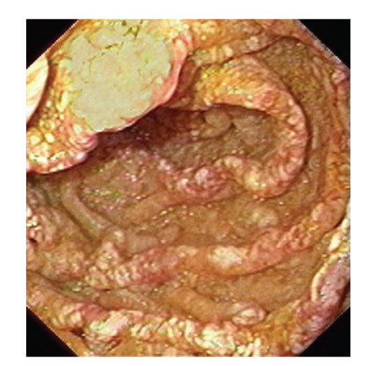
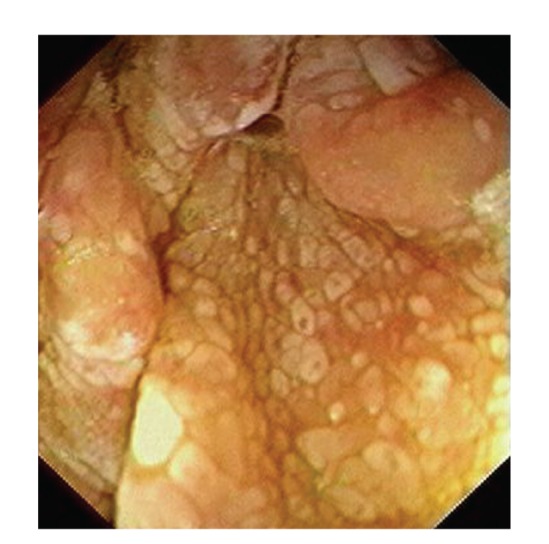
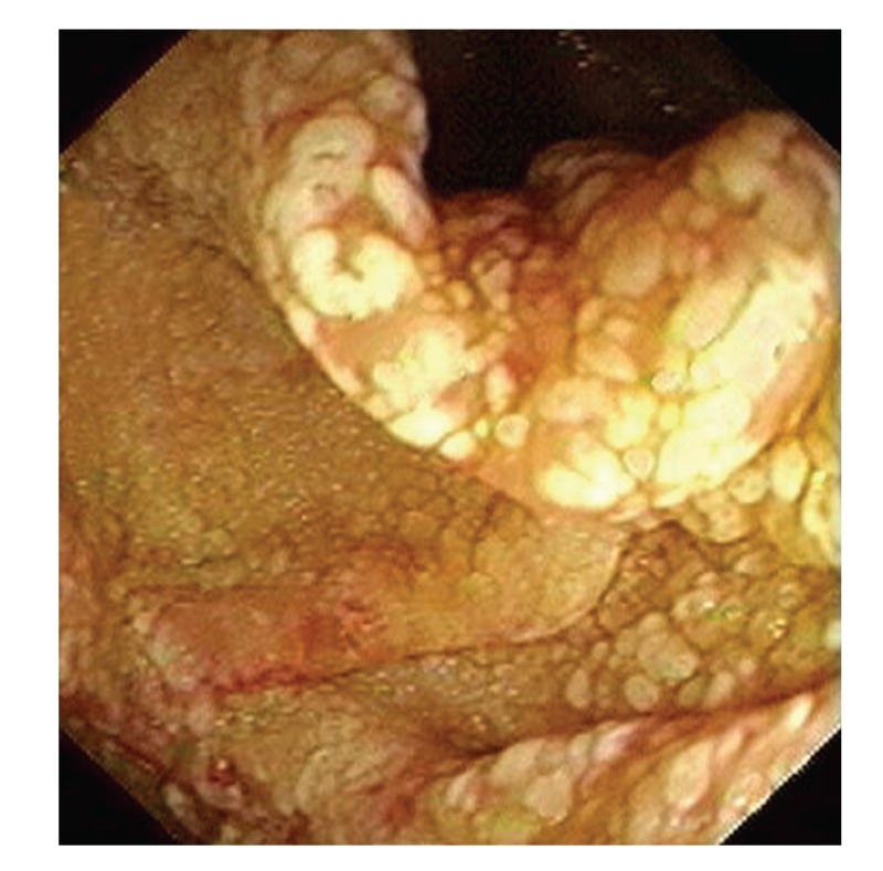
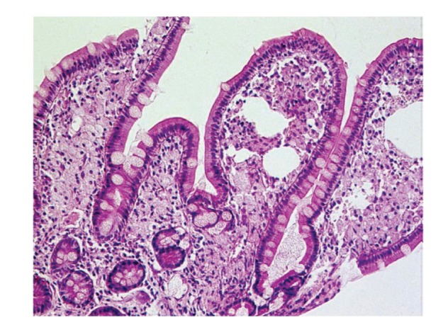
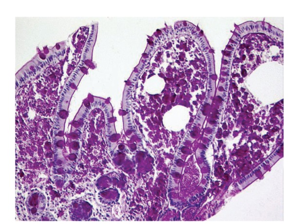
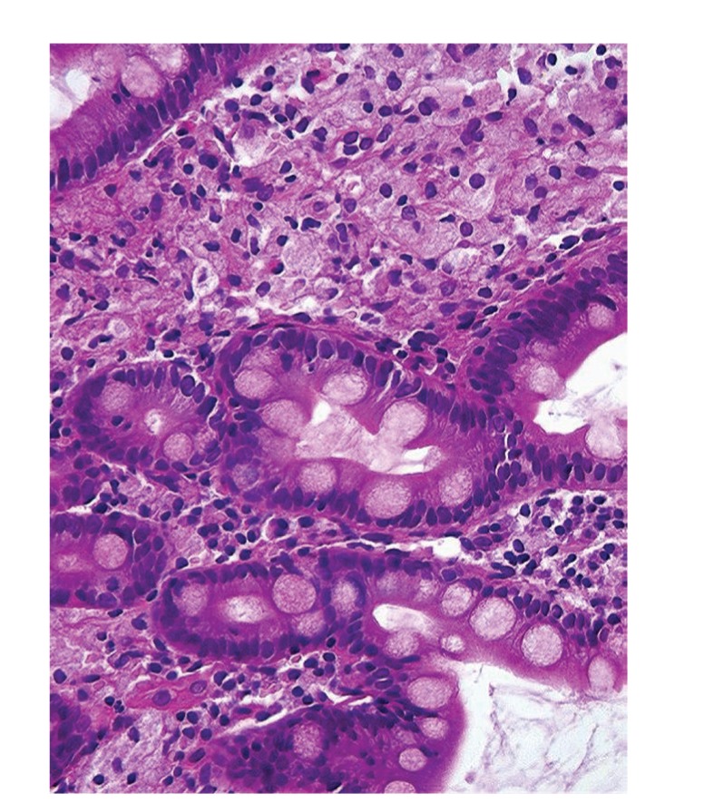
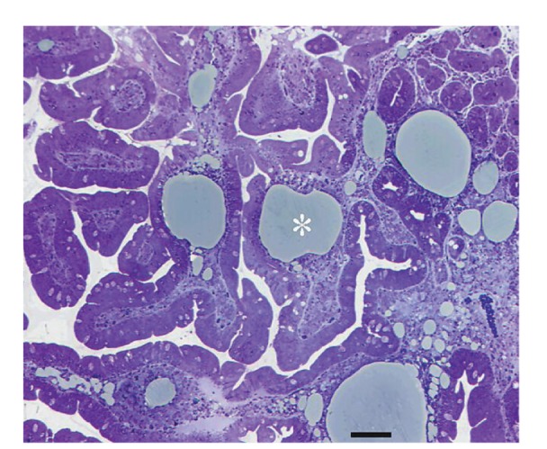
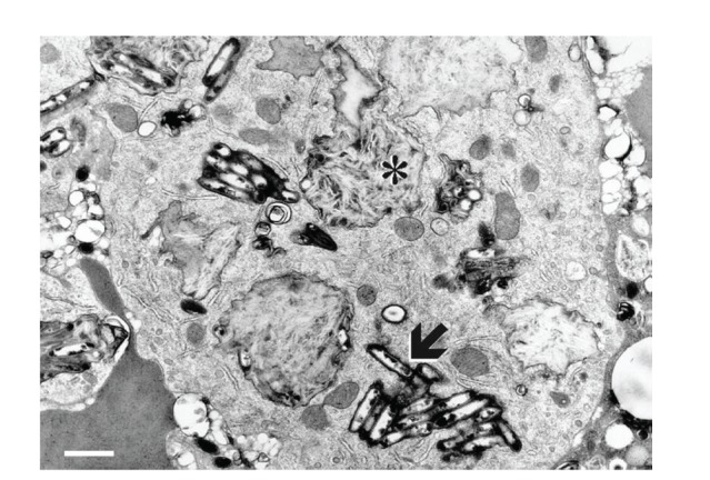
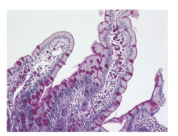
Whipple's disease is a chronic infectious systemic disease caused by the bacterium Tropheryma whipplei. Nondeforming arthritis is frequently an initial complaint. Gastrointestinal and general symptoms include marked diarrhoea (with serious malabsorption), abdominal pain, prominent weight loss, and low-grade fever. Possible neurologic symptoms (up to 20%) might be associated with worse prognosis. Diagnosis is based on the clinical picture and small intestinal histology revealing foamy macrophages containing periodic-acid-Schiff- (PAS-) positive material. Long-term (up to one year) antibiotic therapy provides a favourable outcome in the vast majority of cases. This paper provides review of the literature and an analysis of our 5 patients recorded within a 20-year period at a tertiary gastroenterology centre. Patients were treated using i.v. penicillin G or amoxicillin-clavulanic acid + i.v. gentamicin for two weeks, followed by p.o. doxycycline (100 mg per day) plus p.o. salazopyrine (3 g per day) for 1 year. Full remission was achieved in all our patients.
Figures
Similar articles
-
Whipple's disease: multiple hospital admissions of a man with diarrhoea, fever and arthralgia.J Infect. 2005 Aug;51(2):E35-7. doi: 10.1016/j.jinf.2004.08.010. J Infect. 2005. PMID: 16038747
-
Whipple's disease in a man of North African descent : case report and brief review of the literature.Acta Gastroenterol Belg. 2019 Jan-Mar;82(1):83-86. Acta Gastroenterol Belg. 2019. PMID: 30888759 Review.
-
Whipple's in the valleys: a case of Whipple's with thrombocytopenia and endocarditis.J Clin Pathol. 2014 May;67(5):445-8. doi: 10.1136/jclinpath-2013-201915. Epub 2014 Jan 23. J Clin Pathol. 2014. PMID: 24459171
-
Short article: Relapsing Whipple's disease: a case report and literature review.Eur J Gastroenterol Hepatol. 2016 Mar;28(3):267-70. doi: 10.1097/MEG.0000000000000539. Eur J Gastroenterol Hepatol. 2016. PMID: 26649803 Review.
-
First case of Whipple's disease successfully treated with tigecycline.Germs. 2021 Mar 15;11(1):105-110. doi: 10.18683/germs.2021.1246. eCollection 2021 Mar. Germs. 2021. PMID: 33898347 Free PMC article.
Cited by
-
Whipple's disease review, prevalence, mortality, and characteristics in the United States: A cross-sectional national inpatient study.Medicine (Baltimore). 2022 Dec 9;101(49):e32231. doi: 10.1097/MD.0000000000032231. Medicine (Baltimore). 2022. PMID: 36626499 Free PMC article.
-
Seronegative Celiac Disease - A Challenging Case.Cureus. 2022 Aug 6;14(8):e27730. doi: 10.7759/cureus.27730. eCollection 2022 Aug. Cureus. 2022. PMID: 36106223 Free PMC article.
-
Mesenteric lymphadenitis as a presenting feature of Whipple's disease.IDCases. 2017 Jun 15;9:50-52. doi: 10.1016/j.idcr.2017.06.002. eCollection 2017. IDCases. 2017. PMID: 28660130 Free PMC article.
-
Whipple disease diagnosed by enteroscopy: first case report in Colombia of an underdiagnosed disease and literature review.BMC Gastroenterol. 2020 Jun 23;20(1):197. doi: 10.1186/s12876-020-01302-2. BMC Gastroenterol. 2020. PMID: 32576148 Free PMC article. Review.
-
Drug Response Diversity: A Hidden Bacterium?J Pers Med. 2021 Apr 25;11(5):345. doi: 10.3390/jpm11050345. J Pers Med. 2021. PMID: 33922920 Free PMC article. Review.
References
-
- la Scola B, Fenollar F, Fournier P-E, Altwegg M, Mallet M-N, Raoult D. Description of Tropheryma whipplei gen. nov., sp. nov., the Whipple’s disease bacillus. International Journal of Systematic and Evolutionary Microbiology. 2001;51(4):1471–1479. - PubMed
-
- Whipple GH. A hitherto undescribed disease characterized anatomically by deposits of fat and fatty acids in the intestinal and mesenteric lymphatic tissues. Bulletin of the Johns Hopkins Hospital. 1907;18:382–393.
-
- Apstein MD, Schneider T. Whipple’s disease. Wellesley, 2013, http://www.uptodate.com/
-
- Paulley JW. A case of Whipple’s disease (intestinal lipodystrophy) Gastroenterology. 1952;22(1):128–133. - PubMed
-
- Yardley JH, Hendrix TR. Combined electron and light microscopy in Whipple’s disease. Demonstration of “ bacillary bodies” in the intestine. Bulletin of the Johns Hopkins Hospital. 1961;109:80–98. - PubMed
LinkOut - more resources
Full Text Sources
Other Literature Sources
