

Biochanin a promotes osteogenic but inhibits adipogenic differentiation: evidence with primary adipose-derived stem cells
- PMID: 23843885
- PMCID: PMC3697292
- DOI: 10.1155/2013/846039
Biochanin a promotes osteogenic but inhibits adipogenic differentiation: evidence with primary adipose-derived stem cells
Abstract
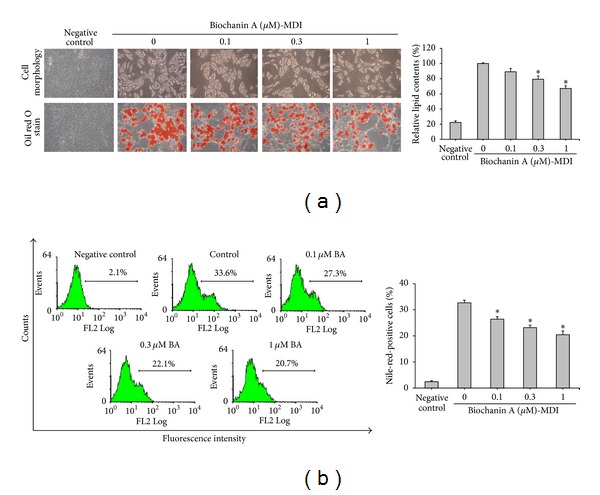
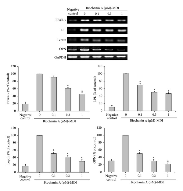
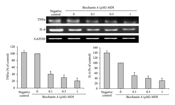
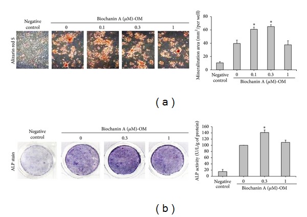
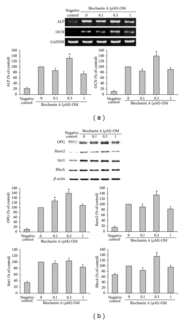
Biochanin A has promising effects on bone formation in vivo, although the underlying mechanism remains unclear yet. This study therefore aimed to investigate whether biochanin A regulates osteogenic and adipogenic differentiation using primary adipose-derived stem cells. The effects of biochanin A (at a physiologically relevant concentration of 0.1-1 μM) were assessed in vitro using various approaches, including Oil red O staining, Nile red staining, alizarin red S staining, alkaline phosphatase (ALP) activity, flow cytometry, RT-PCR, and western blotting. The results showed that biochanin A significantly suppressed adipocyte differentiation, as demonstrated by the inhibition of cytoplasmic lipid droplet accumulation, along with the inhibition of peroxisome proliferator-activated receptor gamma (PPAR γ ), lipoprotein lipase (LPL), and leptin and osteopontin (OPN) mRNA expression, in a dose-dependent manner. On the other hand, treatment of cells with 0.3 μM biochanin A increased the mineralization and ALP activity, and stimulated the expression of the osteogenic marker genes ALP and osteocalcin (OCN). Furthermore, biochanin A induced the expression of runt-related transcription factor 2 (Runx2), osteoprotegerin (OPG), and Ras homolog gene family, member A (RhoA) proteins. These observations suggest that biochanin A prevents adipogenesis, enhances osteoblast differentiation in mesenchymal stem cells, and has beneficial regulatory effects in bone formation.
Figures
Similar articles
-
A novel semisynthetic molecule icaritin stimulates osteogenic differentiation and inhibits adipogenesis of mesenchymal stem cells.Int J Med Sci. 2013 Apr 23;10(6):782-9. doi: 10.7150/ijms.6084. Print 2013. Int J Med Sci. 2013. PMID: 23630444 Free PMC article.
-
[Spermidine enhances osteogenic differentiation and inhibits adipogenic differentiation of bone marrow-derived mesenchymal stem cells from ovariectomized mice].Xi Bao Yu Fen Zi Mian Yi Xue Za Zhi. 2015 Jun;31(6):787-91. Xi Bao Yu Fen Zi Mian Yi Xue Za Zhi. 2015. PMID: 26062423 Chinese.
-
Interleukin-17A promotes osteogenic differentiation by increasing OPG/RANKL ratio in stem cells from human exfoliated deciduous teeth (SHED).J Tissue Eng Regen Med. 2018 Aug;12(8):1856-1866. doi: 10.1002/term.2706. Epub 2018 Jun 25. J Tissue Eng Regen Med. 2018. PMID: 29774992
-
Irradiation alters the differentiation potential of bone marrow mesenchymal stem cells.Mol Med Rep. 2016 Jan;13(1):213-23. doi: 10.3892/mmr.2015.4539. Epub 2015 Nov 10. Mol Med Rep. 2016. PMID: 26572960 Free PMC article.
-
Epimedium-derived flavonoids modulate the balance between osteogenic differentiation and adipogenic differentiation in bone marrow stromal cells of ovariectomized rats via Wnt/β-catenin signal pathway activation.Chin J Integr Med. 2012 Dec;18(12):909-17. doi: 10.1007/s11655-012-1294-2. Epub 2012 Dec 13. Chin J Integr Med. 2012. PMID: 23238999
Cited by
-
Mechanisms Behind the Pharmacological Application of Biochanin-A: A review.F1000Res. 2023 Dec 11;12:107. doi: 10.12688/f1000research.126059.3. eCollection 2023. F1000Res. 2023. PMID: 38106650 Free PMC article. Review.
-
Protective effects of biochanin A on articular cartilage: in vitro and in vivo studies.BMC Complement Altern Med. 2014 Nov 15;14:444. doi: 10.1186/1472-6882-14-444. BMC Complement Altern Med. 2014. PMID: 25398247 Free PMC article.
-
The Role of Isoflavones in Type 2 Diabetes Prevention and Treatment-A Narrative Review.Int J Mol Sci. 2020 Dec 28;22(1):218. doi: 10.3390/ijms22010218. Int J Mol Sci. 2020. PMID: 33379327 Free PMC article. Review.
-
Growth and Osteogenic Differentiation of Discarded Gingiva-Derived Mesenchymal Stem Cells on a Commercial Scaffold.Front Cell Dev Biol. 2020 May 21;8:292. doi: 10.3389/fcell.2020.00292. eCollection 2020. Front Cell Dev Biol. 2020. PMID: 32509773 Free PMC article.
-
The Antiaging Gene Klotho Regulates Proliferation and Differentiation of Adipose-Derived Stem Cells.Stem Cells. 2016 Jun;34(6):1615-25. doi: 10.1002/stem.2305. Epub 2016 Feb 26. Stem Cells. 2016. PMID: 26865060 Free PMC article.
References
-
- Rosen CJ, Bouxsein ML. Mechanisms of disease: is osteoporosis the obesity of bone? Nature Clinical Practice Rheumatology. 2006;2(1):35–43. - PubMed
-
- Justesen J, Stenderup K, Eriksen EF, Kassem M. Maintenance of osteoblastic and adipocytic differentiation potential with age and osteoporosis in human marrow stromal cell cultures. Calcified Tissue International. 2002;71(1):36–44. - PubMed
-
- Nuttall ME, Gimble JM. Controlling the balance between osteoblastogenesis and adipogenesis and the consequent therapeutic implications. Current Opinion in Pharmacology. 2004;4(3):290–294. - PubMed
-
- Rodriguez JP, Montecinos L, Rios S, Reyes P, Martinez J. Mesenchymal stem cells from osteoporotic patients produce a type I collagen-deficient extracellular matrix favoring adipogenic differentiation. Journal of Cellular Biochemistry. 2000;79(4):557–565. - PubMed
-
- Astudillo P, Ríos S, Pastenes L, Pino AM, Rodríguez JP. Increased adipogenesis of osteoporotic human-mesenchymal stem cells (MSCs) is characterized by impaired leptin action. Journal of Cellular Biochemistry. 2008;103(4):1054–1065. - PubMed
LinkOut - more resources
Full Text Sources
Other Literature Sources
Research Materials
