

The many spaces of uPAR: delivery of theranostic agents and nanobins to multiple tumor compartments through a single target
- PMID: 23843897
- PMCID: PMC3706693
- DOI: 10.7150/thno.4953
The many spaces of uPAR: delivery of theranostic agents and nanobins to multiple tumor compartments through a single target
Abstract
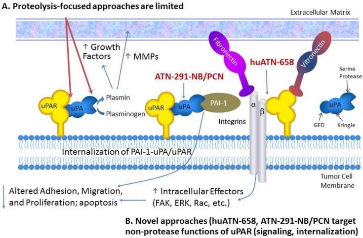
The urokinase plasminogen activator (uPA) system is a proteolytic system comprised of uPA, a cell surface receptor for uPA (uPAR), and an inhibitor of uPA (PAI-1) and is implicated in many aspects of tumor growth and metastasis. The uPA system has been identified in nearly all solid tumors examined to date as well as several hematological malignancies. In adults, transient expression of the uPA system is observed during wound healing and inflammatory processes while only limited expression is identified in healthy, quiescent tissue. Members of the uPA system are expressed not only on cancer cells but also on tumor-associated stromal cells. These factors make the uPA system an ideal therapeutic target for cancer therapies. To date most therapeutics targeted at the uPA system have been inhibitors of either the uPA-uPAR interaction or uPA proteolysis but have not shown robust anti-tumor activity. There is now mounting evidence that uPAR participates in a complex signaling network central to its role in cancer progression, which provides a basis for the hypothesis that uPAR may be a marker for cancer stem cells. Several new uPAR-directed therapies have recently been developed based on this new information. A monoclonal antibody has been developed that disrupts the interactions of uPAR with signaling partners and is poised to enter the clinic. In addition, nanoscale drug delivery vehicles targeted to the uPA system using monoclonal antibodies, without disrupting the normal functioning of the system, are also in development. This review will highlight some of these new discoveries and the new uPA system-based therapeutic approaches that have arisen from them.
Keywords: nanobins; theranostics; urokinase plasminogen activator.
Conflict of interest statement
Competing Interests: The authors have declared that no competing interest exists.
Figures

References
-
- Blasi F, Carmeliet P. uPAR: a versatile signalling orchestrator. Nat Rev Mol Cell Biol. 2002;3:932–43. - PubMed
-
- Dass K, Ahmad A, Azmi AS, Sarkar SH, Sarkar FH. Evolving role of uPA/uPAR system in human cancers. Cancer Treat Rev. 2008;34:122–36. doi:S0305-7372(07)00181-8 [pii]10.1016/j.ctrv.2007.10.005. - PubMed
-
- Mazar AP. Urokinase plasminogen activator receptor choreographs multiple ligand interactions: implications for tumor progression and therapy. Clin Cancer Res. 2008;14:5649–55. - PubMed
-
- Binder BR, Mihaly J, Prager GW. uPAR-uPA-PAI-1 interactions and signaling: a vascular biologist's view. Thromb Haemost. 2007;97:336–42. - PubMed
-
- Hildenbrand R, Gandhari M, Stroebel P, Marx A, Allgayer H, Arens N. The urokinase-system--role of cell proliferation and apoptosis. Histol Histopathol. 2008;23:227–36. - PubMed
Publication types
MeSH terms
Substances
Grants and funding
LinkOut - more resources
Full Text Sources
Other Literature Sources
Miscellaneous
