. 2013 Jul 3;8(7):e65927.
doi: 10.1371/journal.pone.0065927. Print 2013.

The role of the RACK1 ortholog Cpc2p in modulating pheromone-induced cell cycle arrest in fission yeast

Affiliations

The role of the RACK1 ortholog Cpc2p in modulating pheromone-induced cell cycle arrest in fission yeast

Magdalena Mos et al. PLoS One. .

Expression of concern in

Abstract

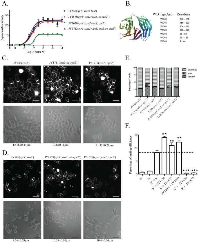
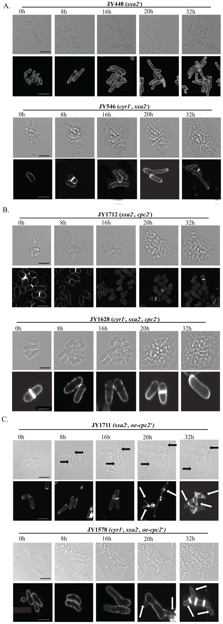
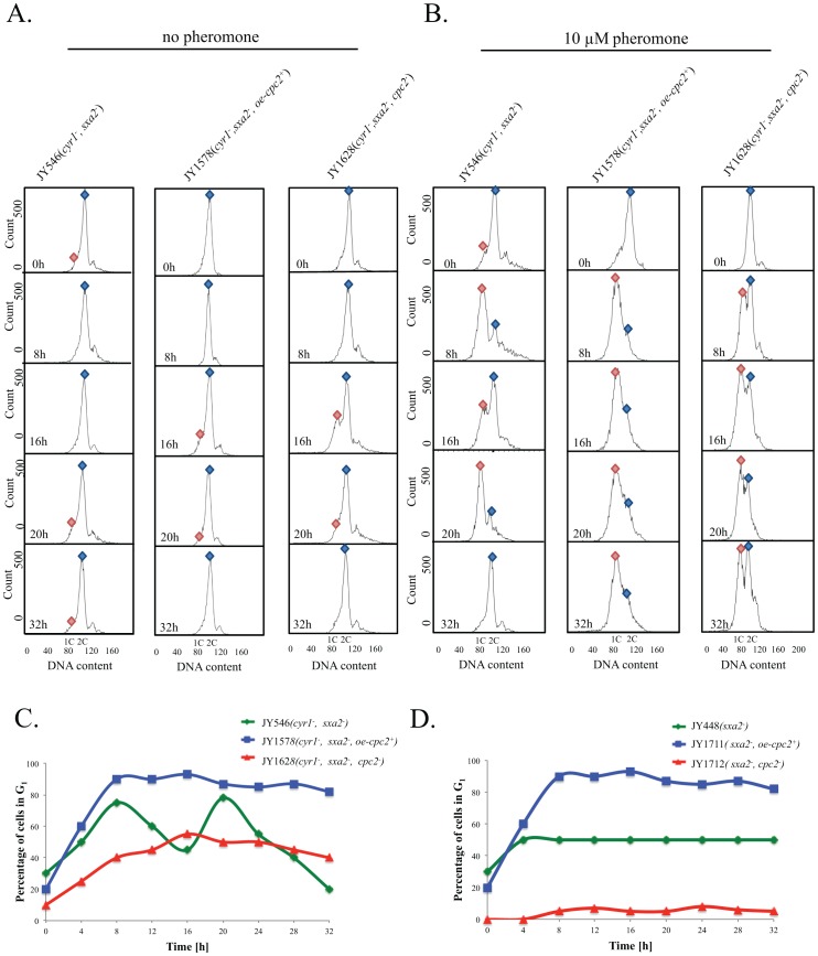
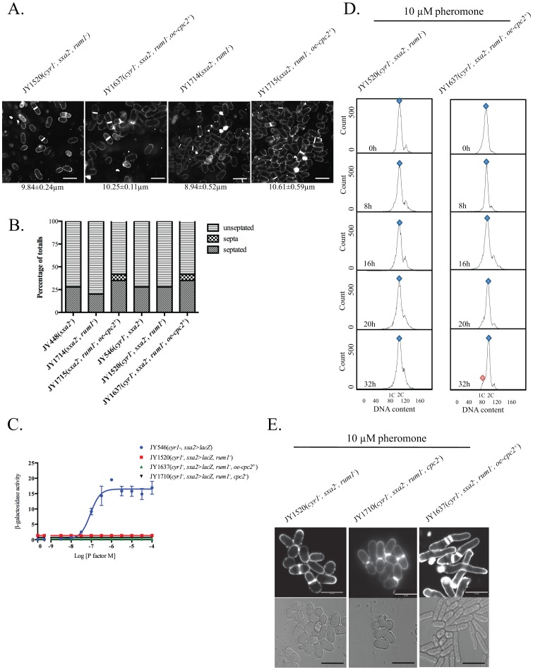
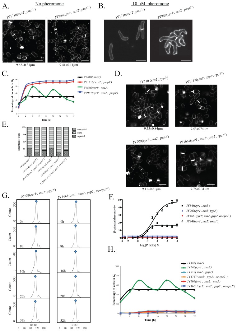
The detection and amplification of extracellular signals requires the involvement of multiple protein components. In mammalian cells the receptor of activated C kinase (RACK1) is an important scaffolding protein for signal transduction networks. Further, it also performs a critical function in regulating the cell cycle by modulating the G1/S transition. Many eukaryotic cells express RACK1 orthologs, with one example being Cpc2p in the fission yeast Schizosaccharomyces pombe. In contrast to RACK1, Cpc2p has been described to positively regulate, at the ribosomal level, cells entry into M phase. In addition, Cpc2p controls the stress response pathways through an interaction with Msa2p, and sexual development by modulating Ran1p/Pat1p. Here we describe investigations into the role, which Cpc2p performs in controlling the G protein-mediated mating response pathway. Despite structural similarity to Gβ-like subunits, Cpc2p appears not to function at the G protein level. However, upon pheromone stimulation, cells overexpressing Cpc2p display substantial cell morphology defects, disorientation of septum formation and a significantly protracted G1 arrest. Cpc2p has the potential to function at multiple positions within the pheromone response pathway. We provide a mechanistic interpretation of this novel data by linking Cpc2p function, during the mating response, with its previous described interactions with Ran1p/Pat1p. We suggest that overexpressing Cpc2p prolongs the stimulated state of pheromone-induced cells by increasing ste11 gene expression. These data indicate that Cpc2p regulates the pheromone-induced cell cycle arrest in fission yeast by delaying cells entry into S phase.


Conflict of interest statement

Competing Interests: The authors have declared that no competing interests exist.

Figures

Figure 1
Figure 1. Characterization of potential Gβ-subunit mimic in the pheromone-response pathway.
(A) Pheromone-dependent transcription for the strains JY546 (h, cyr1, sxa2>lacZ), JY1578 (h, cyr1, sxa2>lacZ+oe-cpc2+), JY1628 (h, cyr1, sxa2>lacZ, cpc2) and JY1713 (h, cyr1, sxa2>lacZ, cpc2+oe-cpc2+) was determined using the sxa2>lacZ reporter. Values are means of triplicate determinations ± S.E.M. (B) Molecular structure as determined using the SWISS-MODEL Repository for Cpc2p. Cpc2p contains seven WD-40 repeat domains with positions of residues as indicated by colors on the molecular structure. (C) Cell morphology and size, at division (micrometers ± S.D.) for strains JY448 (h, cyr1, sxa2), JY1711 (h, sxa2+oe-cpc2+), JY1712 (h, sxa2, cpc2), grown in minimal medium at 29°C and stained with calcofluor white (top panel) or imaged through using bright field microscopy on agar pads (bottom panel). Scale bars 10 µm. (D) As for C except strains JY546 (h, cyr1, sxa2>lacZ), JY1578 (h, cyr1, sxa2>lacZ+oe-cpc2+), and JY1628 (h, cyr1, sxa2>lacZ, cpc2) were used. (E) Numbers of non-septated, septated and multiple septa containing cells for the strains in C and D were determined from 400 individual cells. Values shown correspond to the percentages of the total population. Cells were stained with calcofluor white, to enable visualization of septum material. (F) Mating efficiency was quantitated for cells overexpressing (JY1634, h, +oe-cpc2+; JY1633 h+, +oe-cpc2+) or lacking Cpc2p (JY1636, h, cpc2; JY1635 h+, cpc2) compared to M (JY402, h) and P (JY383, h+) control cells. Statistical significance determined compared to M (h) and P (h+) using a one-way Anova with a tukey multiple comparison post test where ★★★ represents p≤0.001, ★★ represents p≤0.01, and ★ represents p≤0.1.
Figure 2
Figure 2. Cpc2p has profound morphological effects upon pheromone-stimulated cells.
The strains (A) JY448 (h, sxa2) and JY546 (h, cyr1, sxa2>lacZ); (B) JY1712 (h, sxa2, cpc2) and JY1628 (h, cyr1, sxa2>lacZ, cpc2); (C) JY1711 (h, sxa2+oe-cpc2+), JY1578 (h, cyr1, sxa2>lacZ+oe-cpc2+), were grown to mid-exponential phase over 32 h in minimal media. Cells were then imaged using bright field microscopy on pads containing 10 µM of pheromone (see methods). Cells were also stained with calcofluor white (lower panels A-C) to visualize septation. Scale bars 10 µm. Prolonged exposure to pheromone for cells overexpressing Cpc2p (oe-cpc2+) results in multiple projection tips and a failure to undergo cytokinesis. Cells lacking Cpc2p fail to generate the classical shmoo formation as observed for control cells.
Figure 3
Figure 3. Cpc2p control the G1/S transition in pheromone-stimulated cells.
(A) The strains JY546 (h, cyr1, sxa2>lacZ), JY1578 (h, cyr1, sxa2>lacZ, +oe-cpc2+) and JY1628 (h, cyr1, sxa2>lacZ, cpc2) were grown in minimal medium and (B) minimal media containing 10 µM of pheromone for the times indicated. Cells were harvested and fixed prior to staining with propidium iodide prior to analysis using flow cytometry (see methods). The proportion of cells exhibiting 1C or 2C DNA content was determined using FACSDiva v4.1 software for the assigned gates indicated by the blue and red shapes. (C) The percentage of cells containing a 1C content (arrested in G1) as determined from B. Cells containing an additional content of Cpc2 fail to desensitize following pheromone stimulation and remain arrested for the time frame analyzed. Cells lacking Cpc2p fail to generate a significant arrest in G1 following exposure to 10 µM pheromone. (D) The percentage of cells from the strains JY448 (h, sxa2), JY1711 (h, sxa2+oe-cpc2+), JY1712 (h, sxa2, cpc2) containing a 1C content (arrested in G1) following nitrogen starvation and stimulation with 10 µM pheromone.
Figure 4
Figure 4. Overexpression of cpc2 mediates its pheromone effects in a G1-dependent manner.
(A) Cell morphology and size, at division (micrometers ± S.D.) for strains JY1520 (h, cyr1, sxa2>lacZ, rum1), JY1637 (h, cyr1, sxa2>lacZ, rum1+ oe-cpc2+), JY1714 (h, sxa2, rum1) and JY1715 (h, sxa2, rum1+oe-cpc2+) grown in minimal medium at 29°C and stained with calcofluor. Scale bars 10 µm. (B) Number of non-septated, septated and multiple septa containing cells for the strains JY448 (h, sxa2), JY1714 (h, sxa2, rum1), JY1715 (h, sxa2, rum1+oe-cpc2+), JY546 (h, cyr1, sxa2>lacZ), JY1520 (h, cyr1, sxa2>lacZ, rum1 ) and JY1637 (h, cyr1, sxa2>lacZ, rum1+oe-cpc2+) were determined from 400 individual cells. Values shown correspond to the percentage of the total population. Cells were stained with calcofluor white, to enable visualization of septum material. (C) Pheromone-dependent transcription for the strains JY546, JY1520, JY1637 and JY1710 (h, cyr1, sxa2>lacZ, rum1, cpc2) was determined using the sxa2>lacZ reporter. Cells were stimulated with pheromone for 16 h in minimal media and assayed for β-galactosidase production using ONPG. Activity is expressed as OD420 units per 106 cells. Values are means of triplicate determinations ± S.E.M. (D) The strains JY1520 and JY1637 were grown in minimal medium containing 10 µM of pheromone for the times indicated. Cells were harvested and fixed prior to staining with propidium iodide prior to analysis using flow cytometry (see methods). The proportion of cells exhibiting 1C or 2C DNA content was determined using FACSDiva v4.1 software for the assigned gates indicated by the blue and red shapes (E) The strains JY1520, JY1710 and JY1637 were grown to mid-exponential phase over 32 h in minimal media. Cells were then stained with calcofluor white to visualize septation (top panel) and imaged bright field microscopy (bottom panel) after 32 h exposure to pheromone. Scale bars 10 µm.
Figure 5
Figure 5. Overexpression of Cpc2 in pheromone stimulated cells mimics prolonged pheromone stimulation.
(A) Cell morphology and size, at division (micrometers ± S.D.) for the strains JY1716 (h, sxa2, pmp1) and JY948 (h, cyr1, sxa2>lacZ, pmp1) grown in minimal medium at 29°C and stained with calcofluor white. (B) Strains from A strains were exposure to 10 µM of pheromone for 32 h and stained with calcofluor white. (C) The percentage of cells containing a 1C content (arrested in G1) for the strains JY448, JY1716, JY546 and JY948 as determined using flow cytometry. Cells lacking Pmp1p show a failure to exit from a G1 arrest analogous to strains where the cpc2 ORF has been deleted. (D) Cell morphology and size, at division (micrometers ± S.D.) for strains JY710 (h, sxa2 pyp2) and JY1717 (h, sxa2, pyp2+oe-cpc2+) grown in minimal medium at 29°C and stained with calcofluor white (top panel). Cell morphology and size at division (micrometers ± S.D.) for the strains JY709 (h, cyr1, sxa2>lacZ, pyp2) and JY1661 (h, cyr1, sxa2>lacZ, pyp2+oe-cpc2+) grown in minimal medium at 29°C and stained with calcofluor white (bottom panel). (E) Numbers of non-septated, septated and multiple septa containing cells for the strains JY448, JY1714, JY1715, JY546, JY987 and JY1661 were determined from 400 individual cells. Values shown correspond to the percentages of the total population. Cells were stained with calcofluor white, to enable visualization of septum material. (F) Pheromone-dependent transcription for the strains JY546, JY709, JY1661 and JY948 was determined using the sxa2>lacZ reporter. Cells were stimulated with pheromone for 16 h in minimal media and assayed for β-galactosidase production using ONPG. Activity is expressed as OD420 units per 106 cells. Values are means of triplicate determinations ± S.E.M. (G) The strains JY709 and JY1661 were grown in minimal medium containing 10 µM of pheromone for the times indicated. Cells were harvested and fixed prior to staining with propidium iodide prior to analysis using flow cytometry (see methods). The proportion of cells exhibiting 1C or 2C DNA content was determined using FACSDiva v4.1 software for the assigned gates indicated by the blue and red shapes. (H) The percentage of cells containing a 1C content (arrested in G1) as determined for the strains JY448, JY546, JY710, JY1717, JY709 and JY1661.

Similar articles

Cited by

References

    1. Gilman AG (1987) G proteins: transducers of receptor-generated signals. Annu Rev Biochem 56: 615–649. - PubMed
    1. Dupré DJ, Robitaille M, Rebois RV, Hébert TE (2009) The Role of Gβγ Subunits in the Organization, Assembly, and Function of GPCR Signaling Complexes. Annu Rev Pharmacol Toxicol 49: 31–56. - PMC - PubMed
    1. Hill C, Goddard A, Ladds G, Davey J (2009) The cationic region of Rhes mediates its interactions with specific Gbeta subunits. Cell Physiol Biochem 23: 1–8. - PubMed
    1. Hiskens R, Vatish M, Hill C, Davey J, Ladds G (2005) Specific in vivo binding of activator of G protein signalling 1 to the G [beta] 1 subunit. Biochem Biophy Res Commun 2 337: 1038–1046. - PubMed
    1. Blumer JB, Cismowski MJ, Sato M, Lanier SM (2005) AGS proteins: receptor-independent activators of G-protein signaling. Trends Pharmacol Sci 26: 470–476. - PubMed

Publication types

MeSH terms

Substances