

Avian WNT4 in the female reproductive tracts: potential role of oviduct development and ovarian carcinogenesis
- PMID: 23843947
- PMCID: PMC3699571
- DOI: 10.1371/journal.pone.0065935
Avian WNT4 in the female reproductive tracts: potential role of oviduct development and ovarian carcinogenesis
Abstract
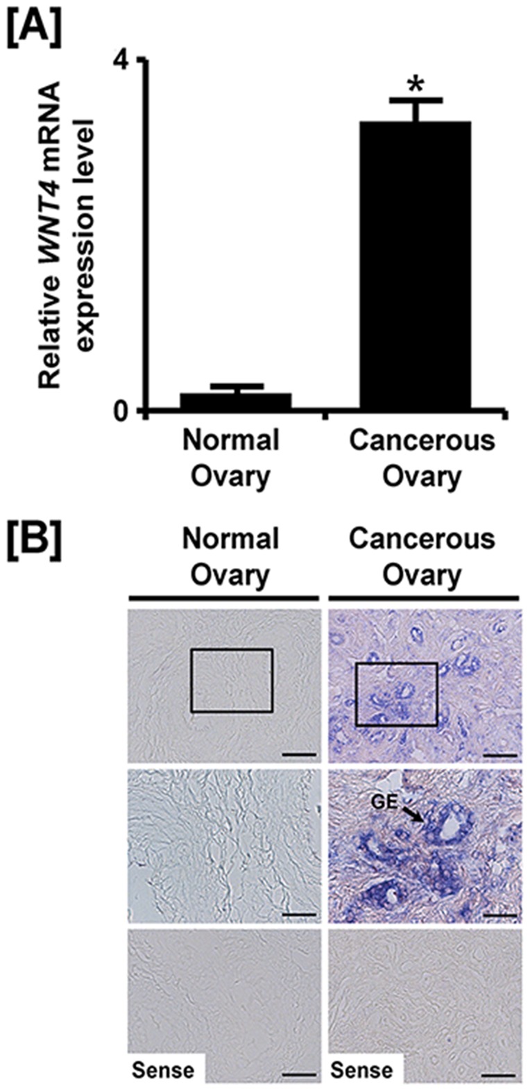
The wingless-type MMTV integration site family of proteins (WNTs) is highly conserved secreted lipid-modified signaling molecules that play a variety of pivotal roles in developmental events such as embryogenesis, tissue homeostasis and cell polarity. Although, of these proteins, WNT4 is known to be involved in genital development in fetuses of mammalian species, its role is unknown in avian species. Therefore, in this study, we investigated expression profiles, as well as hormonal and post-transcriptional regulation of WNT4 expression in the reproductive tract of female chickens. Results of this study demonstrated that WNT4 is most abundant in the stromal and luminal epithelial cells of the isthmus and shell gland of the oviduct, respectively. WNT4 is also most abundant in the glandular epithelium of the shell gland of the oviduct of laying hens at 3 h post-ovulation during the laying cycle. In addition, treatment of young chicks with diethylstilbestrol (DES, a synthetic estrogen agonist) stimulated WNT4 only in the glandular epithelial cells of the isthmus and shell gland of the oviduct. Moreover, results of our study demonstrated that miR-1786 influences WNT4 expression via specific binding sites in its 3'-UTR. On the other hand, our results also indicate that WNT4 is expressed predominantly in the glandular epithelium of cancerous ovaries, but not in normal ovaries of hens. Collectively, these results indicate cell-specific expression of WNT4 in the reproductive tract of chickens and that it likely has crucial roles in development and function of oviduct as well as initiation of ovarian carcinogenesis in laying hens.
Conflict of interest statement
Figures


Similar articles
-
Differential expression of alpha 2 macroglobulin in response to dietylstilbestrol and in ovarian carcinomas in chickens.Reprod Biol Endocrinol. 2011 Oct 7;9:137. doi: 10.1186/1477-7827-9-137. Reprod Biol Endocrinol. 2011. PMID: 21978460 Free PMC article.
-
Hormonal regulation of beta-catenin during development of the avian oviduct and its expression in epithelial cell-derived ovarian carcinogenesis.Mol Cell Endocrinol. 2014 Jan 25;382(1):46-54. doi: 10.1016/j.mce.2013.09.010. Epub 2013 Sep 18. Mol Cell Endocrinol. 2014. PMID: 24055276
-
Expression and regulation of beta-defensin 11 in the oviduct in response to estrogen and in ovarian tumors of chickens.Mol Cell Endocrinol. 2013 Feb 5;366(1):1-8. doi: 10.1016/j.mce.2012.10.031. Epub 2012 Nov 16. Mol Cell Endocrinol. 2013. PMID: 23159989
-
Differential expression of vitelline membrane outer layer protein 1: hormonal regulation of expression in the oviduct and in ovarian carcinomas from laying hens.Mol Cell Endocrinol. 2015 Jan 5;399:250-8. doi: 10.1016/j.mce.2014.10.015. Epub 2014 Oct 18. Mol Cell Endocrinol. 2015. PMID: 25458700
-
WNT4, RSPO1, and FOXL2 in sex development.Semin Reprod Med. 2012 Oct;30(5):387-95. doi: 10.1055/s-0032-1324722. Epub 2012 Oct 8. Semin Reprod Med. 2012. PMID: 23044875 Review.
Cited by
-
Advantages of the avian model for human ovarian cancer.Mol Clin Oncol. 2015 Nov;3(6):1191-1198. doi: 10.3892/mco.2015.619. Epub 2015 Aug 11. Mol Clin Oncol. 2015. PMID: 26807219 Free PMC article.
-
Characterization and miRNA-mediated posttranscriptional regulation of vitelline membrane outer layer protein I in the adult chicken oviduct.In Vitro Cell Dev Biol Anim. 2015 Mar;51(3):222-9. doi: 10.1007/s11626-014-9826-2. Epub 2014 Nov 8. In Vitro Cell Dev Biol Anim. 2015. PMID: 25381035
-
Modulation of folliculogenesis in adult laying chickens by bisphenol A and bisphenol S: Perspectives on ovarian morphology and gene expression.Reprod Toxicol. 2021 Aug;103:181-190. doi: 10.1016/j.reprotox.2021.06.010. Epub 2021 Jun 18. Reprod Toxicol. 2021. PMID: 34147626 Free PMC article.
-
A Review of Principal Studies on the Development and Treatment of Epithelial Ovarian Cancer in the Laying Hen Gallus gallus.Comp Med. 2021 Aug 1;71(4):271-284. doi: 10.30802/AALAS-CM-20-000116. Epub 2021 Jul 29. Comp Med. 2021. PMID: 34325771 Free PMC article. Review.
-
A Novel Single-Nucleotide Polymorphism in WNT4 Promoter Affects Its Transcription and Response to FSH in Chicken Follicles.Genes (Basel). 2022 Oct 1;13(10):1774. doi: 10.3390/genes13101774. Genes (Basel). 2022. PMID: 36292659 Free PMC article.
References
-
- Dougherty DC, Sanders MM (2005) Estrogen action: revitalization of the chick oviduct model. Trends Endocrinol Metab 16: 414–419. - PubMed
-
- Palmiter RD (1972) Regulation of protein synthesis in chick oviduct. I. Independent regulation of ovalbumin, conalbumin, ovomucoid, and lysozyme induction. J Biol Chem 247: 6450–6461. - PubMed
-
- Franco HL, Dai D, Lee KY, Rubel CA, Roop D, et al. (2011) WNT4 is a key regulator of normal postnatal uterine development and progesterone signaling during embryo implantation and decidualization in the mouse. FASEB journal : official publication of the Federation of American Societies for Experimental Biology 25: 1176–1187. - PMC - PubMed
Publication types
MeSH terms
Substances
LinkOut - more resources
Full Text Sources
Other Literature Sources
Medical
Molecular Biology Databases
