

A multivariate surface-based analysis of the putamen in premature newborns: regional differences within the ventral striatum
- PMID: 23843961
- PMCID: PMC3700976
- DOI: 10.1371/journal.pone.0066736
A multivariate surface-based analysis of the putamen in premature newborns: regional differences within the ventral striatum
Abstract
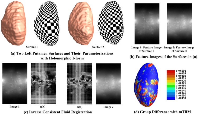
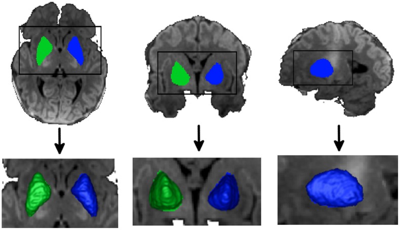
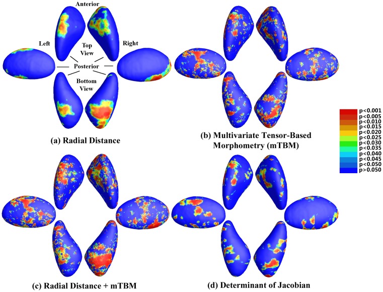
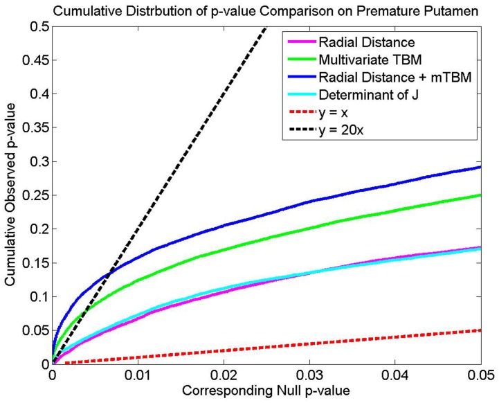
Many children born preterm exhibit frontal executive dysfunction, behavioral problems including attentional deficit/hyperactivity disorder and attention related learning disabilities. Anomalies in regional specificity of cortico-striato-thalamo-cortical circuits may underlie deficits in these disorders. Nonspecific volumetric deficits of striatal structures have been documented in these subjects, but little is known about surface deformation in these structures. For the first time, here we found regional surface morphological differences in the preterm neonatal ventral striatum. We performed regional group comparisons of the surface anatomy of the striatum (putamen and globus pallidus) between 17 preterm and 19 term-born neonates at term-equivalent age. We reconstructed striatal surfaces from manually segmented brain magnetic resonance images and analyzed them using our in-house conformal mapping program. All surfaces were registered to a template with a new surface fluid registration method. Vertex-based statistical comparisons between the two groups were performed via four methods: univariate and multivariate tensor-based morphometry, the commonly used medial axis distance, and a combination of the last two statistics. We found statistically significant differences in regional morphology between the two groups that are consistent across statistics, but more extensive for multivariate measures. Differences were localized to the ventral aspect of the striatum. In particular, we found abnormalities in the preterm anterior/inferior putamen, which is interconnected with the medial orbital/prefrontal cortex and the midline thalamic nuclei including the medial dorsal nucleus and pulvinar. These findings support the hypothesis that the ventral striatum is vulnerable, within the cortico-stiato-thalamo-cortical neural circuitry, which may underlie the risk for long-term development of frontal executive dysfunction, attention deficit hyperactivity disorder and attention-related learning disabilities in preterm neonates.
Conflict of interest statement
Figures
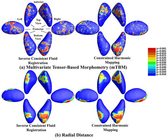
 with the two registration methods. Bottom panel: P-values for the comparison of
  with the two methods. The left column represents the fluid registration, while the right one is for the constrained harmonic mapping. The meaning of the different colors is shown in the colorbar.
Similar articles
-
Thalamic alterations in preterm neonates and their relation to ventral striatum disturbances revealed by a combined shape and pose analysis.Brain Struct Funct. 2016 Jan;221(1):487-506. doi: 10.1007/s00429-014-0921-7. Epub 2014 Nov 1. Brain Struct Funct. 2016. PMID: 25366970 Free PMC article.
-
Surface fluid registration and multivariate tensor-based morphometry in newborns - the effects of prematurity on the putamen.Signal Inf Process Assoc Annu Summit Conf APSIPA Asia Pac. 2012 Dec;2012:https://ieeexplore.ieee.org/stamp/stamp.jsp?arnumber=6411997. Epub 2013 Jan 17. Signal Inf Process Assoc Annu Summit Conf APSIPA Asia Pac. 2012. PMID: 29938710 Free PMC article.
-
Basal ganglia surface morphology and the effects of stimulant medications in youth with attention deficit hyperactivity disorder.Am J Psychiatry. 2010 Aug;167(8):977-86. doi: 10.1176/appi.ajp.2010.09091259. Epub 2010 Jul 1. Am J Psychiatry. 2010. PMID: 20595414 Free PMC article.
-
[Structural and functional neuroanatomy of attention-deficit hyperactivity disorder (ADHD)].Encephale. 2009 Apr;35(2):107-14. doi: 10.1016/j.encep.2008.01.005. Epub 2008 Jul 7. Encephale. 2009. PMID: 19393378 Review. French.
-
Striatal tissue transplantation in non-human primates.Prog Brain Res. 2000;127:381-404. doi: 10.1016/s0079-6123(00)27018-0. Prog Brain Res. 2000. PMID: 11142037 Review.
Cited by
-
Predicting Alzheimer's Disease Using Combined Imaging-Whole Genome SNP Data.J Alzheimers Dis. 2015;46(3):695-702. doi: 10.3233/JAD-150164. J Alzheimers Dis. 2015. PMID: 25869783 Free PMC article.
-
The Relationship between Neurocircuitry Dysfunctions and Attention Deficit Hyperactivity Disorder: A Review.Biomed Res Int. 2016;2016:3821579. doi: 10.1155/2016/3821579. Epub 2016 Sep 1. Biomed Res Int. 2016. PMID: 27689077 Free PMC article. Review.
-
Impact of Early and Late Visual Deprivation on the Structure of the Corpus Callosum: A Study Combining Thickness Profile with Surface Tensor-Based Morphometry.Neuroinformatics. 2015 Jul;13(3):321-336. doi: 10.1007/s12021-014-9259-9. Neuroinformatics. 2015. PMID: 25649876 Free PMC article.
-
Neuroimaging PheWAS (Phenome-Wide Association Study): A Free Cloud-Computing Platform for Big-Data, Brain-Wide Imaging Association Studies.Neuroinformatics. 2021 Apr;19(2):285-303. doi: 10.1007/s12021-020-09486-4. Neuroinformatics. 2021. PMID: 32822005 Free PMC article.
-
Ventricular shape and relative position abnormalities in preterm neonates.Neuroimage Clin. 2017 May 28;15:483-493. doi: 10.1016/j.nicl.2017.05.025. eCollection 2017. Neuroimage Clin. 2017. PMID: 28649491 Free PMC article.
References
-
- Lou HC (1996) Etiology and pathogenesis of attention-de_cit hyperactivity disorder (ADHD): significance of prematurity and perinatal hypoxic-haemodynamic encephalopathy. Acta Paediatr 85(11): 1266–71. - PubMed
-
- Powell KB, Voeller KKS (2004) Prefrontal executive function syndromes in children. J Child Neurol 19: 785–797. - PubMed
-
- Wang Y, Panigrahy A, Shi J, Ceschin R, Nelson MD, et al... (2011a) Surface Multivariate Tensor based Morphometry on premature neonates: a pilot study. Proceedings of the MICCAI workshop on Image Analysis of Human Brain Development (IAHBD 2011).
-
- Peterson BS, Vohr B, Staib LH, Dolberg A, Schneider KC, et al. (2000) Regional brain volume abnormalities and long-term cognitive outcome in preterm infants. JAMA-J Am Med Assoc 284(15): 1939–1947. - PubMed
Publication types
MeSH terms
Grants and funding
LinkOut - more resources
Full Text Sources
Other Literature Sources
Medical
