. 2013 Jul 2;8(7):e67084.
doi: 10.1371/journal.pone.0067084. Print 2013.

Pulmonary abnormalities in animal models due to Niemann-Pick type C1 (NPC1) or C2 (NPC2) disease

Affiliations

Pulmonary abnormalities in animal models due to Niemann-Pick type C1 (NPC1) or C2 (NPC2) disease

Blair R Roszell et al. PLoS One. .

Abstract

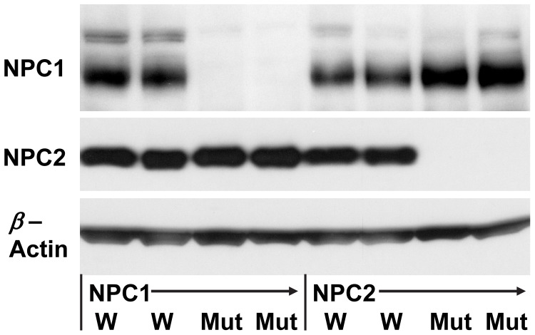
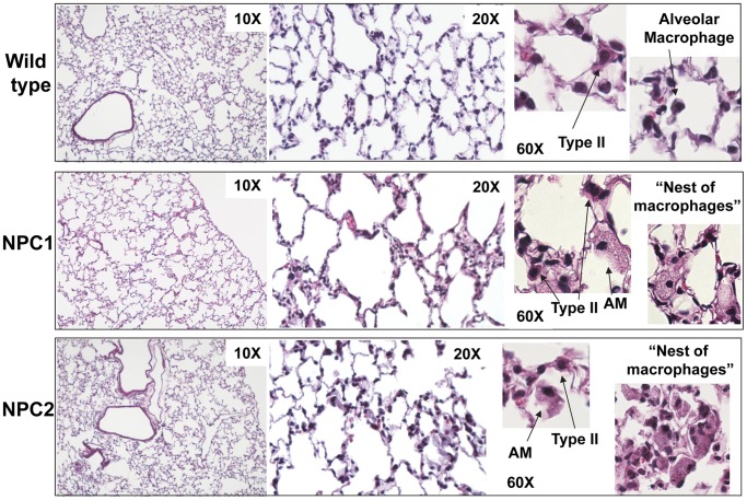
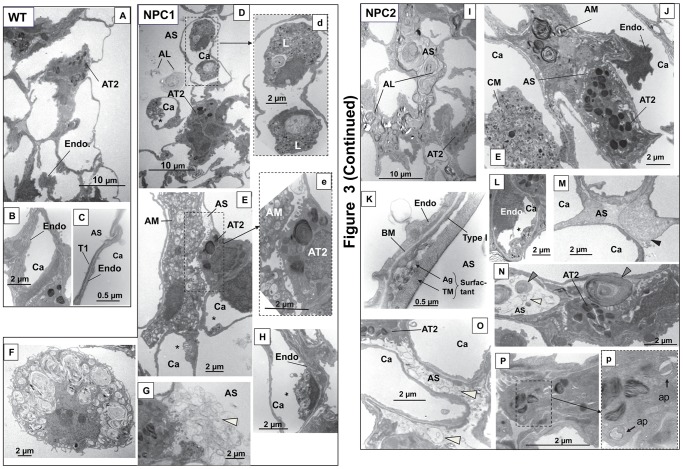
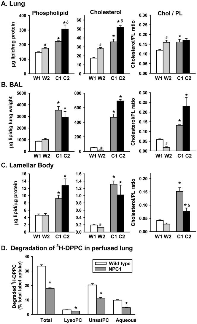
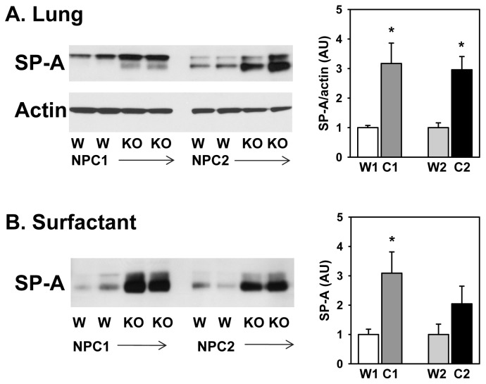
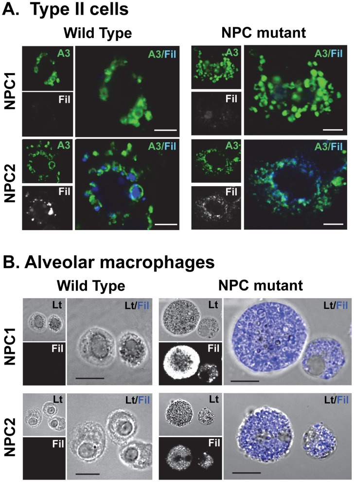
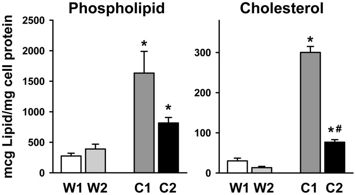
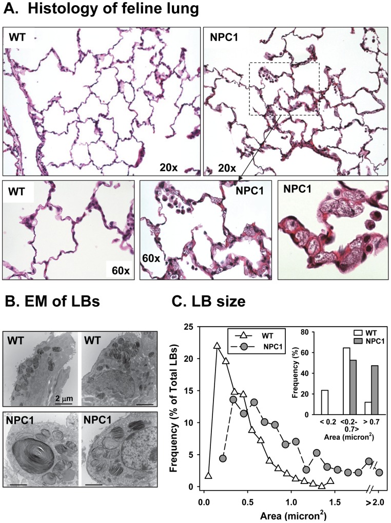
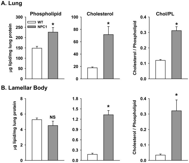
Niemann-Pick C (NPC) disease is due to loss of NPC1 or NPC2 protein function that is required for unesterified cholesterol transport from the endosomal/lysosomal compartment. Though lung involvement is a recognized characteristic of Niemann-Pick type C disease, the pathological features are not well understood. We investigated components of the surfactant system in both NPC1 mutant mice and felines and in NPC2 mutant mice near the end of their expected life span. Histological analysis of the NPC mutant mice demonstrated thickened septae and foamy macrophages/leukocytes. At the level of electron microscopy, NPC1-mutant type II cells had uncharacteristically larger lamellar bodies (LB, mean area 2-fold larger), while NPC2-mutant cells had predominantly smaller lamellar bodies (mean area 50% of normal) than wild type. Bronchoalveolar lavage from NPC1 and NPC2 mutant mice had an approx. 4-fold and 2.5-fold enrichment in phospholipid, respectively, and an approx. 9-fold and 35-fold enrichment in cholesterol, consistent with alveolar lipidosis. Phospholipid and cholesterol also were elevated in type II cell LBs and lung tissue while phospholipid degradation was reduced. Enrichment of surfactant protein-A in the lung and surfactant of the mutant mice was found. Immunocytochemical results showed that cholesterol accumulated in the LBs of the type II cells isolated from the affected mice. Alveolar macrophages from the NPC1 and NPC2 mutant mice were enlarged compared to those from wild type mice and were enriched in phospholipid and cholesterol. Pulmonary features of NPC1 mutant felines reflected the disease described in NPC1 mutant mice. Thus, with the exception of lamellar body size, the lung phenotype seen in the NPC1 and NPC2 mutant mice were similar. The lack of NPC1 and NPC2 proteins resulted in a disruption of the type II cell surfactant system contributing to pulmonary abnormalities.


Conflict of interest statement

Competing Interests: The authors have declared that no competing interests exist.

Figures

Figure 1
Figure 1. NPC proteins in mouse lungs.
Western blot of wild type (W) littermates, NPC1 (Mut) or NPC2 (Mut) mutant mouse lungs using anti-NPC1 or -NPC2 antibody. β-actin used as a loading control. 30 µg protein/lane.
Figure 2
Figure 2. Light micrograph of mutant mice lungs stained with hematoxylin and eosin.
Lungs from BALB/c wild type (95 days), NPC1 mutant (70 days) and NPC2 mutant mice (88 days). NPC1 and NPC2 mutant mice show “nests” of macrophages and alveolar macrophages with large inclusions.
Figure 3
Figure 3. Morphology of wild type and mutant mice lungs by electron microscopy.
Wild type lung (A–C). A. Overview of section of lung with alveolar type II cell (AT2) and endothelial cell (Endo). B. Capillary (Ca). C. Respiratory membrane with type I cell (T1) and endothelial cell (Endo). AS, alveolar space. NPC1 mutant lung (D–H). D. Overview of lung with leukocytes in the capillary and excess surfactant in alveolar space. AL, alveolar lipidosis. d. Enlargement of area in D showing vacuolar leukocyte (L). Similar leukocytes were seen in NPC2 mutant lung. E. Alveolar type II cell (AT2) with a foamy alveolar macrophage (AM) in close proximity in the alveolar space. e. Enlargement of area in E showing type II cell-macrophage contact. *Indicates vacuolar inclusions in endothelial cell. F. Alveolar macrophage with lipid-like material and vacuolar inclusions. G. Type II cells with excess surfactant (white arrowhead). H. Endothelial cell with vesicular inclusions (*). NPC2 mutant lung (I–P). I. Overview of lung with surfactant completely filling the alveolar space characteristic of alveolar lipidosis. J. AT2 with an alveolar macrophage containing multivesicular whirls and a foamy circulating macrophage (CM). K. Respiratory membrane of endothelial cell, basement membrane (BM) and type I cell and demonstrating large amounts of surfactant as tubular myelin (TM) and aggregate (Ag) structures. L. Endothelial cell with vesicular structures. M. Alveolar space with black arrowhead indicating proteinaceous material. Similar material was seen in NPC1 mutant lung. N. Large aggregate structure with tightly packed phospholipid-type whirls (gray arrowheads) or string-like structures (white arrowhead) in alveolar space. O. Surfactant vesicles (white arrowheads) filling the alveolar space. P. Type II cell with inset (p) showing autophagosome-like structures (ap) in enlargement.
Figure 4
Figure 4. Size of mouse lamellar bodies.
Using electron microscopic photographs of type II cells from wild type, NPC1 and NPC2 mutant mice, the size of the lamellar bodies was analyzed using ImageJ. Top. Electron micrographs of typical type II cells from wild type (WT, left), NPC1 (middle) and NPC2 (right) mutant mice. LB, lamellar body. Bottom. Histogram of the lamellar bodies from wild type (white triangles), NPC1 (gray circles) and NPC2 (black circles) type II cells. Frequency of each lamellar body size in micron2 is expressed as a % of the total numbers of lamellar bodies. Inset: size of lamellar bodies in grouped bins. Wild type, 60 type II cells, 451 LBs, 3 mice; NPC1, 53 type II cells, 435 LBs, 3 mice; NPC2, 37 type II cells, 459 LBs, 3 mice. *Statistically significant difference, P<0.05. E. Lamellar body sizes in grouped bins of ranges of areas.
Figure 5
Figure 5. Lipid content and degradation of DPPC in mice lungs.
A–C Cholesterol and phospholipid content of mice lungs. A. Lipid content of the lavaged lung as µg lipid/mg lung protein. Data are mean±SE, n = 4 separate mice. B. Broncho-alveolar lavage (BAL) of mice lungs. Data as µg lipid/gm weight of lung and are mean±SE, n = 4 separate mice. C. Lipid content of lamellar bodies isolated from the lungs of the wild type and mutant mice. Four (NPC1 or wild type littermates) or three (NPC2 or wild type littermates) lamellar body preparation isolated from the lungs of 2–6 mice. *Statistically significant difference versus wild type littermates, #statistically significant difference between wild types. δ Statistically significant difference between mutants, P<0.05. D. Degradation of 3H-labeled DPPC as percentage of total label uptake. 3H-DPPC liposome degradation by the isolated, perfused lungs of three wild type or NPC1 mutant mice after intratracheal instillation. Values are means±SE. *Statistically significant difference from wild type. P<0.001, n = 3. Total degradation is the sum of lysophosphatidyl choline (lysoPC), unsaturated PC (unsatPC), and the aqueous fractions.
Figure 6
Figure 6. Surfactant protein-A (SP-A) content of (A) lungs and (B) surfactant isolated from wild type (W) or NPC mutant (NPC1, NPC2) mice.
Left, Western blots of SP-A or actin. Right, quantitation of Western blots. A. Arbitrary units (AU) of SP-A relative to actin from lungs of NPC1 or NPC2 mutant mice (C1 or C2) or age-matched wild type controls (W1 or W2) (n = 6–8) or B. Arbitrary units of SP-A in surfactant (n = 4–9). All samples were loaded at equal protein values. *Significant difference from wild type, (P<0.05). The two SP-A bands are due to differences in glycosylation.
Figure 7
Figure 7. Immunocytochemistry of (A.) type II cells or (B.) alveolar macrophages isolated from wild type and NPC mutant mice.
A. Type II cells were isolated from NPC1 (Top) or NPC2 (Bottom) mutant mice or their corresponding wild type littermates and placed in culture for 24 hrs. The cells were fixed and stained with anti-ABCA3 antibody (green) to mark the lamellar body limiting membrane or filipin (Fil, gray or blue) to mark cholesterol. The merged pictures with anti-ABCA3 in green and filipin in blue are enlarged. Scale bar = 5 µm. B. Alveolar macrophages from NPC mutant mice contain cholesterol. Alveolar macrophages were isolated from the lung lavage from NPC1 (Top) or NPC2 (Bottom) mutant mice or their corresponding wild type littermates and placed in culture for 2 hrs. The cells were fixed and stained with filipin which labels free unesterified cholesterol. Lt, Phase micrograph; Fil, Filipin stain in gray. Merge of phase and filipin (blue) are enlarged. Scale bar = 10 µm.
Figure 8
Figure 8. Lipid content of NPC mutant macrophages is elevated.
Alveolar macrophages were isolated and cultured as in Fig. 7. The cells were harvested and the phospholipid and cholesterol content analyzed. W1,W2. Wild type littermates from NPC1 or NPC2 mutant mice, respectively. C1, NPC1; C2, NPC2. The data are mean±SE, n = 4 separate mice. *Statistically significant difference from corresponding wild type littermates. #Significant difference between NPC1 and NPC2 mutant macrophages, P<0.05.
Figure 9
Figure 9. Micrograph of feline lung.
A. Histology of feline lung. Wild type (WT) and NPC1 mutant (NPC1) feline lungs (25 weeks old) stained with hematoxylin and eosin. NPC1 mutant feline show thickened septae, enlarged “foamy” macrophages in capillaries and alveolar macrophages in the alveolar space. B. Electron micrographs (EM) of typical type II cells from wild type (WT) and NPC1 mutant (NPC1) feline indicating that the size of NPC1 mutant feline lamellar bodies is enlarged. C. Histogram of the lamellar bodies from wild type (white triangles) and NPC1 (gray circles) type II cells. Using electron microscopic photographs of type II cells from wild type and NPC1 mutant felines, the size of the lamellar bodies was analyzed using ImageJ. C, Inset: size of lamellar bodies in grouped bins in ranges of areas. Frequency of each lamellar body size in micron2 is expressed as a % of the total numbers of lamellar bodies. Wild type (WT): 28 type II cells, 250 LBs, 3 feline; NPC1 mutant: 35 type II cells, 323 LBs, 3 feline.
Figure 10
Figure 10. Phospholipid and cholesterol content of feline lungs.
A. Lipid content of the lung. Data are mean±SE, n = 3 feline. B. Lipid content of lamellar bodies isolated from the lungs of the wild type and NPC1 mutant feline. Data are the mean±SE and range of 5 (WT) or 4 (NPC1 mut) lamellar body preparations isolated from feline lungs analyzed in triplicate. *Significant different from WT.

Similar articles

Cited by

References

    1. Rosenbaum AI, Maxfield FR (2011) Niemann-Pick type C disease: molecular mechanisms and potential therapeutic approaches. Journal of neurochemistry 116: 789–795. - PMC - PubMed
    1. Griese M, Brasch F, Aldana VR, Cabrera MM, Goelnitz U, et al. (2010) Respiratory disease in Niemann-Pick type C2 is caused by pulmonary alveolar proteinosis. Clin Genet 77: 119–130. - PubMed
    1. Meiner V, Shpitzen S, Mandel H, Klar A, Ben-Neriah Z, et al. (2001) Clinical-biochemical correlation in molecularly characterized patients with Niemann-Pick type C. Genetics in medicine : official journal of the American College of Medical Genetics. 3: 343–348. - PubMed
    1. Millat G, Chikh K, Naureckiene S, Sleat DE, Fensom AH, et al. (2001) Niemann-Pick disease type C: spectrum of HE1 mutations and genotype/phenotype correlations in the NPC2 group. Am J Hum Genet 69: 1013–1021. - PMC - PubMed
    1. Vanier MT (2010) Niemann-Pick disease type C. Orphanet journal of rare diseases. 5: 16. - PMC - PubMed

Publication types

MeSH terms

LinkOut - more resources