. 2013 Jul 3;8(7):e67474.
doi: 10.1371/journal.pone.0067474. Print 2013.

Mesenchymal stem cells improve medullary inflammation and fibrosis after revascularization of swine atherosclerotic renal artery stenosis

Affiliations

Mesenchymal stem cells improve medullary inflammation and fibrosis after revascularization of swine atherosclerotic renal artery stenosis

Behzad Ebrahimi et al. PLoS One. .

Abstract

Atherosclerotic renal artery stenosis (ARAS) raises blood pressure and can reduce kidney function. Revascularization of the stenotic renal artery alone does not restore renal medullary structure and function. This study tested the hypothesis that addition of mesenchymal stem cells (MSC) to percutaneous transluminal renal angioplasty (PTRA) can restore stenotic-kidney medullary tubular transport function and attenuate its remodeling. Twenty-seven swine were divided into three ARAS (high-cholesterol diet and renal artery stenosis) and a normal control group. Six weeks after ARAS induction, two groups were treated with PTRA alone or PTRA supplemented with adipose-tissue-derived MSC (10 × 10(6) cells intra-renal). Multi-detector computed tomography and blood-oxygenation-level-dependent (BOLD) MRI studies were performed 4 weeks later to assess kidney hemodynamics and function, and tissue collected a few days later for histology and micro-CT imaging. PTRA effectively decreased blood pressure, yet medullary vascular density remained low. Addition of MSC improved medullary vascularization in ARAS+PTRA+MSC and increased angiogenic signaling, including protein expression of vascular endothelial growth-factor, its receptor (FLK-1), and hypoxia-inducible factor-1α. ARAS+PTRA+MSC also showed attenuated inflammation, although oxidative-stress remained elevated. BOLD-MRI indicated that MSC normalized oxygen-dependent tubular response to furosemide (-4.3 ± 0.9, -0.1 ± 0.4, -1.6 ± 0.9 and -3.6 ± 1.0 s(-1) in Normal, ARAS, ARAS+PTRA and ARAS+PTRA+MSC, respectively, p<0.05), which correlated with a decrease in medullary tubular injury score (R(2) = 0.33, p = 0.02). Therefore, adjunctive MSC delivery in addition to PTRA reduces inflammation, fibrogenesis and vascular remodeling, and restores oxygen-dependent tubular function in the stenotic-kidney medulla, although additional interventions might be required to reduce oxidative-stress. This study supports development of cell-based strategies for renal protection in ARAS.


Conflict of interest statement

Competing Interests: The authors have declared that no competing interests exist.

Figures

Figure 1
Figure 1. BOLD parametric map in Normal, ARAS, ARAS+PTRA and ARAS+PTRA+MSC pigs.
Arrows indicate the medullary regions. Vessels localized from T2*-images have been crossed-out to differentiate from hypoxic regions (A). Medullary R2* was higher both at baseline and after furosemide in all ARAS groups compared to normal (B). The response to furosemide remained blunted in ARAS, and partial response was detected in ARAS+PTRA, but tubular oxygen-dependent function was restored in ARAS+PTRA+MSC (C) (*p≤0.05 vs. Normal, #p≤0.05 ARAS+PTRA+MSC).
Figure 2
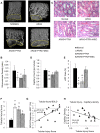
Figure 2. Medullary neovascularization and tubular injury.
Medullary regions outlined in dashed-line were used to calculate vasa recta density from micro-CT (A). The medullary microcirculation in stenotic kidneys. Vasa recta density was calculated from micro-CT as the fraction of tissue volume (B). Representative H&E-stained medullary tubules (x40 images) (C). The number of capillaries per tubule was calculated from H&E stained slides at x100 (D), and vWF quantified from vWF stained slides (x20) (E), Semi-quantitative tubular injury score (1–5 scale) calculated from corresponding H&E images (F) Medullary BOLD response to furosemide (G) and capillary density (H) showed moderate but significant correlation with tubular injury (*p≤0.05 vs. Normal, #p≤0.05 ARAS+PTRA+MSC).
Figure 3
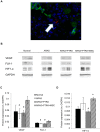
Figure 3. Angiogenic factors and cell engraftment.
MSC, labeled with CM-DiI (red), were localized in medullary slides co-stained with DAPI nuclear stain (blue) and the tubular marker cytokeratin (green). MSC were observed mainly engrafted in the medullary interstitium (A). Representative immunoblots (B) and quantification of medullary expression of the angiogenic factors VEGF, FLK-1 (C) and hypoxia inducible factor, HIF1-α (D). (*p≤0.05 vs. Normal, #p≤0.05 vs. ARAS+PTRA+MSC).
Figure 4
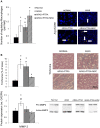
Figure 4. Markers of oxidative stress in the medulla.
Co-staining (x40) of DHE (red) and the nuclear marker DAPI (blue) was used to quantify their ratios (A) in Normal, ARAS, ARAS+PTRA and ARAS+PTRA+MSC. Medullary expression of NAD(P)H oxidase P47 was elevated in all ARAS medullas (B) (*p≤0.05 vs. Normal).
Figure 5
Figure 5. Medullary markers of inflammation.
TNF-α (A) and MCP-1 (B) expression, and CD163+ macrophage count (C). The anti-inflammatory factor IL-10 showed a significantly increased expression in ARAS+PTRA+MSC vs. the two other ARAS groups (D) (*p≤0.05 vs. Normal, #p≤0.05 ARAS+PTRA+MSC).
Figure 6
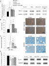
Figure 6. Quantification of regulatory M2 macrophages in CD68/Arg-1 double-stained slides.
The number of M2 macrophages in ARAS+PTRA+MSC was significantly higher than Normal and in ARAS+PTRA groups (A). Medullary fibrosis. Trichrome staining and MMP-2 expression decreased in ARAS+PTRA+MSC compared to ARAS (B) (*P≤0.05 vs. Normal, #p≤0.05 ARAS+PTRA+MSC).

References

    1. de Mast Q, Beutler JJ (2009) The prevalence of atherosclerotic renal artery stenosis in risk groups: a systematic literature review. J Hypertens 27: 1333–1340. - PubMed
    1. Lerman LO, Textor SC, Grande JP (2009) Mechanisms of tissue injury in renal artery stenosis: ischemia and beyond. Prog Cardiovasc Dis 52: 196–203. - PMC - PubMed
    1. Daghini E, Sanna M, Versari D, Di Paco I, Ghiadoni L, et al. (2010) Long Term Follow up of Hypertensive Patients with Atherosclerosis Renal Artery Stenosis after Percutaneous Renal Artery Angioplasty or Stenting: Blood Pressure Outcome. Journal of Hypertension 28: E210–E210.
    1. Eirin A, Zhu XY, Urbieta-Caceres VH, Grande JP, Lerman A, et al. (2011) Persistent kidney dysfunction in swine renal artery stenosis correlates with outer cortical microvascular remodeling. Am J Physiol Renal Physiol 300: F1394–1401. - PMC - PubMed
    1. Wheatley K, Ives N, Gray R, Kalra PA, Moss JG, et al. (2009) Revascularization versus Medical Therapy for Renal-Artery Stenosis. New England Journal of Medicine 361: 1953–1962. - PubMed

Publication types

MeSH terms

LinkOut - more resources