

BOLD fMRI in the white matter as a marker of aging and small vessel disease
- PMID: 23844047
- PMCID: PMC3699638
- DOI: 10.1371/journal.pone.0067652
BOLD fMRI in the white matter as a marker of aging and small vessel disease
Abstract
Purpose: Determine whether white matter signal fluctuation on T2* weighted BOLD contrast images are associated with aging and cerebral small vessel disease (SVD).
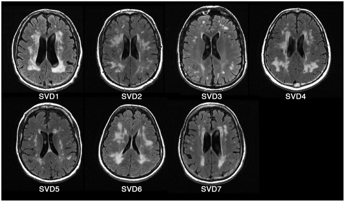
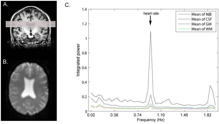
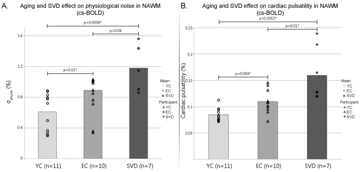
Methodology: Resting state BOLD data were collected with a 250 ms repetition time (TR) to achieve unaliased, ungated cardiac sampled BOLD (cs-BOLD) images on 11 young adult controls, 10 healthy older adult controls and 7 adults with extensive white matter hyperintensities (WMH) from SVD. Tissue classes (WM and GM) were segmented on T1 images. WMH were identified on FLAIR images in the SVD group. Raw physiological noise (σphysio) and cardiac pulsatility (i.e. fluctuations at the cardiac frequency) were calculated voxel wise and group differences were tested by ANOVA. It was also possible to calculate σphysio in 2s TR cardiac aliased whole-brain BOLD (wb-BOLD) data (N = 84) obtained from the International Consortium for Brain Mapping.
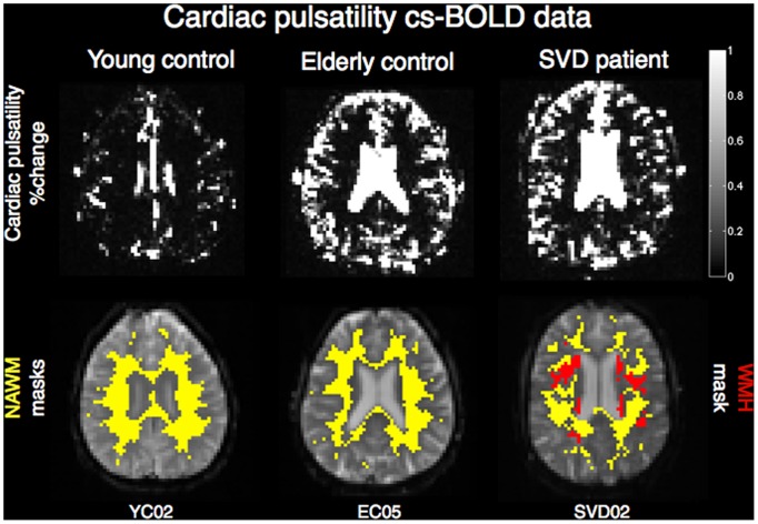
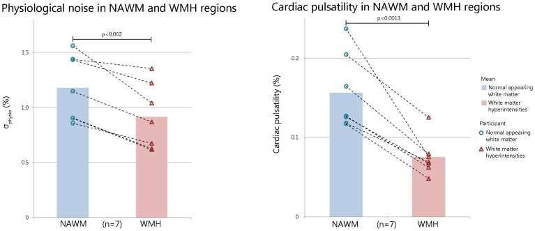
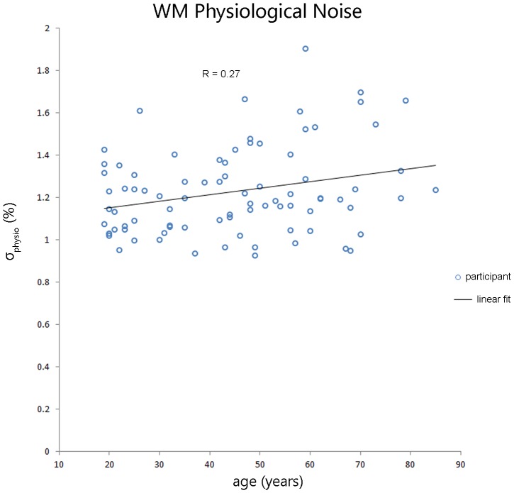
Results: CS-BOLD metrics showed an aging and SVD effects (p<0.0005). Covariates such as thermal noise, WM volume and partial volume did not influence the significant aging effect seen on the cardiac pulsatility metric (p<0.017) but did influence the σphysio (p = 0.184). As a verification of the cs-BOLD findings, the wb-BOLD also showed a linear aging effect of σphysio in WM. In the SVD adults, cardiac pulsatility and σphysio were lower in WMH regions compared to normal appearing white matter (NAWM) regions (p<0.0013 and p<0.002, respectively). Cardiac pulsatility was better able to distinguish WMH regions from NAWM than σphysio as measured by effect size (Cohen's d 2.2 and 0.88, respectively).
Conclusion: NAWM was found to have graded increases in cardiac pulsations due to age and SVD, independently. Within SVD participants, WMH lesions had reduced physiological noise compared to NAWM. Cardiac pulsatility in resting BOLD data may provide a complementary dynamic measure of WM integrity to add to static FLAIR anatomical images.
Conflict of interest statement
Figures
References
-
- Kruger G, Glover GH (2001) Physiological noise in oxygenation-sensitive magnetic resonance imaging. Magn Reson Med 46: 631–637. - PubMed
-
- Birn RM, Diamond JB, Smith MA, Bandettini PA (2006) Separating respiratory-variation-related fluctuations from neuronal-activity-related fluctuations in fMRI. Neuroimage 31: 1536–1548. - PubMed
-
- Biswal B, DeYoe AE, Hyde JS (1996) Reduction of physiological fluctuations in fMRI using digital filters. Magn Reson Med 35: 107–113. - PubMed
-
- Glover GH, Li TQ, Ress D (2000) Image-based method for retrospective correction of physiological motion effects in fMRI: RETROICOR. Magn Reson Med 44: 162–167. - PubMed
-
- Hu X, Le TH, Parrish T, Erhard P (1995) Retrospective estimation and correction of physiological fluctuation in functional MRI. Magn Reson Med 34: 201–212. - PubMed
Publication types
MeSH terms
Substances
Grants and funding
LinkOut - more resources
Full Text Sources
Other Literature Sources
Medical
Research Materials
