. 2013 Jul 3;8(7):e67660.
doi: 10.1371/journal.pone.0067660. Print 2013.

The bi-functional organization of human basement membranes

Affiliations

The bi-functional organization of human basement membranes

Willi Halfter et al. PLoS One. .

Abstract

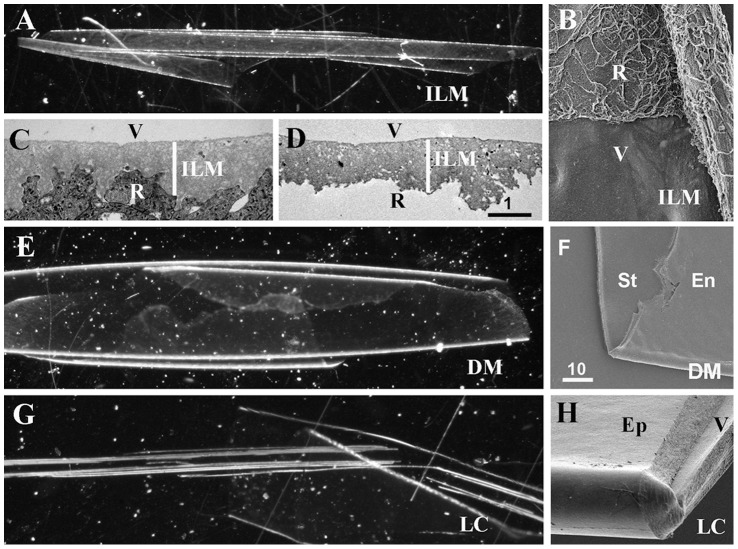
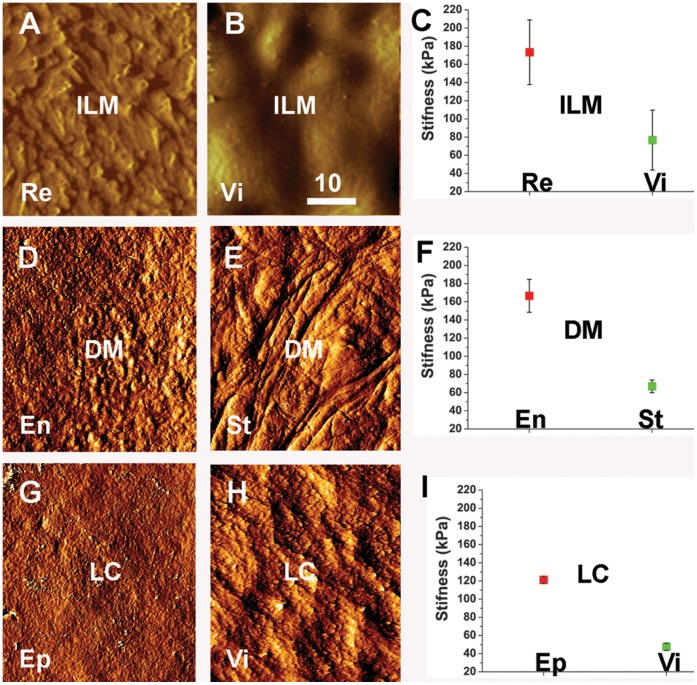
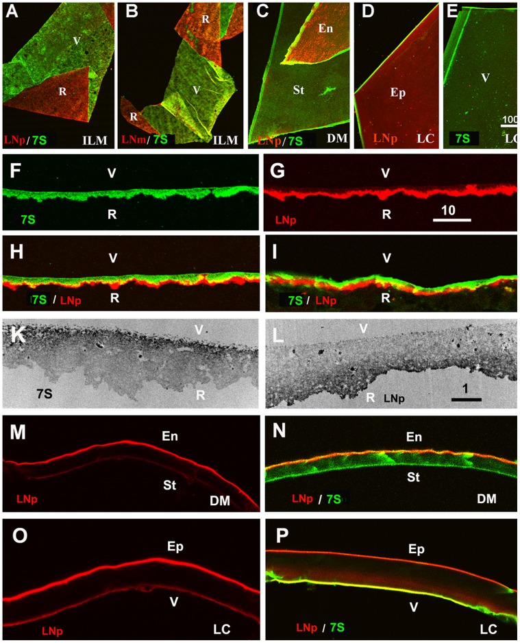
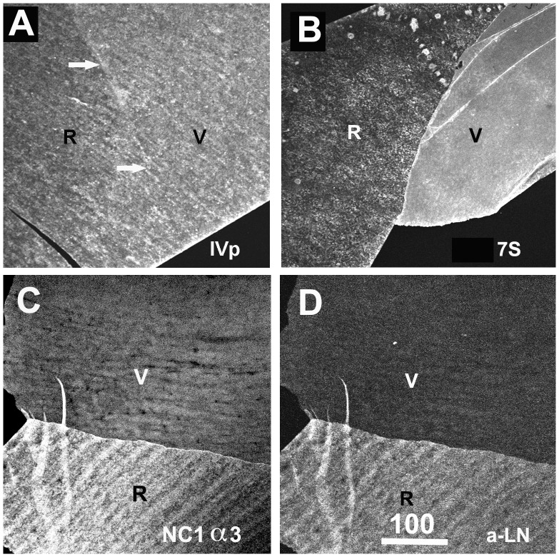
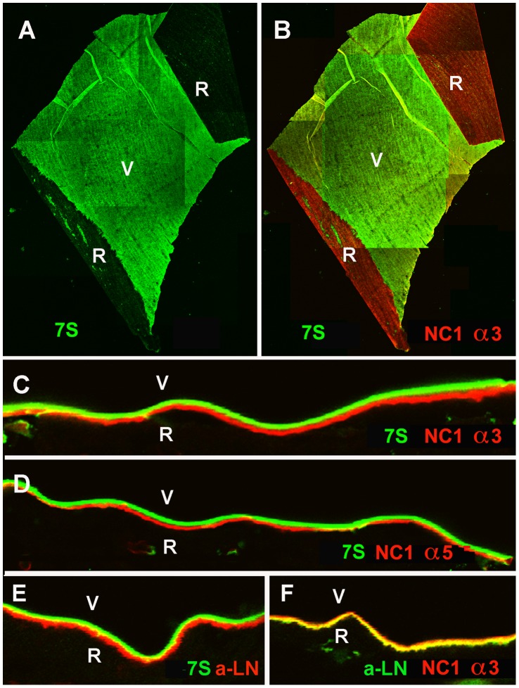
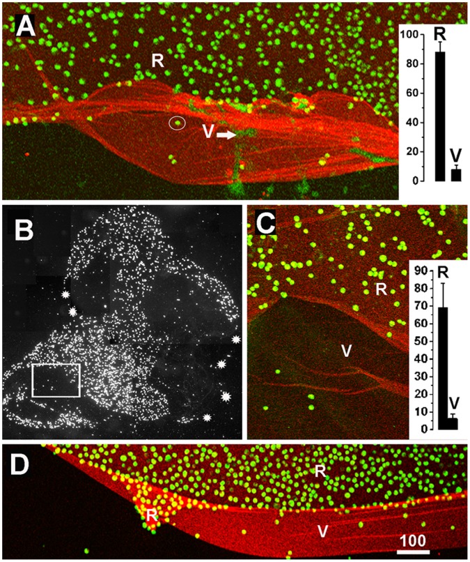
The current basement membrane (BM) model proposes a single-layered extracellular matrix (ECM) sheet that is predominantly composed of laminins, collagen IVs and proteoglycans. The present data show that BM proteins and their domains are asymmetrically organized providing human BMs with side-specific properties: A) isolated human BMs roll up in a side-specific pattern, with the epithelial side facing outward and the stromal side inward. The rolling is independent of the curvature of the tissue from which the BMs were isolated. B) The epithelial side of BMs is twice as stiff as the stromal side, and C) epithelial cells adhere to the epithelial side of BMs only. Side-selective cell adhesion was also confirmed for BMs from mice and from chick embryos. We propose that the bi-functional organization of BMs is an inherent property of BMs and helps build the basic tissue architecture of metazoans with alternating epithelial and connective tissue layers.


Conflict of interest statement

Competing Interests: The authors have declared that no competing interest exist.

Figures

Figure 1
Figure 1. Isolated human ILM (A), Descemet’s membrane (DM, E) and lens capsule (LC, G) as they appear under a dissecting microscope using dark field.
All BMs are transparent and rolled up, whereby the epithelial surfaces are facing the exterior and the stromal surfaces is facing the interior of the rolled-up BM sheets. SEM micrographs of a folded ILM (B), a DM (F) and a LC (H) show the two surfaces of the BMs. As shown in panel (B), the irregular retinal (R) surface of the ILM can be readily distinguished from the smooth vitreal surface (V), consistent with the morphological differences seen in TEM micrographs of crossections of ILMs in situ (C) and isolated ILMs (D). The endothelial (En) surface of the DM and epidermal (Ep) surface of the LC are indistinguishable from their stromal or vitreal (V) surfaces (F, H). Scale bars: C, D: 1 µm; B, F; H: 10 µm.
Figure 2
Figure 2. AFM testing of the two surfaces of the ILM (A–C), the DM (D–F) and LC (G–I).
The AFM imaging mode shows the morphological differences between the retinal (Re)/epithelial (Ep)/endothelial (En) surfaces and the vitreal (Vi)/stromal (St) surfaces of the ILM, DM and the LC. The graphs in (C, F, I) show the quantification of the stiffness measurements obtained by AFM “forced indentation”. The measurements were obtained by probing five ILMs, three DMs and three LCs. The epithelial surfaces of all tested BMs were about twice stiffer than the stromal surfaces. The differences were statistically significant. Scale bar: 10 µm.
Figure 3
Figure 3. The asymmetric structure of human BMs as shown by double labeling of folded ILMs (A, B), folded Descemet’s membranes (DM, C) and differently mounted lens capsule segments (LC, D, E).
The retinal surface (R) of the ILM and the endothelial (En) and epithelial (Ep) sides of DM and LC were stained with a polyclonal (red, LNp; A, C, D) and a monoclonal antibody to laminin (red, LNm, B), whereas the vitreal (V) or stromal side (St) of the BMs were labeled with an antibody to the 7S domain of collagen IV α3/4/5 (green; A-C, E). The asymmetry of BMs was also detected by single and double labeling of crossections of an isolated ILM (F-H) and an ILM in situ (I). The TEM micrographs in panel (K, L) show crossections of isolated ILMs stained for 7S collagen IV α3/4/5 (K) and laminin (L). The dark label shows the localization of 7S collagen IV on the vitreal side (K) and laminin on retinal side of the ILM (L). An asymmetric distribution for laminin and collagen 7S was also detected for the Descemet’s membranes (M, N) and the lens capsule (O, P). The sections were stained for laminin (red; M-P) and collagen IV 7S (green; N, P). Scale Bars: A-E: 100 µm; F-I and M-P: 10 µm; K, L, I: 1 µm.
Figure 4
Figure 4. Domain andα chain-specificity of Mab J3-2, a monoclonal antibody that recognizes the 7S domain of α3/4/α5 collagen IV.
The domain specificity was determined by Western blotting (lanes 1–6, panel A) and immunoprecipitation (lanes 7, 8, panel A) using adult human lens capsules as sample. The western blot of lanes 1 and 2, stained with a polyclonal antiserum to human collagen IV, shows the complex peptide banding pattern of intact collagen IV from human lens capsules age 24 (lane 1) and age 65 (lane 2). Staining of the 65 year-old lens capsule sample with the J3-2 Mab (lane 3) shows a similar, but not entirely identical banding pattern. Red bars mark corresponding bands. The non-collagenous (NC1 and 7S) domains of collagen IV were detected in western blots after digestion of lens capsules with collagenase: western blots with the soluble supernatant from digested lens capsule samples, stained with polyclonal collagen IV antiserum, shows the NC-domains of collagen IV as a long smear at 300 kD and a ladder of lower molecular weight peptides (lane 4, panel A). The NC1 domain of α3 collagen IV appears as a sharp band below 49 kD (lane 5, red bar; panel A) as shown by staining with a monoclonal antibody specific to the collagen IV α3 NC1 domain. The high molecular weight smear stained by Mab J3-2 correspond to the variably crosslinked 7S domain (Lane 6, panel A). The smear is due to the glycosylation of the 7S domain , . For immunoprecipitation, the collagenase-digest of lens capsule was incubated with Mab J3-2 or anti-laminin as a control followed by anti-mouse IgM or anti-rabbit IgG agarose. The beads were washed and the bound peptide released by boiling in high molar urea/SDS sample buffer. The released peptides were separated by SDS PAGE and western blotted. The immuno-precipitated peptides were visualized in the blot by a polyclonal anti-collagen IV antibody/alkaline phosphatase-conjugated secondary antibody. For the J3-2 pull-down, a smear of approximately 300 kD was detected (lane 7, panel A), similar to the smear detected by Western blotting (lane 6). A control pull-down with anti-laminin is shown in lane 8. The blots shows that Mab J3-2 i) recognizes human collagen IV, that it ii) recognizes an NC domain of collagen IV and that iii) the molecular weight of the detected peptides are different from the molecular weight of the NC1 domain but identical to the molecular weight of the 7S domain of collagen IV from glomerular BM and lens capsule. To determine the chain-specificity of the antibody, sections of adult human retina were stained with Mab J3-2 (B), to the NC1 domain of collagen IV α3 (C), to the NC1 domain of collagen IV α1 (D) and to the NC1 domain of collagen α5 (E). A3, α4 (not shown) and α5 collagen IV are very prominent in the ILM but sparse in the BMs of the retinal blood vessels (C, E), whereas α1/2 was very sparse in the ILM but abundant in the blood vessels (D). The staining with Mab J3-2 (A) resembles most closely the staining of collagen α3 α4 and α5, and it is very different from the distribution of collagen IV α1/α2. Scale Bar: 50 µm.
Figure 5
Figure 5. Collagen IV distribution in the embryonic human kidney.
Sections of 20-week fetal human kidney were stained with 7S-specfic Mab J3-2 (7S, A, E) and antibodies to the NC1 domain of collagen IV α1 (B), to the NC1 domain of collagen IV α3 (C), to the NC1 domain of collagen IV α5 (F) and to the NC1 domain of collagen IV α6 (D). A3/4/5 collagen IV is very prominent in the glomerular BMs (C, F), whereas α1/2 was equally abundant in all BMs of the kidney (B). The α6 chain is present in the BM of Bowman’s capsule (D). The staining with Mab J3-2 (7S, A, E) resembles most closely the staining for collagen α3 (C) and α5 (F), and it is different from the distribution of collagen IV α1/2 (B) and α6 (D). Double labeling with Mab J3-2 and the anti-NC1 α5 (G) or the α6 (H) shows the overlap with of 7S with α5 staining and the different distribution with α6 (H). Bar: 50 µm.
Figure 6
Figure 6. Detection of collagen IV in human ILM whole mounts.
Staining with polyclonal antibodies resulted in a uniform and even labeling of the retinal (R) and vitreal (V) side of the ILM. In contrast, staining of ILM with antibodies specific to the 7S (B) or the NC1 domain (C) of collagen IV alpha3/4/5 resulted in the selective labeling of the vitreal (B) or the retinal side (C). The distribution of laminin in the ILM (D) is very similar to that of collagen IV NC1 at the epithelial side. The ILM sample in panel C and D was double-labeled; the NC1 domain was labeled with a Cy3 (red; C) secondary antibody, whereas laminin was detected with a Cy2 (green; D) secondary antibody. Scale Bar: 100 µm.
Figure 7
Figure 7. Distinct localization of the N and C-terminal domains of collagen IV in the human ILM.
Labeling of ILM flat mounts with an antibody specific for the 7S domain of collagen IV α3/4/5 (7S) stained only the vitreal side (V) of the BM (green, A). The epithelial/retinal side of the ILM was prominently labeled with an antibody to NC1 domain of collagen IV α3 (B, red, NC1 α3) as shown by double labeling. Double labeling of retinal crossections with antibodies to the 7S domain of collagen IV α3/4/5 (green, C-E) and the NC1 domain of collagen IV α3 (NC1 α3, red, C) or α5 (NC1 α5, red, D) confirmed the distinct distribution of the C and N-terminal domains of collagen IV in this BM. Adjacent sections were stained for 7S of collagen IV α3/4/5 (green) and laminin (LN; red; E), and laminin (red) and NC-1 collagen IV α3 (green; F). Panel (E) shows the distinct localization of laminin and the 7S domain of collagen IV, and the yellow label in panel (F) the co-localization of laminin with the NC1 domain of collagen IV. Bar: A, B: 100 µm; C-F: 10 µm.
Figure 8
Figure 8. Staining of human DM and LC with domain-specific antibodies to collagen IV.
Whole mounts and cross sections of DM (A, B) and LC (C-F) were stained for the NC1 domain (A, C, E) and (or) the 7S domain of collagen IV α3/4/5 (B, D, F). A labeled DM whole mount showed that the NC1 domain of collagen IV is prominently localized at the epithelia side of this BM (A). Double labeling of a DM crossection (B) showed a selective distribution of the NC1 domain at the epithelial side (red) and a prominent labeling for the 7S domain at the stromal side. Whole mounts (C, D) and crossections (E, F) of LC stained for the NC1 domain of collagen IV showed a strong labeling of the vitreal side and a weaker labeling of the epithelial side (C, E). The same whole mount and crossection stained for the 7S domain showed an almost exclusive labeling of the vitreal side (D, F). The data combined showed that the NC1 and 7S domain of collagen IV have a side-selective distribution in the DM, but do not have this side-selectivity in the LC. Scale Bar: A, C, D: 100 µm; B, E, F: 10 µm.
Figure 9
Figure 9. Cell adhesion assays revealing the sidedness of BMs.
MDCK cells (A, B) and fibroblasts (C) were incubated on top of ILM (A, C) and DM (B). The BMs were folded to expose both the retinal/endothelial (R/E) and the vitreal/stromal (V/St) surface of the BMs. The non-adherent cells were washed off, and the firmly attached cells were visualized by nuclear dye staining (green). The BMs were stained for collagen IV 7S α3/4/5 (red) to visualize the V/St surface. The MDCK cells adhered almost exclusively to the R/E side of the folded BMs, and very few cells were detected on the 7S collagen IV-rich V/St side (A, B). Fibroblasts (Fibro) did not show this selective adhesion (C). Counting cells per unit BM surface area showed the strong preference of MDCK cells for the R/E side of the ILM, DM and LC. A similar adhesion preference was detected for corneal epithelial cells (Cor), embryonic chick retinal cells (Ret) and human vascular endothelial cells (Endo; p<0.001). Fibroblasts did not have a preference for either of the two BM sides. When a retinal explant strip was placed over a flat-mounted and folded DM (E), retinal axons only grew out on the endothelial (E) surface of the BM and not on the stromal surface (St). Scale bars: A-C, E: 250 µm.
Figure 10
Figure 10. Cell adhesion assays showing the functional sidedness of mouse and embryonic chick ILMs.
Mouse (A, two-week-old) and embryonic chick ILMs (B-D, embryonic day 9) were prepared and flat-mounted. When MDCK cells (green) were incubated on top of the ILM preparations, the cells prominently adhered to the retinal but not to the vitreal side of these BMs flat-mounts (A, D). The quantification of MDCK cell adhesion to either the retinal (R) or the vitreal (V) side of these BMs are shown in the inserted graphs (n = 10; A for mouse, C for chick). The white arrow in (A) points to DNA released from squashed retinal cells during the ILM flat-mount preparation. Its staining is very different from the crisp, circular DNA labeling of the adherent MDCK cells (circled). Chick ILMs were also flat-mounted by centrifugation (B, C). The dark field image of an entire chick BM preparation (B) shows that MDCK cells (white spots) adhered to only segments of the entire ILM (outlined by the white stars). The boxed area in (B) is shown at higher magnification in (C) confirming the selective adhesion of cells (green) to only one side of the ILM. The mouse ILM was stained for collagen IV (A), the chick ILMs were stained for laminin (C, D). Scale bar: A, C, D: 100 µm.

References

    1. Yurchenco PD, Patton BL (2009) Developmental and pathogenic mechanisms of basement membrane assembly. Curr Pharm Des 15: 1277–1294. - PMC - PubMed
    1. Timpl R (1996) Brown,JC (1996) Supramolecular assembly of basement membranes. Bioassays 18: 123–132. - PubMed
    1. Erickson AC, Couchman JR (2000) Still more complexity in mammalian basement membranes. J Histochem and Cytochem 48: 1291–1306. - PubMed
    1. Stephens LE, Sutherland AE, Klimanskaya IV, Andrieux A, Meneses J, et al. (1995) Deletion of beta 1 integrins in mice results in inner cell mass failure and peri-implantation lethality. Genes Dev 9: 1896–908. - PubMed
    1. Fassler R, Meyer M (1995) Consequences of lack of β1 integrin gene expression in mice. Genes Dev 9: 1896–908. - PubMed

Publication types