

Differentiation of breast cancer from fibroadenoma with dual-echo dynamic contrast-enhanced MRI
- PMID: 23844077
- PMCID: PMC3699626
- DOI: 10.1371/journal.pone.0067731
Differentiation of breast cancer from fibroadenoma with dual-echo dynamic contrast-enhanced MRI
Abstract
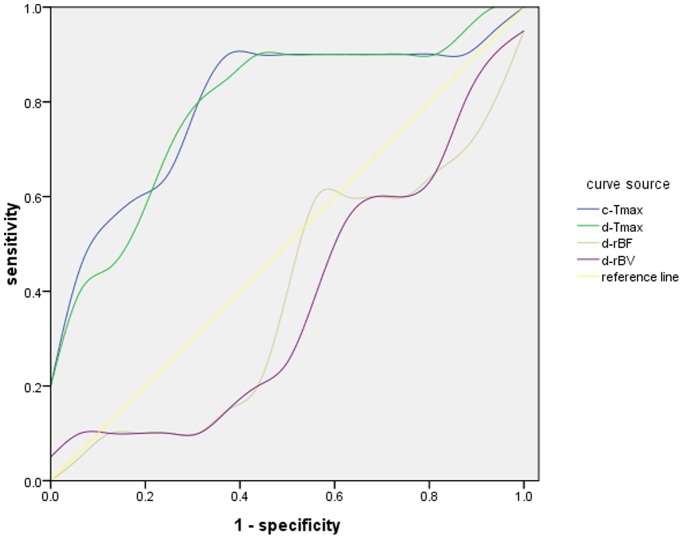
Dynamic contrast-enhanced magnetic resonance imaging (DCE MRI) of the breast is a routinely used imaging method which is highly sensitive for detecting breast malignancy. Specificity, though, remains suboptimal. Dynamic susceptibility contrast magnetic resonance imaging (DSC MRI), an alternative dynamic contrast imaging technique, evaluates perfusion-related parameters unique from DCE MRI. Previous work has shown that the combination of DSC MRI with DCE MRI can improve diagnostic specificity, though an additional administration of intravenous contrast is required. Dual-echo MRI can measure both T1W DCE MRI and T2*W DSC MRI parameters with a single contrast bolus, but has not been previously implemented in breast imaging. We have developed a dual-echo gradient-echo sequence to perform such simultaneous measurements in the breast, and use it to calculate the semi-quantitative T1W and T2*W related parameters such as peak enhancement ratio, time of maximal enhancement, regional blood flow, and regional blood volume in 20 malignant lesions and 10 benign fibroadenomas in 38 patients. Imaging parameters were compared to surgical or biopsy obtained tissue samples. Receiver operating characteristic (ROC) curves and area under the ROC curves were calculated for each parameter and combination of parameters. The time of maximal enhancement derived from DCE MRI had a 90% sensitivity and 69% specificity for predicting malignancy. When combined with DSC MRI derived regional blood flow and volume parameters, sensitivity remained unchanged at 90% but specificity increased to 80%. In conclusion, we show that dual-echo MRI with a single administration of contrast agent can simultaneously measure both T1W and T2*W related perfusion and kinetic parameters in the breast and the combination of DCE MRI and DSC MRI parameters improves the diagnostic performance of breast MRI to differentiate breast cancer from benign fibroadenomas.
Conflict of interest statement
Figures
Similar articles
-
Breast lesions: evaluation with dynamic contrast-enhanced T1-weighted MR imaging and with T2*-weighted first-pass perfusion MR imaging.Radiology. 2000 Aug;216(2):545-53. doi: 10.1148/radiology.216.2.r00au36545. Radiology. 2000. PMID: 10924584
-
Intravoxel incoherent motion MR imaging for breast lesions: comparison and correlation with pharmacokinetic evaluation from dynamic contrast-enhanced MR imaging.Eur Radiol. 2016 Nov;26(11):3888-3898. doi: 10.1007/s00330-016-4241-6. Epub 2016 Feb 10. Eur Radiol. 2016. PMID: 26863896
-
Automatic identification and classification of characteristic kinetic curves of breast lesions on DCE-MRI.Med Phys. 2006 Aug;33(8):2878-87. doi: 10.1118/1.2210568. Med Phys. 2006. PMID: 16964864
-
Ultrafast Dynamic Contrast-Enhanced MRI of the Breast: From Theory to Practice.J Magn Reson Imaging. 2024 Aug;60(2):401-416. doi: 10.1002/jmri.29082. Epub 2023 Dec 12. J Magn Reson Imaging. 2024. PMID: 38085134 Review.
-
Ultrafast Dynamic Contrast-enhanced MRI of the Breast: How Is It Used?Magn Reson Med Sci. 2022 Mar 1;21(1):83-94. doi: 10.2463/mrms.rev.2021-0157. Epub 2022 Feb 25. Magn Reson Med Sci. 2022. PMID: 35228489 Free PMC article. Review.
Cited by
-
Diagnostic value of dynamic contrast-enhanced magnetic resonance imaging in rectal cancer and its correlation with tumor differentiation.Mol Clin Oncol. 2016 Apr;4(4):500-506. doi: 10.3892/mco.2016.762. Epub 2016 Feb 3. Mol Clin Oncol. 2016. PMID: 27073650 Free PMC article.
-
Development of a Non-invasive Assessment of Hypoxia and Neovascularization with Magnetic Resonance Imaging in Benign and Malignant Breast Tumors: Initial Results.Mol Imaging Biol. 2019 Aug;21(4):758-770. doi: 10.1007/s11307-018-1298-4. Mol Imaging Biol. 2019. PMID: 30478507 Free PMC article.
References
-
- Kuhl CK (2007) Current status of breast MR imaging. Part 2. Clinical applications. Radiology 244: 672–691. - PubMed
-
- Bluemke DA, Gatsonis CA, Chen MH, DeAngelis GA, DeBruhl N, et al. (2004) Magnetic resonance imaging of the breast prior to biopsy. JAMA 292: 2735–2742. - PubMed
-
- Peters NH, Borel RI, Zuithoff NP, Mali WP, Moons KG, et al. (2008) Meta-analysis of MR imaging in the diagnosis of breast lesions. Radiology 246: 116–124. - PubMed
-
- Moy L, Elias K, Patel V, Lee J, Babb JS, et al. (2009) Is breast MRI helpful in the evaluation of inconclusive mammographic findings? AJR Am J Roentgenol 193: 986–993. - PubMed
-
- Vassiou K, Kanavou T, Vlychou M, Poultsidi A, Athanasiou E, et al. (2009) Characterization of breast lesions with CE-MR multimodal morphological and kinetic analysis: comparison with conventional mammography and high-resolution ultrasound. Eur J Radiol 70: 69–76. - PubMed
Publication types
MeSH terms
Substances
LinkOut - more resources
Full Text Sources
Other Literature Sources
Medical
