. 2013 Jul 2;8(7):e67735.
doi: 10.1371/journal.pone.0067735. Print 2013.

Dectin-1/TLR2 and NOD2 agonists render dendritic cells susceptible to infection by X4-using HIV-1 and promote cis-infection of CD4(+) T cells

Affiliations

Dectin-1/TLR2 and NOD2 agonists render dendritic cells susceptible to infection by X4-using HIV-1 and promote cis-infection of CD4(+) T cells

Sandra C Côté et al. PLoS One. .

Abstract

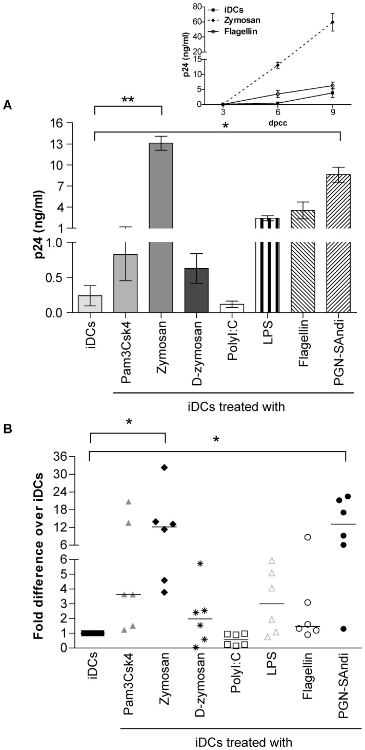
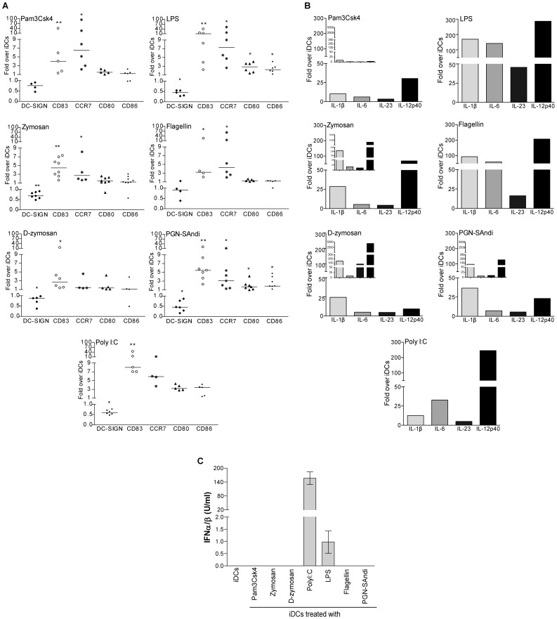
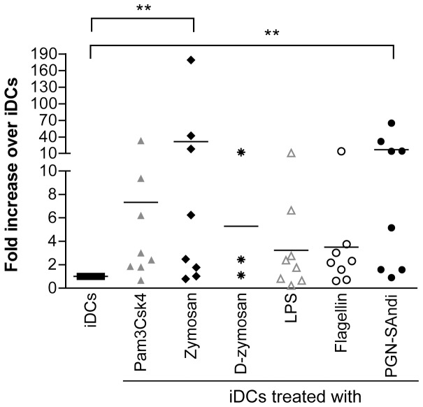
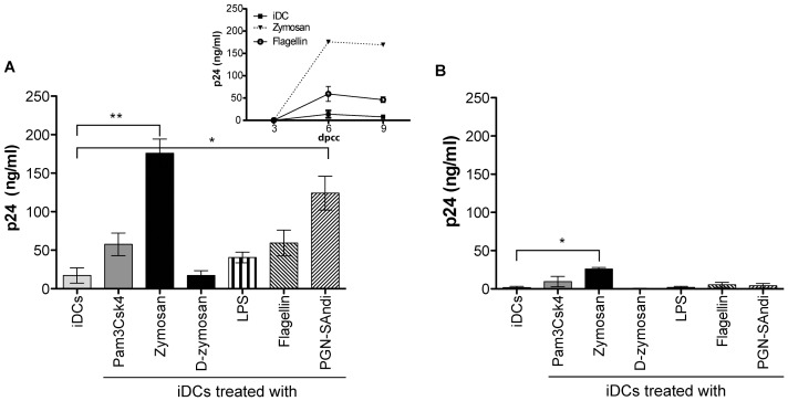
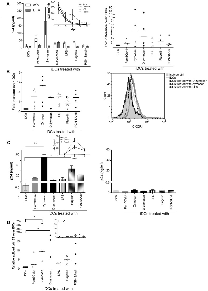
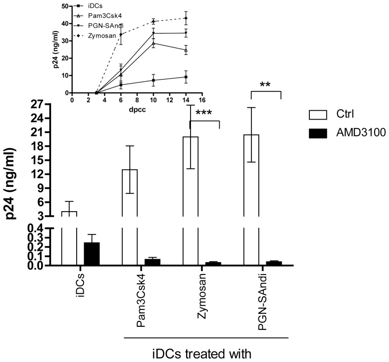
HIV-1 pathogenesis is intimately linked with microbial infections and innate immunity during all stages of the disease. While the impact of microbial-derived products in transmission of R5-using virus to CD4(+) T cells by dendritic cells (DCs) has been addressed before, very limited data are available on the effect of such compounds on DC-mediated dissemination of X4-tropic variant. Here, we provide evidence that treatment of DCs with dectin-1/TLR2 and NOD2 ligands increases cis-infection of autologous CD4(+) T cells by X4-using virus. This phenomenon is most likely associated with an enhanced permissiveness of DCs to productive infection with X4 virus, which is linked to increased surface expression of CXCR4 and the acquisition of a maturation profile by DCs. The ensuing DC maturation enhances susceptibility of CD4(+) T cells to productive infection with HIV-1. This study highlights the crucial role of DCs at different stages of HIV-1 infection and particularly in spreading of viral strains displaying a X4 phenotype.


Conflict of interest statement

Competing Interests: The authors have declared that no competing interests exist.

Figures

Figure 1
Figure 1. Replication of X4 virus in DC-T cell co-cultures is influenced by the nature of PAMPs.
iDCs were first either left untreated or treated for 24 hours with the listed PAMPs. Next, cells were exposed to X4-using NL4-3 for 1 hour at 37°C before initiation of a co-culture with autologous resting CD4+ T cells. Cell-free supernatants were harvested at 3, 6 and 9 days after initiation of the co-culture and the viral content was assessed by performing a p24 ELISA test. (A) Data shown represent the means ± SEM of quadruplicate samples from one representative donor out of six at day 6 after initiation of the co-culture (days post-coculture/dpcc). The small insert shows kinetics of virus production for this representative donor (only iDCs either left untreated or treated with flagellin or zymosan are illustrated). (B) This panel illustrates data for all six independent donors. Each point represents the mean of quadruplicate samples for each donor. The horizontal line represents median results of all donors. Asterisks denote statistically significant data (*: p<0.05; **: p<0.01).
Figure 2
Figure 2. The nature of PAMPs modulates both maturation profile and cytokine expression pattern of DCs.
(A) iDCs were either left untreated or treated for 72 hours with the indicated PAMPs. Afterwards, cell surface expression of DC-SIGN, CD83, CCR7, CD80 and CD86 was evaluated by flow cytometry. Data are expressed as the percentage of positive cells multiplied by the mean fluorescence intensity for the PAMP-stimulated DCs compared to untreated DCs (i.e iDCs). Each point represents a different donor. The horizontal line represents the median of the values for all donors. Asterisks denote statistically significant data (*: p<0.05; **: p<0.01). (B) iDCs were treated with the indicated PAMPs for 4 or 24 hours (small inserts) and total RNA was extracted. The variation of IL-1β, IL-6, IL-23 and IL-12p40 mRNA levels was evaluated by real-time PCR. After normalization on 18S ribosomal RNA subunit, the variation of mRNA levels for each cytokine was compared to that of untreated cells. Data depicted represent results obtained from one representative donor out of four. (C) iDCs were either left untreated or stimulated for 6 hours with the listed microbial-derived products. Cell-free supernatants were harvested and the levels of IFNα/β were quantified using HEK-Blue™ IFNα/β cells. Data shown represent the mean ± standard deviations of quadruplicate samples from one donor representative of two.
Figure 3
Figure 3. PAMP-treated DCs sensitize resting CD4+ T cells to productive HIV-1 infection.
iDCs were either left untreated or treated with the indicated PAMPs for 48 hours. Autologous resting CD4+ T cells were infected with NL4-3 for 48 hours, extensively washed and co-cultured with iDCs or PAMP-stimulated DCs. Thereafter, cell-free supernatants were collected at day 6 following initiation of the co-culture and the p24 content was assessed by ELISA. Each point represents the mean of quadruplicate samples for one donor. The horizontal line represents mean result of all donors. Asterisks denote statistically significant data (*: p<0.05; **: p<0.01).
Figure 4
Figure 4. Treatment of DCs with PAMPs promotes cis-infection of CD4+ T cells with X4 virus.
iDCs were left untreated or treated with the indicated PAMPs for 24 hours. Cells were next either left untreated (A) or treated with EFV (B) to block productive infection of DCs prior to loading with NL4-3 for 4 hours at 37°C. Cells were extensively washed and incubated for 16 hours before addition of autologous resting CD4+ T cells. Cell-free supernatants were collected at 3, 6 and 9 days following initiation of the co-culture. Data shown represent the mean ± SEM of quadruplicate samples from one representative donor out of four at 6 days following initiation of the co-culture. (*: p<0.05; **: p<0.01). The small insert shows kinetics of virus production for this donor (only iDCs either left untreated or treated with zymosan or flagellin are illustrated).
Figure 5
Figure 5. PAMPs enhance CXCR4 expression on DCs and susceptibility to productive infection with X4 virus.
(A) iDCs were left untreated or treated for 24 hours with the listed PAMPs. Next, DCs were washed and pretreated, or not, with EFV for 15 minutes, and then exposed to NL4-3 for another 24 hours. Cell-free supernatants were harvested at 3, 6, 9, 12 and 15 days post-infection (dpi). Data depicted in the upper left panel represent the means ± SEM of quadruplicate samples from one representative donor out of four at 6 days post-infection. The small insert shows kinetics of virus production for this donor (only iDCs either left untreated or treated with zymosan, LPS, or flagellin are illustrated). Each point depicted in the right panel represents the mean of quadruplicate samples for each of the four donors tested. The horizontal line represents median results of all four distinct donors. (B) iDCs were left untreated or treated for 72 hours with the indicated PAMPs. Cell surface expression of CXCR4 was analyzed by flow cytometry. Expression of CXCR4 is depicted as the percentage of positive cells multiplied by the mean fluorescence intensity for the treated condition, over that of untreated cells. Each point represents a different donor while the horizontal bar represents the mean of all donors. One representative CXCR4 staining is depicted in the left panel. (C) iDCs were left untreated or treated with the indicated PAMPs for 72 hours to allow CXCR4 surface expression. Cells were next either left untreated (left panel) or treated with EFV (right panel) prior to loading with NL4-3 for 4 hours at 37°C. Cells were extensively washed and incubated for 16 hours before addition of resting CD4+ T cells. Supernatants were collected at 3, 6 and 9 days following initiation of the co-culture. Data represents the mean ± SEM of quadruplicates from one representative donor out of four at 6 days following initiation of the co-culture. The small insert on the left panel represents the kinetics of virus infection for this donor. (D) iDCs were either left untreated or treated with the indicated PAMPs for 72 hours. Cells were either left untreated or treated with EFV (small insert) and next incubated with NL4-3 for 72 hours. Total RNA was extracted and the relative amount of spliced tat mRNA was evaluated by semi-quantitative RT-PCR. After normalization on 18S ribosomal RNA subunit, the amount of spliced tat mRNA in PAMP-treated DCs was expressed as folds of that of untreated cells. Each point represents a different donor and the horizontal bar represents the median of all donors. Asterisks denote statistically significant data (*: p<0.05; **: p<0.01).
Figure 6
Figure 6. Replication of X4 virus in DC-T cell co-cultures in presence of different PAMPs and AMD3100.
iDCs were first either left untreated or treated for 24 hours with the listed PAMPs. Next, cell were extensively washed 3 times with PBS and treated for 30 min with AMD3100 (20 µg/ml), washed again extensively to remove the residual drug and inoculated with NL4-3 (10 ng of p24 per 105 cells) for 1 hour at 37°C. Cells were again extensively washed before initiation of co-culture with autologous resting CD4+ T cells. Cell-free supernatants were harvested at different days following initiation of the co-culture (day 10 is depicted). The viral content was assessed by performing a p24 ELISA test. Data shown represent the means ± SEM of quadruplicate samples from two distinct donors. Asterisks denote statistically significant data (**: p<0.01). The small insert shows kinetics of virus production for one donor.

References

    1. Galvin SR, Cohen MS (2004) The role of sexually transmitted diseases in HIV transmission. Nat Rev Microbiol 2: 33–42. - PubMed
    1. Fleming DT, Wasserheit JN (1999) From epidemiological synergy to public health policy and practice: the contribution of other sexually transmitted diseases to sexual transmission of HIV infection. Sex Transm Infect 75: 3–17. - PMC - PubMed
    1. Sewankambo N, Gray RH, Wawer MJ, Paxton L, McNaim D, et al. (1997) HIV-1 infection associated with abnormal vaginal flora morphology and bacterial vaginosis. Lancet 350: 546–550. - PubMed
    1. Taha TE, Hoover DR, Dallabetta GA, Kumwenda NI, Mtimavalye LA, et al. (1998) Bacterial vaginosis and disturbances of vaginal flora: association with increased acquisition of HIV. Aids 12: 1699–1706. - PubMed
    1. Joffre O, Nolte MA, Sporri R, Reis e Sousa C (2009) Inflammatory signals in dendritic cell activation and the induction of adaptive immunity. Immunol Rev 227: 234–247. - PubMed

Publication types

MeSH terms