. 2013 Jul 3;8(7):e67765.
doi: 10.1371/journal.pone.0067765. Print 2013.

Differential expression of three members of the multidomain adhesion CCp family in Babesia bigemina, Babesia bovis and Theileria equi

Affiliations

Differential expression of three members of the multidomain adhesion CCp family in Babesia bigemina, Babesia bovis and Theileria equi

Reginaldo G Bastos et al. PLoS One. .

Abstract

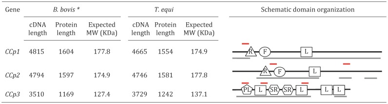
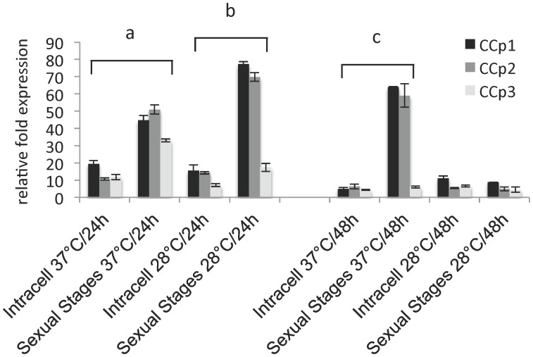
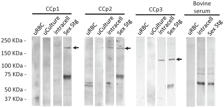
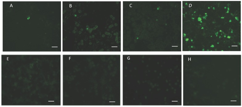
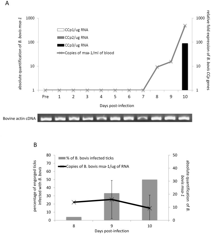
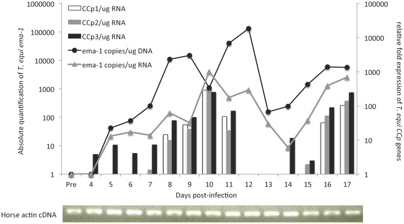
Members of the CCp protein family have been previously described to be expressed on gametocytes of apicomplexan Plasmodium parasites. Knocking out Plasmodium CCp genes blocks the development of the parasite in the mosquito vector, making the CCp proteins potential targets for the development of a transmission-blocking vaccine. Apicomplexans Babesia bovis and Babesia bigemina are the causative agents of bovine babesiosis, and apicomplexan Theileria equi causes equine piroplasmosis. Bovine babesiosis and equine piroplasmosis are the most economically important parasite diseases that affect worldwide cattle and equine industries, respectively. The recent sequencing of the B. bovis and T. equi genomes has provided the opportunity to identify novel genes involved in parasite biology. Here we characterize three members of the CCp family, named CCp1, CCp2 and CCp3, in B. bigemina, B. bovis and T. equi. Using B. bigemina as an in vitro model, expression of all three CCp genes and proteins was demonstrated in temperature-induced sexual stages. Transcripts for all three CCp genes were found in vivo in blood stages of T. equi, and transcripts for CCp3 were detected in vivo in blood stages of B. bovis. However, no protein expression was detected in T. equi blood stages or B. bovis blood stages or B. bovis tick stages. Collectively, the data demonstrated a differential pattern of expression of three orthologous genes of the multidomain adhesion CCp family by B. bigemina, B. bovis and T. equi. The novel CCp members represent potential targets for innovative approaches to control bovine babesiosis and equine piroplasmosis.


Conflict of interest statement

Competing Interests: The authors have declared that no competing interests exist.

Figures

Figure 1
Figure 1. Gene identification, cDNA length, protein length, expected protein size and schematic domain representation of CCp1-3 in Babesia bovis and Theileria equi.
Black lines represent the full-length of the CCp proteins in B. bovis and T. equi. Grey lines represent the regions of the CCp proteins available in B. bigemina. Red lines above CCp1-3 indicate the regions used to design synthetic peptides. R: Ricin B lectin domain. F: F5/F8 type C domain. L: LCCL domain. PL: PLAT domain. SR: Scavenger receptor domain. * In silico data of B. bovis CCp1-3 has been previously shown by Becker et al .
Figure 2
Figure 2. Relative expression of B. bigemina CCp1-3 by extracellular sexual stages incubated either at 37°C or 28°C for 24 h and 48 h is compared to gene expression by intracellular parasites (Intracell) incubated under identical conditions for each temperature and time point.
Gene expression was normalized to the total amount of RNA used to generate the cDNA. The transcription level was calculated as a relative expression using the formula: Relative expression (sample) = 2 [Cq (control) – Cq (sample)], where the control is the highest Cq value for a given gene of interest. Results represent the means of three experiments, each containing three technical replicates. Differences in gene expression were determined by the Student’s t-test. “a” indicates the expression of CCp1-3 was significantly higher (P<0.001) by sexual stages incubated for 24 h at 37°C than by Intracell. “b” indicates the expression of CCp1and CCp2 was significantly higher (P<0.001) by sexual stages incubated for 24 h at 28°C than by Intracell. “c” indicates the expression of CCp1and CCp2 was significantly higher (P<0.001) by sexual stages incubated for 37 h at 48°C than by Intracell.
Figure 3
Figure 3. Immunoblot analysis demonstrating the expression of CCp1-3 by temperature-induced B. bigemina parasites.
Expression of CCp1-3 proteins was investigated in B. bigemina cultures incubated for 24 h at 28°C. Expression of CCp1 was detected only by extracellular sexual stages (Sex Stg) whereas CCp2 and CCp3 were detected in both Sex Stg and intracellular parasites (Intracell) populations. No bands with the expected molecular mass of the CCp proteins were detected in non-induced B. bigemina cultures (uCulture) or in uninfected red blood cells (uRBC). Polyclonal mouse anti-CCp1 peptide, rabbit anti-CCp2-peptides or CCp3-peptides, and bovine serum from an animal chronically infected with B. bigemina were used at a 1∶50 dilution. Anti-mouse, anti-rabbit, or anti-bovine HRP conjugates were used at a 1∶4,000 dilution. Black arrows indicate the expected size of CCp1-3.
Figure 4
Figure 4. Immunofluorescence assays demonstrating the expression of CCp1-3 in temperature-induced B. bigemina cultures.
Antigen for immunofluorescence was prepared from parasite culture adjusted to 20% pack cell volume (PCV). Five µl of the 20% PCV solution was used to make a uniform thin film on glass microscope slides. Expression of CCp1-3 proteins in temperature-induced B. bigemina gametocyte cultures incubated for 24 h at 28°C is shown in panels A, B and C, respectively. CCp expression was not detected in non-induced B. bigemina cultures kept at 37°C as shown in panels E, F, and G. Panels D and H are representative results of immunofluorescence using sera from either B. bigemina-infected or uninfected bovine, respectively. Polyclonal mouse anti-CCp1 peptide, rabbit anti-CCp2 or CCp3 peptides, and bovine sera were used at a 1∶20 dilution. Anti-mouse, anti-rabbit, or anti-bovine FITC conjugates were used at a 1∶80 dilution. The fields shown are representative of the majority of the samples. The panels present a magnification of 100x and white bars indicate 10 µm.
Figure 5
Figure 5. Expression of CCp1-3 during acute B. bovis infection.
In panel A, relative fold expression of CCp1-3 transcripts in bovine blood during acute B. bovis infection is shown in the right y-axis. Absolute quantification of B. bovis msa-1 DNA in bovine blood is shown in the left y-axis. CCp gene expression was normalized to the total amount of RNA used to generate the cDNA and the transcription level was calculated as a relative expression using the formula: Relative expression (sample) = 2 [Cq (control) – Cq (sample)], where the control is the highest Cq value for a given gene of interest. To confirm the presence of amplifiable cDNA in all tested samples a standard PCR for bovine actin was performed. In panel B, absolute expression of B. bovis msa-1 RNA in bovine blood is shown in the right y-axis and the percentage of engorged ticks infected with B. bovis is shown in the left y-axis.
Figure 6
Figure 6. Expression of CCp1-3 during acute T. equi infection.
Relative fold expression of CCp1-3 transcripts in horse peripheral blood during acute T. equi infection is shown in the right y-axis. Absolute quantification of T. equi emai-1 DNA (black line) and cDNA (grey line) is shown in the left y-axis. CCp gene expression was normalized to the total amount of RNA used to generate the cDNA and the transcription level was calculated as a relative expression using the formula: Relative expression (sample) = 2 [Cq (control) – Cq (sample)], where the control is the highest Cq value for a given gene of interest. To confirm the presence of amplifiable cDNA in all tested samples a standard PCR for horse actin was performed.

Similar articles

Cited by

References

    1. Hay SI, Guerra CA, Tatem AJ, Noor AM, Snow RW (2004) The global distribution and population at risk of malaria: past, present, and future. Lancet Infect Dis 4: 327–336. - PMC - PubMed
    1. Bishop R, Musoke A, Morzaria S, Gardner M, Nene V (2004) Theileria: intracellular protozoan parasites of wild and domestic ruminants transmitted by ixodid ticks. Parasitol 129: S271–S283. - PubMed
    1. Hunfeld KP, Hildebrandt A, Gray JS (2008) Babesiosis: recent insights into an ancient disease. Int J Parasitol 38: 1219–1237. - PubMed
    1. Mehlhorn H, Schein E (1984) The piroplasm: Life cycle and sexual stages. Adv Parasitol 23: 37–103. - PubMed
    1. Delrieu I, Waller CC, Mota MM, Grainger M, Langhorne J, et al. (2002) PSLAP, a protein with multiple adhesive motifs, is expressed in Plasmodium falciparum gametocytes. Mol Biochem Parasitol 121: 11–20. - PubMed

Publication types

LinkOut - more resources