. 2013 Jul 2;8(7):e67773.
doi: 10.1371/journal.pone.0067773. Print 2013.

The non-canonical Wnt/PKC pathway regulates mitochondrial dynamics through degradation of the arm-like domain-containing protein Alex3

Affiliations

The non-canonical Wnt/PKC pathway regulates mitochondrial dynamics through degradation of the arm-like domain-containing protein Alex3

Román Serrat et al. PLoS One. .

Abstract

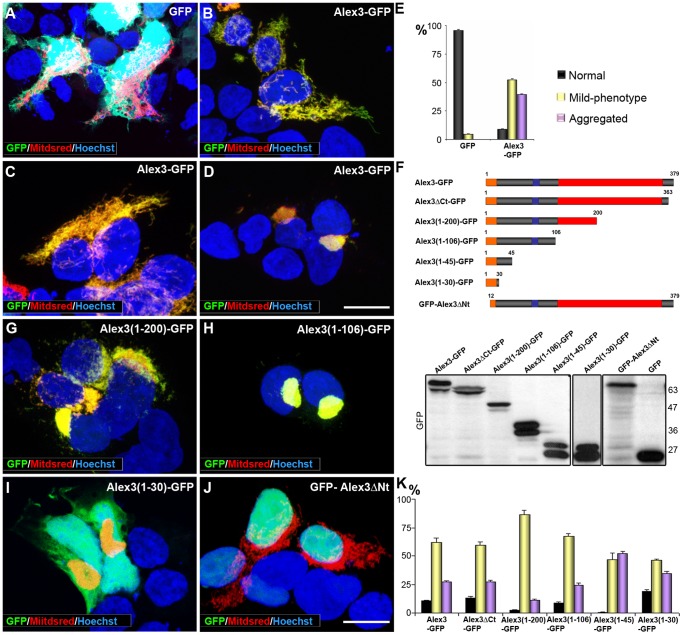
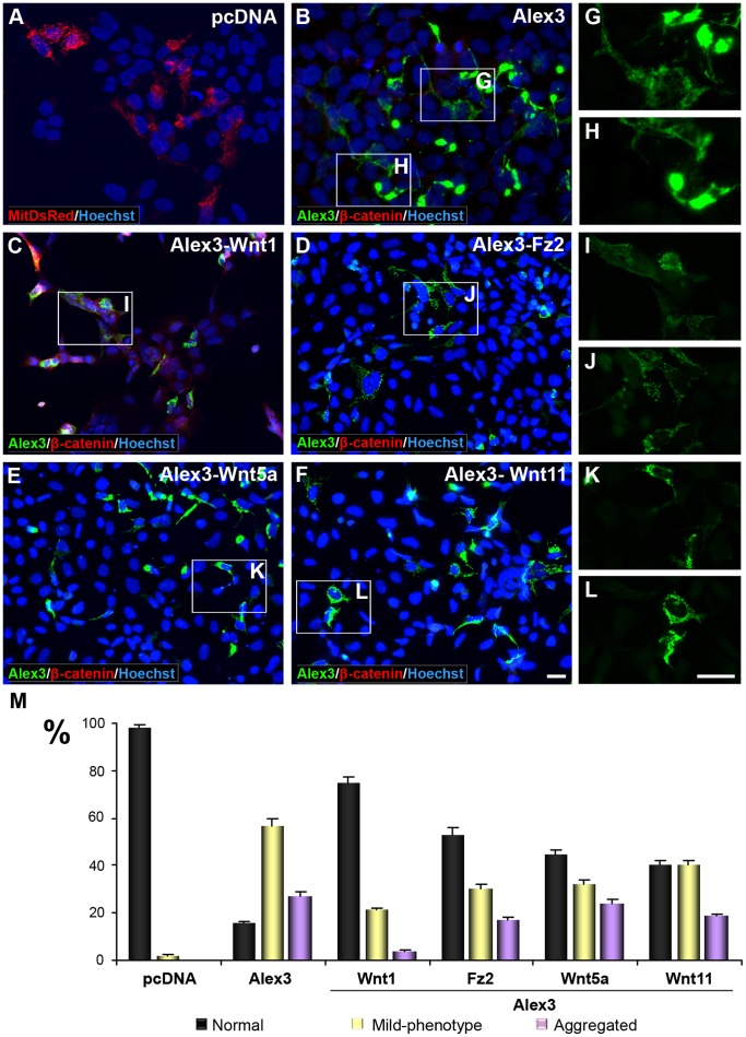
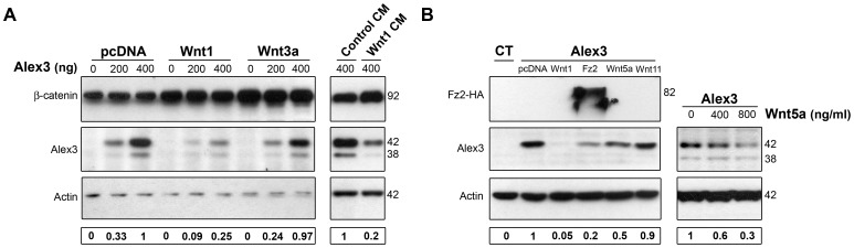
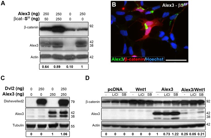
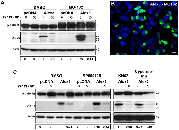
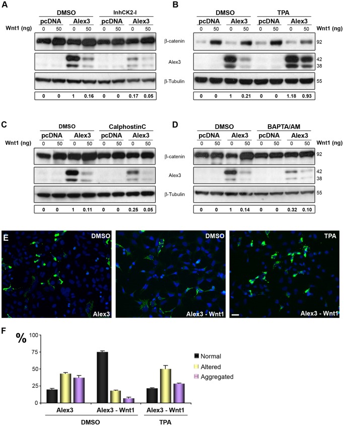
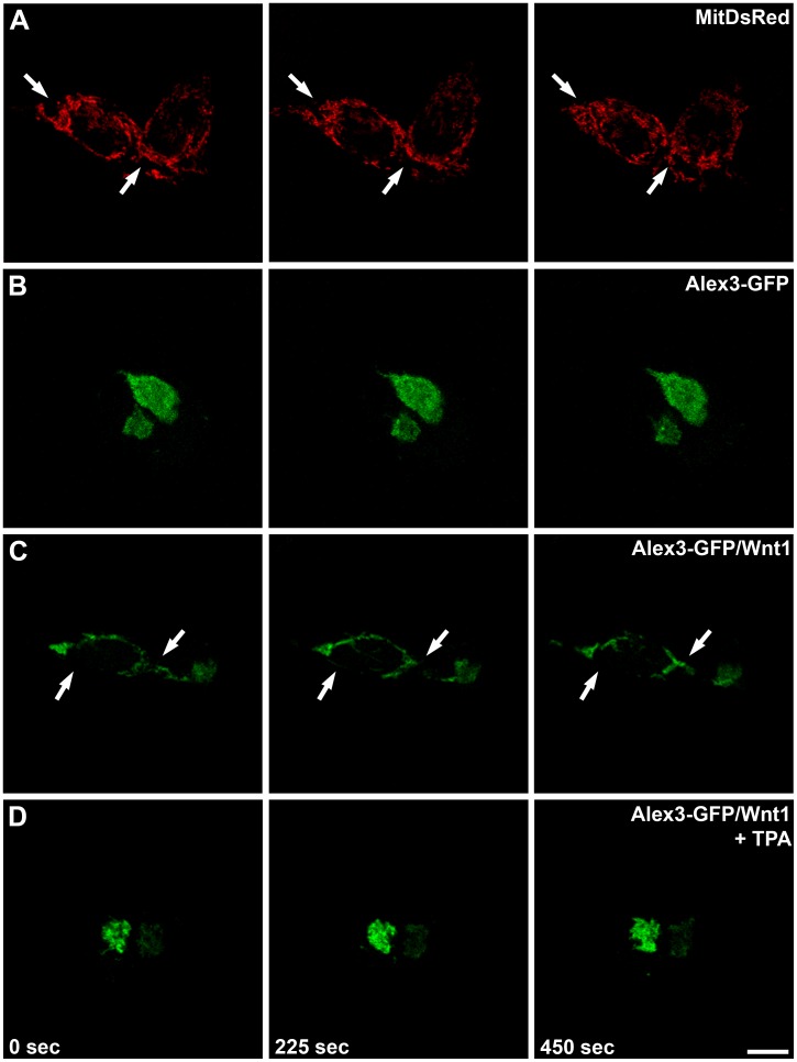
The regulation of mitochondrial dynamics is vital in complex cell types, such as neurons, that transport and localize mitochondria in high energy-demanding cell domains. The Armcx3 gene encodes a mitochondrial-targeted protein (Alex3) that contains several arm-like domains. In a previous study we showed that Alex3 protein regulates mitochondrial aggregation and trafficking. Here we studied the contribution of Wnt proteins to the mitochondrial aggregation and dynamics regulated by Alex3. Overexpression of Alex3 in HEK293 cells caused a marked aggregation of mitochondria, which was attenuated by treatment with several Wnts. We also found that this decrease was caused by Alex3 degradation induced by Wnts. While the Wnt canonical pathway did not alter the pattern of mitochondrial aggregation induced by Alex3, we observed that the Wnt/PKC non-canonical pathway regulated both mitochondrial aggregation and Alex3 protein levels, thereby rendering a mitochondrial phenotype and distribution similar to control patterns. Our data suggest that the Wnt pathway regulates mitochondrial distribution and dynamics through Alex3 protein degradation.


Conflict of interest statement

Competing Interests: The authors have declared that no competing interests exist.

Figures

Figure 1
Figure 1. Schematic representation of Alex3 protein.
Predicted domains are annotated on the basis of databases such as Pfam, Smart or Wolfpsort and bibliographic references. The stars show the position of putative phosphorylation sites in serine or threonine residues by CK2, PKC and PKA kinases.
Figure 2
Figure 2. The N-terminal domain of Alex3 is sufficient to induce mitochondrial aggregation.
(A–D) Overexpression of Alex3-GFP (green) in HEK293T cells induces severe alterations of the mitochondrial network when compared with the expression of control GFP (A). (B) Illustrates an Alex3-transfected cell displaying normal mitochondrial morphology; (C,D) Alex3-overexpressing cells showing mild aggregating phenotypes (C) and severe aggregating mitochondrial phenotypes (D); Alex3 protein was visualized in green, mitochondria in red (MitDsRed), and nuclei were labeled with bisbenzimide (blue). (E) Quantification and graphical representation (mean ± standard deviation) of mitochondrial phenotypes in control (GFP) and Alex3-GFP-overexpressing cells. (F) Top: Scheme of the Alex3-GFP deletion constructs used for transfection. Bottom: Western Blot showing representative truncated Alex3-GFP constructs at the predicted protein sizes. (G–J) Photomicrographs illustrating that expression of the Alex3(1–200)-GFP (G), Alex3(1–106)-GFP (H) and Alex3(1–30)-GFP (I) constructs leads to mitochondrial aggregation; in contrast, deletion of the first N terminal 12 aa (GFP-Alex3ΔNt) targets Alex3 protein to the nucleus (J). Note that the 30 aa N-terminus deletion construct has a truncated outer mitochondrial membrane localization sequence, which may interfere with its mitochondrial targeting, thereby leading to nuclear localization. (K) Quantification and graphical representation (mean ± standard deviation) of mitochondrial phenotypes in HEK293T cells after transfection with several truncated Alex3-GFP constructs; the data show that all the constructs containing the N terminal region cause mitochondrial aggregation. Alex3 protein was visualized in green (GFP), mitochondria in red (MitDsRed) and nuclei in blue (bisbenzimide). Scale bar: 10 µm.
Figure 3
Figure 3. Wnt/Frizzled signaling restores the normal mitochondrial phenotype in Alex3-overexpressing cells.
(A–F) Co-expression of Alex3 (green) and several members of the Wnt/Frizzled signaling pathway; Wnt1, Fz2, Wnt5a and Wnt11 reverse the aggregated mitochondrial phenotypes induced by Alex3 overexpression in HEK293AD cells. (G–L) High magnifications of boxed areas shown in (A–F). Note that the aggregated phenotype induced by the expression of Alex3 (G,H) is reversed by the co-expression of different members of the Wnt pathway (I–L). (M) Quantification and graphical representation (mean ± standard deviation) of mitochondrial phenotypes resulting after transfection with distinct Wnts and Fz2, demonstrating different degrees of mitochondrial aggregation reversion by the constructs used. Alex3 protein was visualized in green, control mitochondrial distribution (A) in red (MitDsRed), β-catenin in red (B–F) and nuclei in blue (bisbenzimide). Scale bar: 10 µm.
Figure 4
Figure 4. The Wnt/Frizzled pathway induces the degradation of Alex3 protein.
(A) WBs showing that Wnt1 co-transfection (left) and incubation with Wnt1-conditioned media (CM, right), but not treatment with Wnt3a (200 ng/ml) (middle), induces the degradation of Alex3 protein. (B) WB showing that co-transfection with Wnt1, Fz2, Wnt5a and Wnt11 lead to different reductions in Alex3 protein levels (left). Recombinant Wnt5a also induces Alex3 degradation in a concentration-dependent manner (right).
Figure 5
Figure 5. Alex3 degradation is independent of the canonical Wnt/β-catenin pathway.
(A,B) Constitutively active β-catenin (red) neither induces Alex3 protein degradation, as seen in WB (A), nor reverts the aggregated mitochondrial phenotypes induced by Alex3 overexpression (green in B). Nuclei were visualized in blue (bisbenzimide) (B). (C,D) Neither co-transfection with Dvl2 (C) nor the inhibition of GSK3β with 10 mM LiCl or with 10 µM SB212763 (D) induces Alex3 protein degradation. Wnt1 transfection was used as a control for Alex3 degradation. Scale bar: 10 µm.
Figure 6
Figure 6. Alex3 degradation by Wnt1 is independent of the proteasome, JNK, CAMKII and Calcineurin pathways.
(A) Proteasome inhibition with 10 µM MG-132 treatment blocks the normal turnover of Alex3 protein but not its Wnt1-induced degradation. (B) Numerous Alex3-overexpressing HEK293AD cells treated with the proteasomal inhibitor MG132 show the most severe mitochondrial aggregating phenotype. (C) Inhibition of JNK with 10 µM SP600125 (downstream effector of the Wnt/PCP pathway), CAMKII with 25 µM KN62 or Calcineurin with 10 µM Cypermetrin (downstream effectors of the Wnt/Ca2+ pathway) do not induce Alex3 protein degradation. Scale bar: 10 µm.
Figure 7
Figure 7. PKC and CKII phosphorylation protects against Wnt/Frizzled degradation of Alex3.
(A) Inhibition of CKII (with 100 µM casein kinase II inhibitor I), downstream effector of the Wnt signaling pathway, is sufficient to trigger Alex3 degradation. (B) In contrast, PKC activation with 1 µM TPA protects against Wnt1-induced degradation of Alex3 protein. (C,D) Inhibition of PKC (with 1 µM Calphostin C) and treatment with 20 µM BAPTA/AM, an intracellular calcium chelator, also reproduces Wnt1 degradation. (E) Photomicrographs demonstrating that treatment with TPA prevents Alex3 degradation induced by Wnt1 and the reversion to normal mitochondrial phenotypes. (F) Quantification and graphical representation (mean ± standard deviation) of mitochondrial phenotypes in HEK293AD cells in the conditions shown in (E); note that incubation with TPA prevents the rescue of mitochondrial phenotypes induced by Wnt1. Scale bar: 10 µm. The quantification of Alex3 protein levels is shown at the bottom.
Figure 8
Figure 8. Wnt1 increases mitochondrial motility and dynamics.
Series of representative confocal images, taken every 225 sec, from live HEK293T cells overexpressing the mitochondrial tagged protein MitDsRed (A), Alex3-GFP fusion protein (B), or Alex3-GFP and Wnt1 cDNAs (4∶1) (C,D). In (D) TPA treatment was used to activate PKC. Arrows identify areas with highly dynamic mitochondria. While mitochondrial motility is high in control (A) and Alex3-GFP/Wnt1 (C) conditions, it is severely reduced in Alex3-GFP-overexpressing cells (B) and in Alex3-GFP/Wnt1/TPA-treated cells (D). (See also Videos S1, S2, S3, and S4). Scale bar: 10 µm.

References

    1. Nicholls DG, Budd SL (2000) Mitochondria and neuronal survival. Physiol Rev 80: 315–360. - PubMed
    1. Chen H, Chan DC (2009) Mitochondrial dynamics–fusion, fission, movement, and mitophagy–in neurodegenerative diseases. Hum Mol Genet 18: R169–176. - PMC - PubMed
    1. Han XJ, Tomizawa K, Fujimura A, Ohmori I, Nishiki T, et al. (2010) Regulation of mitochondrial dynamics and neurodegenerative diseases. Acta Med Okayama 65: 1–10. - PubMed
    1. Schon EA, Przedborski S (2011) Mitochondria: the next (neurode)generation. Neuron 70: 1033–1053. - PMC - PubMed
    1. Lopez-Domenech G, Serrat R, Mirra S, D'Aniello S, Somorjai I, et al. (2012) The Eutherian Armcx genes regulate mitochondrial trafficking in neurons and interact with Miro and Trak2. Nat Commun 3: 814. - PubMed

Publication types

MeSH terms