Interleukin-6 mediates angiotensinogen gene expression during liver regeneration
- PMID: 23844114
- PMCID: PMC3700864
- DOI: 10.1371/journal.pone.0067868
Interleukin-6 mediates angiotensinogen gene expression during liver regeneration
Abstract
Background: Angiotensinogen is the precursor of angiotensin II, which is associated with ischemia-reperfusion injury. Angiotensin II reduces liver regeneration after hepatectomy and causes dysfunction and failure of reduced-size liver transplants. However, the regulation of angiotensinogen during liver regeneration is still unclear.
Aims: To investigate the regulation of angiotensinogen during liver regeneration for preventing angiotensin II-related ischemia-reperfusion injury during liver regeneration.
Methods: A mouse in vitro partial hepatectomy animal model was used to evaluate the expression of interleukin-6 (IL-6) and angiotensinogen during liver regeneration. Serum IL-6 and angiotensinogen were detected by enzyme immunoassay (EIA). Angiotensinogen mRNA was detected by RT-PCR. Tissue levels of angiotensinogen protein were detected by Western blot analysis. Primary cultures of mouse hepatocytes were used to investigate IL-6-induced angiotensinogen. Chemical inhibitors were used to perturb signal transduction pathways. Synthetic double-stranded oligodeoxynucleotides (ODNs) were used as 'decoy' cis-elements to investigate transcription. Ki 67 staining and quantification were used to verify liver regeneration.
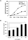
Results: In the in vivo model, the levels of serum IL-6 and angiotensinogen correlated. In the in vitro model, IL-6 transcriptionally regulated angiotensinogen expression. Additionally, IL-6 mediated angiotensinogen expression through the Janus kinase (JAK)/signal transducer and activator of transcription 3 (STAT3) and JAK/p38 signaling. Decoy ODN analyses revealed that STAT3 and nuclear factor-kB (NF-kB) also played critical roles in the transcriptional regulation of angiotensinogen by IL-6. IL-6-mediated signaling, JAK2, STAT3 and p38 inhibitors reduced angiotensinogen expression in the partially hepatectomized mice.
Conclusion: During liver regeneration, IL-6-enhanced angiotensinogen expression is dependent on the JAK/STAT3 and JAK/p38/NF-kB signaling pathways. Interruption of the molecular mechanisms of angiotensinogen regulation may be applied as the basis of therapeutic strategies for preventing angiotensin II-related ischemia-reperfusion injury during liver regeneration.
Conflict of interest statement
Figures


Similar articles
-
IL-6 regulates Mcl-1L expression through the JAK/PI3K/Akt/CREB signaling pathway in hepatocytes: implication of an anti-apoptotic role during liver regeneration.PLoS One. 2013 Jun 25;8(6):e66268. doi: 10.1371/journal.pone.0066268. Print 2013. PLoS One. 2013. PMID: 23825534 Free PMC article.
-
A20 promotes liver regeneration by decreasing SOCS3 expression to enhance IL-6/STAT3 proliferative signals.Hepatology. 2013 May;57(5):2014-25. doi: 10.1002/hep.26197. Epub 2013 Apr 5. Hepatology. 2013. PMID: 23238769 Free PMC article.
-
Red nucleus interleukin-1β evokes tactile allodynia through activation of JAK/STAT3 and JNK signaling pathways.J Neurosci Res. 2018 Dec;96(12):1847-1861. doi: 10.1002/jnr.24324. Epub 2018 Sep 14. J Neurosci Res. 2018. PMID: 30216497
-
The IL-6/JAK/STAT3 pathway: potential therapeutic strategies in treating colorectal cancer (Review).Int J Oncol. 2014 Apr;44(4):1032-40. doi: 10.3892/ijo.2014.2259. Epub 2014 Jan 15. Int J Oncol. 2014. PMID: 24430672 Review.
-
The research development of STAT3 in hepatic ischemia-reperfusion injury.Front Immunol. 2023 Jan 24;14:1066222. doi: 10.3389/fimmu.2023.1066222. eCollection 2023. Front Immunol. 2023. PMID: 36761734 Free PMC article. Review.
Cited by
-
Biomarkers during COVID-19: Mechanisms of Change and Implications for Patient Outcomes.Diagnostics (Basel). 2022 Feb 16;12(2):509. doi: 10.3390/diagnostics12020509. Diagnostics (Basel). 2022. PMID: 35204599 Free PMC article. Review.
-
Association of elevated inflammatory markers and severe COVID-19: A meta-analysis.Medicine (Baltimore). 2020 Nov 20;99(47):e23315. doi: 10.1097/MD.0000000000023315. Medicine (Baltimore). 2020. PMID: 33217868 Free PMC article. Review.
-
Transcriptomic and epigenetic analyses reveal a gender difference in aging-associated inflammation: the Vitality 90+ study.Age (Dordr). 2015 Aug;37(4):9814. doi: 10.1007/s11357-015-9814-9. Epub 2015 Jul 19. Age (Dordr). 2015. PMID: 26188803 Free PMC article.
-
Prognostic value of interleukin-6, C-reactive protein, and procalcitonin in patients with COVID-19.J Clin Virol. 2020 Jun;127:104370. doi: 10.1016/j.jcv.2020.104370. Epub 2020 Apr 14. J Clin Virol. 2020. PMID: 32344321 Free PMC article.
-
Lysophosphatidic acid alters the expression profiles of angiogenic factors, cytokines, and chemokines in mouse liver sinusoidal endothelial cells.PLoS One. 2015 Mar 30;10(3):e0122060. doi: 10.1371/journal.pone.0122060. eCollection 2015. PLoS One. 2015. PMID: 25822713 Free PMC article.
References
-
- Fausto N (2000) Liver regeneration. J Hepatol 32: 19–31. - PubMed
-
- Gilgenkrantz H, Collin de l’Hortet A (2011) New insights into liver regeneration. Clin Res Hepatol Gastroenterol 35: 623–629. - PubMed
-
- Togo S, Makino H, Kobayashi T, Morita T, Shimizu T, et al. (2004) Mechanism of liver regeneration after partial hepatectomy using mouse cDNA microarray. J Hepatol 40: 464–471. - PubMed
-
- Lai HS, Chen Y, Lin WH, Chen CN, Wu HC, et al. (2005) Quantitative gene expressions analysis by cDNA microarray during liver regeneration after partial hepatectomy in rats. Surg Today 35: 396–403. - PubMed
Publication types
MeSH terms
Substances
LinkOut - more resources
Full Text Sources
Other Literature Sources
Miscellaneous

