. 2013 Jul 2;8(7):e67949.
doi: 10.1371/journal.pone.0067949. Print 2013.

Intracranial aneurysm formation in type-one diabetes rats

Affiliations

Intracranial aneurysm formation in type-one diabetes rats

Tao Yan et al. PLoS One. .

Abstract

Background & objective: Diabetes mellitus (DM) plays an important role in the pathogenesis of vascular complications including arteriosclerosis and ischemic stroke. Whether DM impacts intracranial aneurysm (IA) formation has not been extensively investigated. In this study, we tested the underlying mechanism of type one DM (T1DM) induced IA formation in rats.

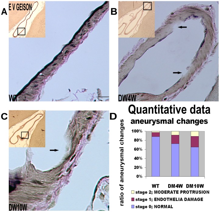
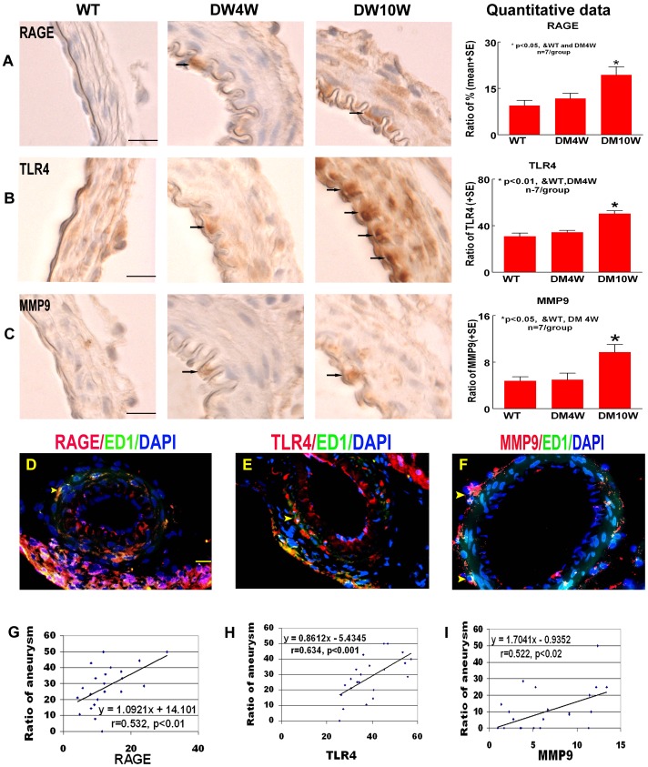
Experimental approaches: T1DM was induced by streptozotocin injection. Rats were euthanized at 0, 4 and 10 weeks after T1DM induction. To evaluate cerebral vascular perfusion, Fluorescein isothiocyanate - dye was injected at 5 min prior to euthanasia. Vascular perfusion was measured by laser scanning confocal microscopy. Trichrome, Elastica van Gieson, alpha-smooth muscle actin (a-SMA) and receptor of advanced glycation end-products (RAGE), toll-like receptor 4 (TLR4) and matrix metalloproteinase 9 (MMP9) immunostaining were performed. The IA formation was classified by 0-3 stages: 0: Normal; 1: Endothelial damage; 2: Moderate protrusion; and 3: Saccular aneurysm formation.

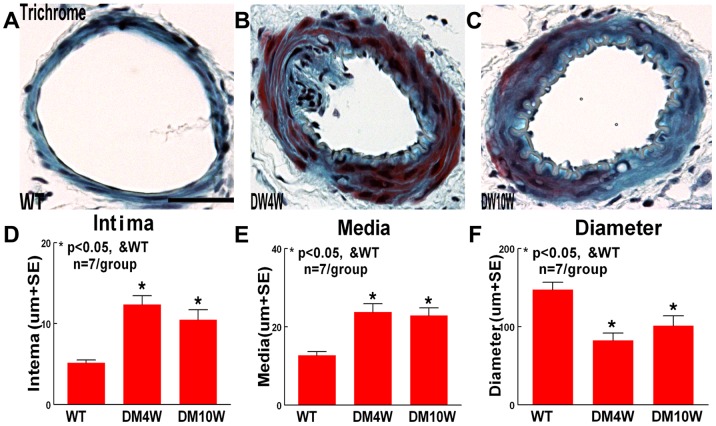
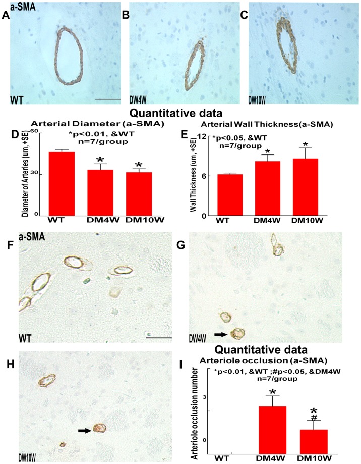
Results: T1DM significantly increased IA formation identified by the classification of aneurysmal changes compared with non-DM rats (p<0.05). However, T1DM induced IA formations were classified as stage 1 and stage 2, but not stage 3. Cerebral vascular perfusion was significantly decreased in T1DM rats compared to non-DM rats (p<0.01). DM10W rats exhibited a significant decrease of cerebral vascular perfusion compared to DM4W rats (p<0.05). T1DM rats also significantly increased the internal carotid artery (ICA) intimae and media thickness, and decreased the internal carotid artery diameter compared to non-DM rats. RAGE, MMP9 and TLR4 expression were significantly increased in T1DM rats compared to non-DM rats. The increased RAGE, TLR4 and MMP9 significantly correlated with IA formation (p<0.05).

Conclusion: T1DM increases IA formation. The increased RAGE, MMP9 and TLR4 expressions might contribute to IA formation in T1DM rats.


Conflict of interest statement

Competing Interests: The authors have declared that no competing interests exist.

Figures

Figure 1
Figure 1. T1DM increases IA formation identified by the classification of aneurysmal changes compared with non-DM rats.
A–C: Elastica Van Geison staining: T1DM increases IA formation compared with non-DM rats. D: Quantitative data: The ratio of stage 1 and stage 2 of aneurysmal formations to total arterial number was significantly increased in T1DM rats (p<0.05). Arrows show a slight focal thinning and bulging of the arterial wall (Fig. 1B and C). Scale bar in A, 20 µm.
Figure 2
Figure 2. T1DM increases RAGE, MMP9 and TLR4 expression.
RAGE, TLR4 and MMP9 is correlated with aneurysm formation in T1DM rats. A–C: RAGE (A), TLR4 (B) and MMP9 (C) immunostaining and quantitative data. T1DM significantly increased RAGE, MMP9 and TLR4 expression compared to non-DM rats. Arrows indicate the positive cells in the arterial wall. D–F: Double immunostaining ED1 with RAGE (D), TLR4 (E) and MMP9 (F). MMP9, TLR4 and RAGE expression is colocalized with ED1. Arrow heads indicate the positive cells in the arterial wall. G–I: Correlation analysis of aneurysm formation with RAGE (G), TLR4 (H) and MMP9 (I). Aneurysm formation significantly correlated with RAGE, TLR4 and MMP9 expression. Scale bar in A, D, 20 µm.
Figure 3
Figure 3. T1DM accelerates arteriosclerosis-like changes in ICA.
A–C: Trichrome staining in WT (A), 4 week T1DM (B) and 10 week T1DM (C) rats. D–F: Quantitative data. T1DM increases the ICA intimae (D) and media thickness (E), and decreased the ICA diameter (F) (p<0.05). Scale bar in A, 0.1 mm.
Figure 4
Figure 4. T1DM decreases cerebral vascular perfusion in the cerebral parenchyma compared to non-DM rats.
A–C: FITC-dye vascular perfusion in WT (A), 4 weeks T1DM (B) and 10 weeks T1DM (C) rats. D: Cerebral vascular perfusion quantitative data. Scale bar in A, 0.1 mm.
Figure 5
Figure 5. T1DM accelerates arteriosclerosis-like changes in cerebral arteries compared to non-DM rats.
T1DM decreases the cerebral arterial diameter while increases the cerebral arterial wall thickness and arterioles occlusion in the cerebral parenchyma compared to non-DM rats. a-SMA immunostaining and quantitative data: A–C: Cerebral artery wall diameter and thickness in WT (A), 4 week T1DM (B) and 10 week T1DM (C) rats, D: quantitative data of arterial diameter, E: quantitative data of arterial wall thickness. F–H: Cerebral arterioles occlusion in WT (F), 4 week T1DM (G) and 10 week T1DM (H) rats. I: Cerebral arterioles occlusion quantitative data. Scale bar in A, F, 0.05 mm. Arrows indicate the occluded arterioles (G, H).

References

    1. Li W, Prakash R, Kelly-Cobbs AI, Ogbi S, Kozak A, et al. (2010) Adaptive cerebral neovascularization in a model of type 2 diabetes: relevance to focal cerebral ischemia. Diabetes 59: 228–235. - PMC - PubMed
    1. Fox CS, Coady S, Sorlie PD, Levy D, Meigs JB, et al. (2004) Trends in cardiovascular complications of diabetes. JAMA 292: 2495–2499. - PubMed
    1. Ye X, Chopp M, Cui X, Zacharek A, Cui Y, et al. (2011) Niaspan enhances vascular remodeling after stroke in type 1 diabetic rats. Exp Neurol 232: 299–308. - PMC - PubMed
    1. Haley AP, Gonzales MM, Tarumi T, Tanaka H (2012) Subclinical vascular disease and cerebral glutamate elevation in metabolic syndrome. Metab Brain Dis. - PMC - PubMed
    1. Tamura T, Jamous MA, Kitazato KT, Yagi K, Tada Y, et al. (2009) Endothelial damage due to impaired nitric oxide bioavailability triggers cerebral aneurysm formation in female rats. J Hypertens 27: 1284–1292. - PubMed

Publication types

MeSH terms