. 2013 Jul 3;8(7):e68640.
doi: 10.1371/journal.pone.0068640. Print 2013.

TLR-2/TLR-4 TREM-1 signaling pathway is dispensable in inflammatory myeloid cells during sterile kidney injury

Affiliations

TLR-2/TLR-4 TREM-1 signaling pathway is dispensable in inflammatory myeloid cells during sterile kidney injury

Gabriela Campanholle et al. PLoS One. .

Abstract

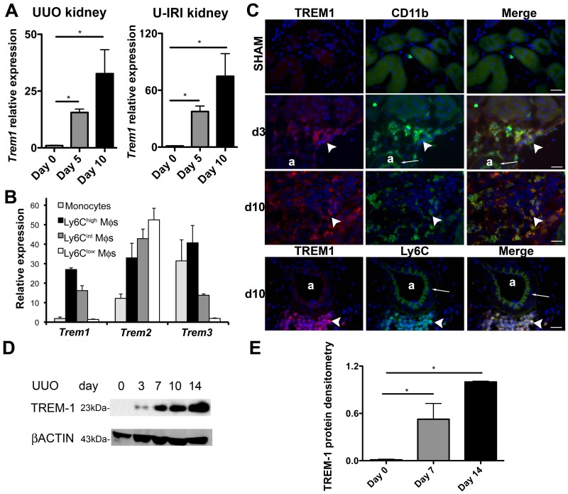
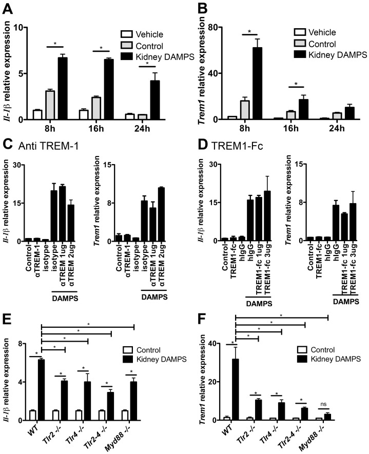
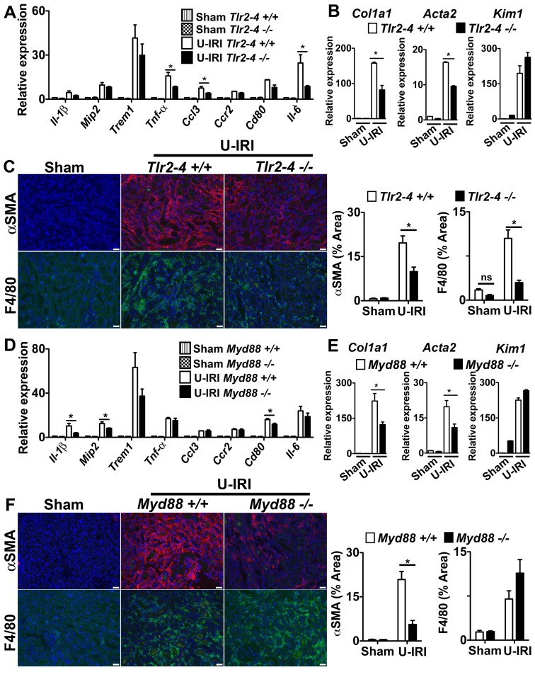
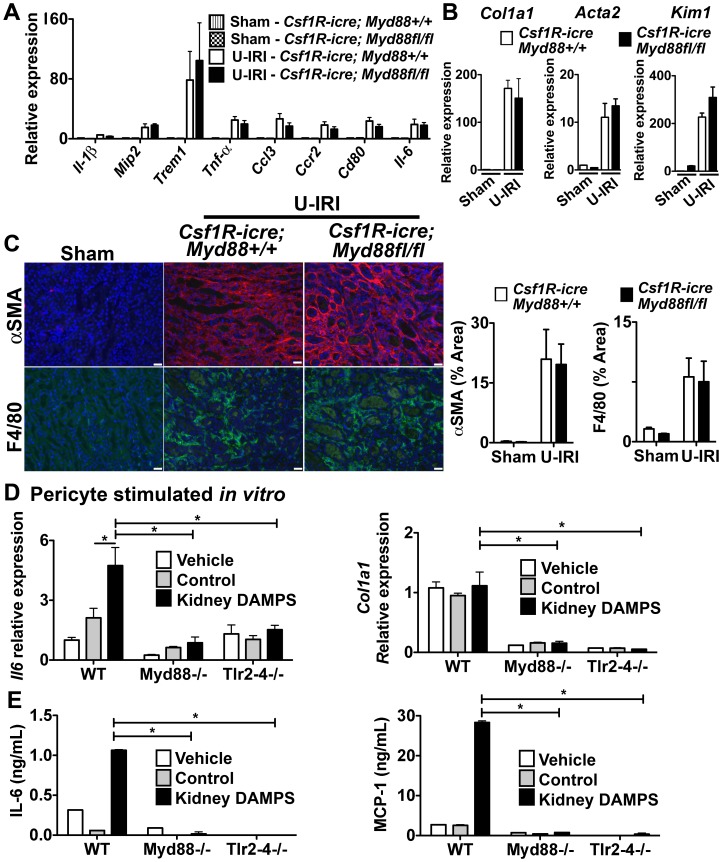
Inflammatory macrophages are abundant in kidney disease, stimulating repair, or driving chronic inflammation and fibrosis. Damage associated molecules (DAMPs), released from injured cells engage pattern recognition receptors (PRRs) on macrophages, contributing to activation. Understanding mechanisms of macrophage activation during kidney injury may lead to strategies to alleviate chronic disease. We identified Triggering-Receptor-in-Myeloid-cells (TREM)-1, a regulator of TLR signaling, as highly upregulated in kidney inflammatory macrophages and tested the roles of these receptors in macrophage activation and kidney disease. Kidney DAMPs activated macrophages in vitro, independently of TREM-1, but partially dependent on TLR-2/-4, MyD88. In two models of progressive interstitial kidney disease, TREM-1 blockade had no impact on disease or macrophage activation in vivo, but TLR-2/-4, or MyD88 deficiency was anti-inflammatory and anti-fibrotic. When MyD88 was mutated only in the myeloid lineage, however, there was no bearing on macrophage activation or disease progression. Instead, TLR-2/-4 or MyD88 deficiency reduced activation of mesenchyme lineage cells resulting in reduced inflammation and fibrosis, indicating that these pathways play dominant roles in activation of myofibroblasts but not macrophages. To conclude, TREM-1, TLR2/4 and MyD88 signaling pathways are redundant in myeloid cell activation in kidney injury, but the latter appear to regulate activation of mesenchymal cells.


Conflict of interest statement

Competing Interests: This study was partly funded by a Genzyme research in progress (GRIP award to JSD). JSD is on the Scientific Advisory Board for Regulus Therapeutics and Promedior Inc. JSD is a co-founder of Muregen LLC. There are no patents, products in development or marketed products to declare. This does not alter the authors' adherence to all the PLOS ONE policies on sharing data and materials, as detailed online in the guide for authors.

Figures

Figure 1
Figure 1. TREMs are highly expressed in macrophages during kidney injury.
(A) Q-PCR for Trem1 expression in whole kidney 0, 5 and 10 days after UUO and U-IRI. (B) Q-PCR for TREM family transcript expression in blood monocytes, and different sub-populations of kidney macrophages purified at day 5 after UUO. (C) Fluorescence images showing CD11b (green), Ly6C (green) and TREM-1 (red) expression in tissue sections from control kidney (sham), day 3 and 10 after UUO (a, arteriole; Bar = 25 µm; arrowhead shows interstitial CD11b+ and TREM-1+ cells; arrow shows autofluorescent arteriole internal elastic lamina). (D) Western blot of whole kidney lysates detecting TREM-1 (23kD) and β-Actin (43kD), 0, 3, 7, 10 and 14 days after UUO. (E) TREM-1 protein densitometry normalized to endogenous control β-Actin. (n = 3–5/group, 3 independent experiments; *P<0.05).
Figure 2
Figure 2. TLR-2 and TLR-4 but not TREM-1, regulate activation of macrophages in vitro by kidney DAMPs.
(A–B) Q-PCR showing Il-1β and Trem1 expression in BMDMφ stimulated for 8, 16 or 24h with soluble factors prepared from normal kidney (control) or disease kidney (kidney DAMPs). (C–D) Graphs showing Il-1β and Trem1 expression by Q–PCR, 16h after BMDMφ were stimulated with kidney DAMPs and (C) activating anti-TREM-1 antibodies, or (D) TREM1-Fc, which blocks TREM-1 receptor by competing for ligands. (E–F) Q-PCR showing (E) Il-1β and (F) Trem1 expression in BMDMφ isolated from WT, Tlr2−/−, Tlr4−/−, Tlr2–4−/−, and Myd88−/− mice stimulated with kidney DAMPs for 16h. Q-PCR results were normalized to their respective control group. (*P<0.05, n = 5–7/group, 3 independent experiments; ns, p is not significant).
Figure 3
Figure 3. Treatment with soluble TREM1-Fc does not prevent macrophage activation, injury and fibrosis in sterile kidney injury.
(A) Schema showing experimental design. Mice were subjected to unilateral ischemia and reperfusion injury (U-IRI) and treated daily with 40 µg/mouse of TREM1-Fc or hIgG, as control. (B) Western blot showing presence of TREM1-Fc (approximately 56kD) in 2 µl of plasma collected at day 2 from mice treated daily with 40 µg of TREM1-Fc. Anti-mouse IgG was used as endogenous control. (C) Q-PCR for different inflammatory transcripts (left) or pro-fibrotic transcripts, Collagen1a1 (Col1a1) and alpha smooth muscle actin (Acta2), from whole kidney day 5 after U-IRI. (D) Representative images (left) and quantitative graphs (right) showing+F4/80 cells (green),+αSMA (red) or collagen deposition (Sirius Red staining) day 5 after U-IRI. (*P<0.05, n = 5–7/group, 3 independent experiments; Bar marker = 50 µm; Q-PCR results were normalized to sham +hIgG).
Figure 4
Figure 4. The TLR-2/TLR-4/MyD88 pathways play a role in fibrosis in the U-IRI model of sterile kidney injury.
(A–C) Tlr2–4−/− (D–F) Myd88−/− mice or respective controls were subjected to U-IRI and kidney harvested for tissue analysis 5 days later. Q-PCR (A,D) for different inflammatory transcripts, (B,E) pro-fibrotic transcripts, collagen1a1 (col1a1) and alpha smooth muscle actin (Acta2), and the tubule injury marker, kidney injury molecule-1 (Kim-1) from whole kidney day 5 after U-IRI. (C,F) Representative fluorescent images (left) and quantitative graphs (right) showing+αSMA (red) cells and+F4/80 cells (green). (*P<0.05, n = 5–7/group, 3 independent experiments; ns, p is not significant; Bar = 50 µm; Q-PCR results were normalized to wild type control).
Figure 5
Figure 5. The TLR-2/TLR-4/MyD88 pathway is dispensable in macrophage activation in kidney fibrosis, but important in mesenchyme cell activation.
(A–C) Csf1R-icre; Myd88fl/fl mice or respective controls were subjected to U-IRI and kidney harvested for tissue analysis 5 days later. Q-PCR (A) for different inflammatory transcripts, (B) pro-fibrotic transcripts, collagen1a1 (col1a1) and alpha smooth muscle actin (Acta2), and the tubule injury marker, kidney injury molecule-1 (Kim-1) from whole kidney day 5 after U-IRI. (C) Representative fluorescent images (left) and quantitative graphs (right) showing+αSMA (red) cells and+F4/80 cells (green). (D–E) Primary pericytes were isolated from Myd88−/− and Tlr2–4−/− mice and stimulated in vitro for 8h with kidney DAMPs. (D) Graph showing Il-6 and Col1a1 transcript expression by Q-PCR. (E) Graph showing IL-6 and MCP-1 concentration in supernatant by ELISA. (*P<0.05, n = 5–7/group, 3 independent experiments; ns, p is not significant; Bar = 50 µm; Q-PCR results were normalized to wild type control).

References

    1. Nelson PJ, Rees AJ, Griffin MD, Hughes J, Kurts C, et al. (2012) The renal mononuclear phagocytic system. Journal of the American Society of Nephrology 23: 194–203 doi:10.1681/ASN.2011070680 - DOI - PMC - PubMed
    1. Duffield JS (2010) Macrophages and Immunologic Inflammation of the Kidney. Seminars in Nephrology 30: 234–254. - PMC - PubMed
    1. Lin SL, Castano AP, Nowlin BT, Lupher MLJ, Duffield JS (2009) Bone Marrow Ly6Chigh Monocytes Are Selectively Recruited to Injured Kidney and Differentiate into Functionally Distinct Populations. The Journal of Immunology 183: 6733–6743. - PubMed
    1. Rosin DL, Okusa MD (2011) Dangers Within: DAMP Responses to Damage and Cell Death in Kidney Disease. J Am Soc Nephrol 22: 416–425. - PMC - PubMed
    1. Anders HJ (2010) Toll-like receptors and danger signaling in kidney injury. Journal of the American Society of Nephrology 21: 1270–1274 doi:10.1681/ASN.2010030233 - DOI - PubMed

Publication types

MeSH terms

Substances