. 2013 Jul 3;8(7):e69069.
doi: 10.1371/journal.pone.0069069. Print 2013.

Rotavirus replication in the cholangiocyte mediates the temporal dependence of murine biliary atresia

Affiliations

Rotavirus replication in the cholangiocyte mediates the temporal dependence of murine biliary atresia

Sujit K Mohanty et al. PLoS One. .

Abstract

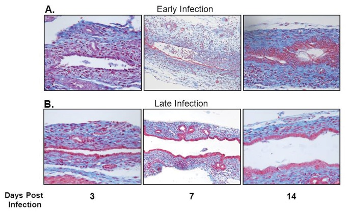
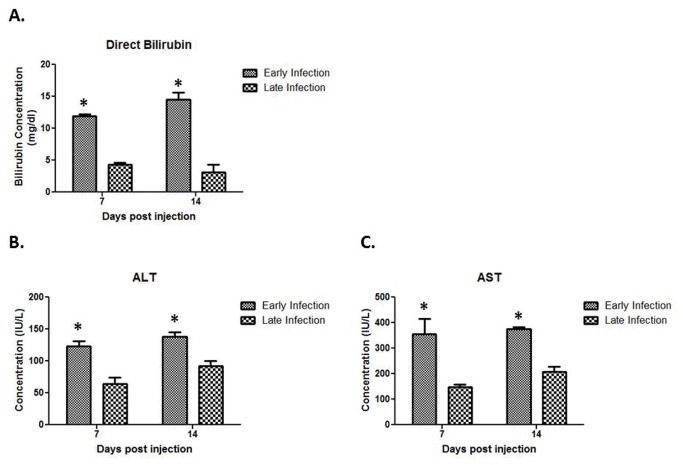
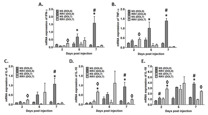
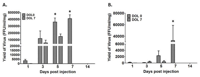
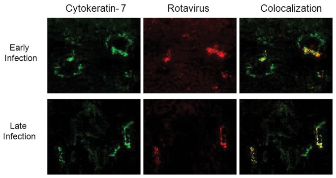
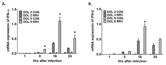
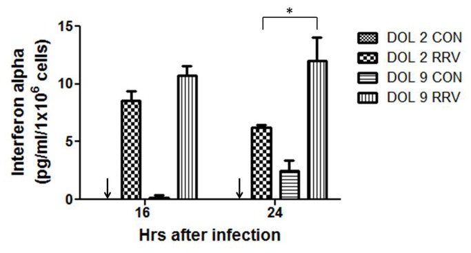
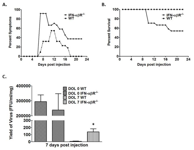
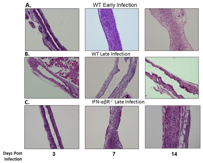
Biliary atresia (BA) is a neonatal disease that results in obliteration of the biliary tree. The murine model of BA, which mirrors the human disease, is based upon infection of newborn mice with rhesus rotavirus (RRV), leading to an obstructive cholangiopathy. The purpose of this study was to characterize the temporal relationship between viral infection and the induction of this model. BALB/c mice were infected with RRV on day of life (DOL) 0, 3, 5, and 7. Groups were characterized as early-infection (infection by DOL 3) or late-infection (infection after DOL 5). Early RRV infection induced symptoms in 95% of pups with a mortality rate of 80%. In contrast, late infection caused symptoms in only 50% of mice, and 100% of pups survived. The clinical findings correlated with histological analysis of extrahepatic biliary trees, cytokine expression, and viral titers. Primary murine cholangiocytes isolated, cultured, and infected with RRV yielded higher titers of infectious virus in those harvested from DOL 2 versus DOL 9 mice. Less interferon alpha and beta was produced in DOL 2 versus DOL 9 RRV infected primary cholangiocytes. Injection of BALB/c interferon alpha/beta receptor knockout (IFN-αβR(-/-)) pups at DOL 7 showed increased symptoms (79%) and mortality (46%) when compared to late infected wild type mice. In conclusion, the degree of injury sustained by relatively immature cholangiocytes due to more robust RRV replication correlated with more severe clinical manifestations of cholangiopathy and higher mortality. Interferon alpha production by cholangiocytes appears to play a regulatory role. These findings confirm a temporal dependence of RRV infection in murine BA and begin to define a pathophysiologic role of the maturing cholangiocyte.


Conflict of interest statement

Competing Interests: The authors have declared that no competing interests exist.

Figures

Figure 1
Figure 1. Symptoms and survival after RRV infection.
The top graph illustrates morbidity from viral infection on the corresponding day of infection. Significant resolution of symptoms in the late infection groups occurs by day of life (DOL) 21. The bottom graph illustrates mortality. * p<0.05 when compared with both infections on DOL 5 and DOL 7 to infections on DOL 0 and DOL 3.
Figure 2
Figure 2. Histologic appearance of the extrahepatic biliary tract following infection with RRV in early versus late injected mice.
Mason’s trichrome staining of extrahepatic bile ducts after infection with RRV leads to complete obstruction of extrahepatic biliary duct in early infection group (A). In contrast, the late infection group had some mild inflammation with a patent bile duct (B). In each group, the images are magnified 40X.
Figure 3
Figure 3. Quantification of Liver Enzymes.
Serum harvested from early and late infected pups demonstrates significantly higher concentrations of direct bilirubin, ALT, and AST at both 7 and 14 days post injection *p<0.05.
Figure 4
Figure 4. Cytokine mRNA expression after RRV infection on DOL 0 or 7.
Expression of IFN-γ (A), TNF-α (B), IL-6 (C), IL-10 (D), and IL-18 (E) mRNA in livers harvested from newborn mice injected with RRV on DOL 0 increases over time as compared to control. In contrast, cytokine levels from livers extracted from mice infected on DOL 7 are more often significantly elevated over control, 2 days post-infection with a general decrease in level as compared to DOL 0 injected mice. Finally, by 7 days post-infection, mRNA expression of all 4 cytokines is higher in mice injected with RRV on DOL 0 as compared to those infected on DOL 7 (* p<0.05 DOL 0 infected vs. DOL 7, # p<0.05 DOL 0 infected vs. uninfected, ◊ p<0.05 DOL 7 infected vs. uninfected).
Figure 5
Figure 5. Quantification of infectious RRV post infection.
The two graphs illustrate the quantity of live RRV present in the extrahepatic biliary tree (A) and in the liver (B) 7 days post injection. * p<0.05.
Figure 6
Figure 6. Immunofluorescence of liver 5 days post-RRV infection.
Column A demonstrates staining of biliary epithelium (cytokeratin 7 positive, green). Column B demonstrates staining for rotavirus (red). Column C demonstrates overlay images where co localization corresponds to yellow-orange staining. Staining of samples from Day 3 infection is similar to Day 0 and samples from Day 5 infection is similar to Day 7 (data not shown). All images were magnified 20X.
Figure 7
Figure 7. INF-α and INF-β mRNA expression after RRV infection in DOL 2 and DOL 9 primary cholangiocytes.
Following RRV infection in the primary cholangiocytes there was a significant increased expression of IFN-α (more than 3 fold) (A) and of IFN-β (2 fold) (B) in the DOL 9 cholangiocytes when compared to DOL 2 cholangiocytes *p<0.05.
Figure 8
Figure 8. Quantification of IFN-α protein in primary cholangiocytes after RRV infection.
Supernatants collected at 16 and 24 hours from DOL 2 and 9 primary cholangiocyte after RRV infection demonstrates greater quantities of IFN-α protein at 16 hr PI with a significantly higher quantity at 24 hours in the DOL 9 cholangiocytes compared to the DOL 2 cholangiocytes *p<0.05. Arrows indicate undetectable levels of interferon alpha.
Figure 9
Figure 9. Symptoms, survival and viral titer in IFN-αβR-/ -mice following RRV infection.
Graph A illustrates morbidity from viral DOL 7 infected IFN-αβR−/−vs WT mice. Graph B illustrates mortality. Graph C demonstrates the quantity of live RRV present in the extrahepatic biliary tree seven days post infection. *p<0.05 when compared between RRV infected IFN-αβR−/−mice and WT mice.
Figure 10
Figure 10. Bile duct histology of WT and IFN-αβR-/ -mice following RRV infection.
Extrahepatic bile ducts were harvested from WT and IFN-αβR−/−mice 3, 7 and 14 days post inoculation at early and late time points with RRV and stained with H and E. RRV injection led to obstruction of the lumens of the extrahepatic bile ducts in the early but not the late infected WT mice. In contrast, the lumens of the late infected IFN-αβR−/−mice were obstructed following RRV injection. Magnification: 10X.

References

    1. Sokol RJ, Shepherd RW, Superina R, Bezerra JA, Robuck P et al. (2007) Screening and outcomes in biliary atresia: summary of a National Institutes of Health workshop. Hepatology 46: 566-581. doi:10.1002/hep.21790. PubMed: 17661405. - DOI - PMC - PubMed
    1. Bangaru B, Morecki R, Glaser JH, Gartner LM, Horwitz MS (1980) Comparative studies of biliary atresia in the human newborn and reovirus-induced cholangitis in weanling mice. Lab Invest 43: 456-462. PubMed: 7421127. - PubMed
    1. Morecki R, Glaser JH, Johnson AB, Kress Y (1984) Detection of reovirus type 3 in the porta hepatis of an infant with extrahepatic biliary atresia: ultrastructural and immunocytochemical study. Hepatology 4: 1137-1142. doi:10.1002/hep.1840040608. PubMed: 6389303. - DOI - PubMed
    1. Riepenhoff-Talty M, Gouvea V, Evans MJ, Svensson L, Hoffenberg E et al. (1996) Detection of group C rotavirus in infants with extrahepatic biliary atresia. J Infect Dis 174: 8-15. doi:10.1093/infdis/174.1.8. PubMed: 8656017. - DOI - PubMed
    1. Mack CL, Tucker RM, Sokol RJ, Karrer FM, Kotzin BL et al. (2004) Biliary atresia is associated with CD4+ Th1 cell-mediated portal tract inflammation. Pediatr Res 56: 79-87. doi:10.1203/01.PDR.0000130480.51066.FB. PubMed: 15128911. - DOI - PMC - PubMed

Publication types

MeSH terms