. 2013;15(4):R55.
doi: 10.1186/bcr3449.

Autocrine IGF-I/insulin receptor axis compensates for inhibition of AKT in ER-positive breast cancer cells with resistance to estrogen deprivation

Autocrine IGF-I/insulin receptor axis compensates for inhibition of AKT in ER-positive breast cancer cells with resistance to estrogen deprivation

Emily M Fox et al. Breast Cancer Res. 2013.

Abstract

Introduction: Estrogen receptor α-positive (ER+) breast cancers adapt to hormone deprivation and acquire resistance to antiestrogen therapies. Upon acquisition of hormone independence, ER+ breast cancer cells increase their dependence on the phosphatidylinositol-3 kinase (PI3K)/AKT pathway. We examined the effects of AKT inhibition and its compensatory upregulation of insulin-like growth factor (IGF)-I/InsR signaling in ER+ breast cancer cells with acquired resistance to estrogen deprivation.

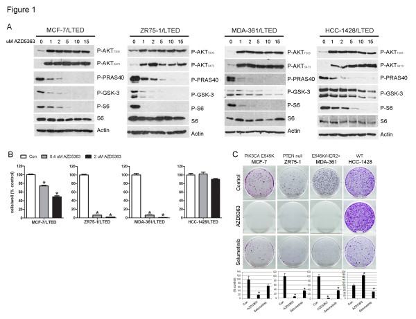
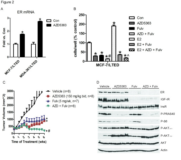
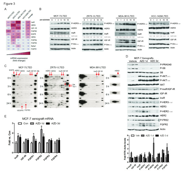
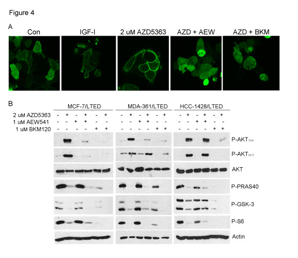
Methods: Inhibition of AKT using the catalytic inhibitor AZD5363 was examined in four ER+ breast cancer cell lines resistant to long-term estrogen deprivation (LTED) by western blotting and proliferation assays. Feedback upregulation and activation of receptor tyrosine kinases (RTKs) was examined by western blotting, real-time qPCR, ELISAs, membrane localization of AKT PH-GFP by immunofluorescence and phospho-RTK arrays. For studies in vivo, athymic mice with MCF-7 xenografts were treated with AZD5363 and fulvestrant with either the ATP-competitive IGF-IR/InsR inhibitor AZD9362 or the fibroblast growth factor receptor (FGFR) inhibitor AZD4547.

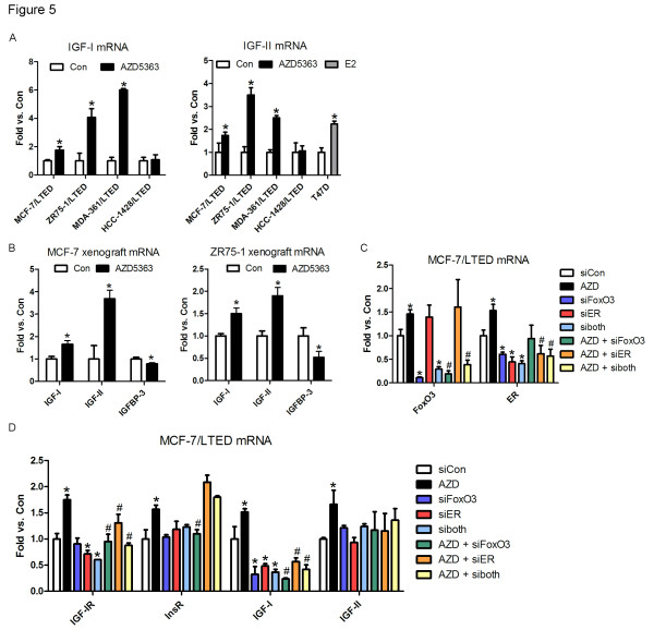
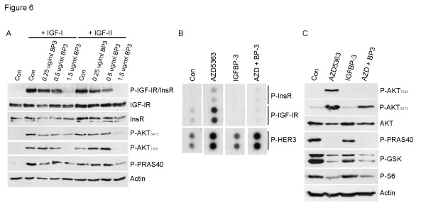
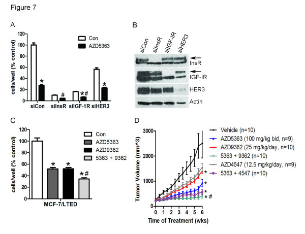
Results: Treatment with AZD5363 reduced phosphorylation of the AKT/mTOR substrates PRAS40, GSK3α/β and S6K while inducing hyperphosphorylation of AKT at T308 and S473. Inhibition of AKT with AZD5363 suppressed growth of three of four ER+ LTED lines and prevented emergence of hormone-independent MCF-7, ZR75-1 and MDA-361 cells. AZD5363 suppressed growth of MCF-7 xenografts in ovariectomized mice and a patient-derived luminal B xenograft unresponsive to tamoxifen or fulvestrant. Combined treatment with AZD5363 and fulvestrant suppressed MCF-7 xenograft growth better than either drug alone. Inhibition of AKT with AZD5363 resulted in upregulation and activation of RTKs, including IGF-IR and InsR, upregulation of FoxO3a and ERα mRNAs as well as FoxO- and ER-dependent transcription of IGF-I and IGF-II ligands. Inhibition of IGF-IR/InsR or PI3K abrogated AKT PH-GFP membrane localization and T308 P-AKT following treatment with AZD5363. Treatment with IGFBP-3 blocked AZD5363-induced P-IGF-IR/InsR and T308 P-AKT, suggesting that receptor phosphorylation was dependent on increased autocrine ligands. Finally, treatment with the dual IGF-IR/InsR inhibitor AZD9362 enhanced the anti-tumor effect of AZD5363 in MCF-7/LTED cells and MCF-7 xenografts in ovariectomized mice devoid of estrogen supplementation.

Conclusions: These data suggest combinations of AKT and IGF-IR/InsR inhibitors would be an effective treatment strategy against hormone-independent ER+ breast cancer.


Figures

Figure 1
Figure 1
Catalytic AKT inhibitor suppresses LTED growth and prevents emergence of hormone-independent breast cancer cells. A) LTED cells were treated with 10% DCC-FBS ± 0 to 15 µM AZD5363 for 24 hours. Protein lysates were analyzed by immunoblot using the indicated antibodies. B) LTED cells were treated with 10% DCC-FBS ± 0.4 or 2 µM AZD5363. Media and drugs were replenished every three days. Cells were counted after five to ten days. Data are presented as percent of control; each bar, mean ± SEM (n = 3; *P <0.0001 versus Con, one-way ANOVA). C) Parental cells in 10% DCC-FBS were treated with ± 2 µM AZD5363 or 1 µM selumetinib. Media and drugs were replenished every three days. When control monolayers reached 60% to 80% confluency (after 15 (MCF-7), 36 (ZR75-1 and MDA-361) or 38 (HCC-1428) days, respectively), cells were fixed and stained with crystal violet. Representative images and quantification of integrated intensity (% control) are shown (*P <0.05 versus control, t-test). ANOVA, analysis of variance; DCC-FBS, dextran/charcoal-treated fetal bovine serum; LTED, long-term estrogen deprivation; SEM, standard error of the mean.
Figure 2
Figure 2
Combined inhibition of AKT and ER suppresses hormone-independent tumor growth. A) LTED cells were treated with 10% DCC-FBS ± 2 µM AZD5363 for 24 hours. RNA was extracted, reverse transcribed to cDNA and analyzed by real-time PCR. Threshold cycle values were normalized for actin. Data are presented as fold versus control; each bar, mean ± SEM (n = 2; *P <0.0001 versus Con, two-way ANOVA). B) LTED cells were treated with 10% DCC-FBS ± 2 µM AZD5363, 1 µM fulvestrant or 1 nM E2. Media and drugs were replenished every three days. Cells were counted after five days. Data are presented as percent of control; each bar, mean ± SEM (n = 3; *P <0.0001 versus Con or E2; #P <0.01 versus AZD or fulvestrant; ^P <0.01 versus E2 + fulvestrant, one-way ANOVA). C) MCF-7 cells were injected s.c. into athymic mice supplemented with 14-day release 17β-estradiol pellets. Mice bearing tumors ≥150 mm3 were randomized to vehicle, AZD5363 (150 mg/kg/day bid), fulvestrant (5 mg/wk), or AZD5363 + fulvestrant for six weeks. Data are presented as mean tumor volume ± SEM (*P <0.01 versus vehicle; #P <0.001 versus AZD or fulvestrant, t-test). D) Xenografts from C) were homogenized one hour after the last dose and tumor lysates were analyzed by immunoblot using the indicated antibodies. ANOVA, analysis of variance; DCC-FBS, dextran/charcoal-treated fetal bovine serum; LTED, long-term estrogen deprivation; SEM, standard error of the mean.
Figure 3
Figure 3
Inhibition of AKT results in feedback upregulation of RTKs. A) LTED cells were treated with 10% DCC-FBS ± 2 µM AZD5363 for 24 hours. RNA was extracted, reverse transcribed to cDNA and analyzed by real-time PCR. Threshold cycle values were normalized for actin. Average fold changes over control (Con) were calculated and used to generate a heatmap. B) LTED cells were treated for 24 hours in 10% DCC-FBS ± 15 µM AZD5363. Protein lysates were analyzed by immunoblot. Densitometric analysis was performed and the ratio of InsR:actin is shown below the InsR blots. C) LTED cells in 10% DCC-FBS were treated ± 2 µM AZD5363 for 0 to 24 hours. Cell lysates (0.5 mg) were prepared and analyzed by phospho-RTK arrays. D) Mice bearing MCF-7 xenografts ≥150 mm3 were treated with vehicle or AZD5363 (150 mg/kg bid p.o.) for one or three days. Xenografts were harvested four hours after the last dose; tumor lysates were prepared and analyzed by immunoblot. Densitometric analysis was performed and a graphical representation of the average RTK:actin levels is shown below the blot. E) Xenografts from D) were homogenized and RNA was extracted and analyzed by real-time PCR as in A. Data are presented as fold versus control; each bar, mean ± SEM (n = 3; *P <0.05 versus Con, t-test). DCC-FBS, dextran/charcoal-treated fetal bovine serum; LTED, long-term estrogen deprivation; RTK, receptor tyrosine kinase; SEM, standard error of the mean.
Figure 4
Figure 4
Inhibition of IGF-IR/InsR or PI3K abrogates AZD5363-induced AKT membrane localization and phosphorylation. A) MCF-7/LTED cells were transfected with an AKT PH-GFP plasmid. On day four, cells were treated with 100 ng/ml IGF-I in serum-free medium for 15 minutes, or pre-incubated with 10% DCC-FBS ± 1 µM AEW541 or 1 µM BKM120 for 30 minutes followed by treatment with 2 µM AZD5363 for four hours. Cells were viewed in a LSM 510Meta confocal microscope at 40x magnification. B) LTED lines were treated with 10% DCC-FBS ± 1 µM AEW541 or BKM120 for 1 hour, followed by the addition of 2 µM AZD5363 for 24 hours. Protein lysates were analyzed by immunoblot using the indicated antibodies. DCC-FBS, dextran/charcoal-treated fetal bovine serum; IGF-I, insulin-like growth factor-I; LTED, long-term estrogen deprivation.
Figure 5
Figure 5
AZD5363-induced upregulation of IGF-IR, IGF-I and IGF-II is dependent on ER and FoxO3. A) LTED cells were treated with 10% DCC-FBS ± 2 µM AZD5363 for 24 hours. T47D cells were serum starved for 24 hours and then treated with 10% DCC-FBS ± 2 µM AZD5363 for 24 hours. RNA was extracted, reverse transcribed to cDNA and analyzed by real-time PCR. Threshold cycle values were normalized for actin. qPCR reactions with no input RNA were used as negative controls. Data are presented as fold versus control; each bar, mean ± SEM (n = 4; *P <0.05 versus Con, t-test). B) Mice bearing MCF-7 or ZR75-1 xenografts ≥150 mm3 were treated with vehicle or AZD5363 (150 mg/kg bid) for one day. Xenografts were harvested four hours after the last dose and homogenized. RNA was extracted, reverse transcribed to cDNA and analyzed by real-time PCR. Threshold cycle values were normalized for actin (MCF-7) or 36B4 (ZR75-1). Data are presented as fold versus control; each bar, mean ± SEM (n = 3; *P <0.05 versus Con, t-test). C, D) MCF-7/LTED cells were transfected with siRNA targeting FoxO3, ER, both (siboth) or a non-silencing control (siCon). Two days later the cells were treated with 10% DCC-FBS ± 2 µM AZD5363 for 24 hours. RNA was extracted and analyzed by real-time PCR as in A. Data are presented as fold versus control; each bar, mean ± SEM (n = 3). C) *P <0.01 versus siCon, t-test. D) *P <0.05 versus siCon; #P <0.05 versus AZD, t-test. DCC-FBS, dextran/charcoal-treated fetal bovine serum; ER, estrogen receptor; FoxO, forkhead box class O; LTED, long-term estrogen deprivation.
Figure 6
Figure 6
IGFBP-3 blocks AZD5363-induced phosphorylation of IGF-IR/InsR and AKT. A) MCF-7/LTED cells were treated overnight with serum-free medium ± 1.5 µg/ml IGFBP-3, followed by treatment for 15 minutes ± 100 ng/ml IGF-I or IGF-II. Protein lysates were analyzed by immunoblot using the indicated antibodies. B) MCF-7/LTED cells in 10% DCC-FBS were treated ± 1.5 µg/ml recombinant IGFBP-3 for 1 hour followed by 2 µM AZD5363 for 24 hours. Cell lysates (0.5 mg) were prepared and analyzed by phospho-RTK arrays. C) MCF-7/LTED cells in 10% DCC-FBS were treated ± 1.5 µg/ml IGFBP-3 for 1 hour followed by 2 µM AZD5363 for 24 hours. Protein lysates were prepared and analyzed by immunoblot using the indicated antibodies. DCC-FBS, dextran/charcoal-treated fetal bovine serum; IGF, insulin-like growth factor; LTED, long-term estrogen deprivation.
Figure 7
Figure 7
Inhibition of IGF-IR/InsR enhances the anti-tumor effect of the AKT inhibitor AZD5363. A) MCF-7 cells were transfected with siRNA specific for a non-silencing control (siCon), InsR, IGF-IR or HER3 and re-seeded the next day for assessment of growth in monolayer (A) or immunoblot analyses (B). In A, cells were treated with 10% DCC-FBS ± 2 µM AZD5363 and counted after five days. Data are presented as % of control; mean ± SEM (n = 3; *P <0.0001 versus each Con, #P <0.01 versus siCon + AZD; two-way ANOVA). In B, cells were grown in 10% DCC-FBS and harvested three days later; lysates were analyzed by immunoblot using the indicated antibodies. C) MCF-7/LTED cells in 10% DCC-FBS were treated ± 2 µM AZD5363 or 1 µM AZD9362. Media and drugs were replenished every three days. Cells were counted after five days. Data are presented as % of control; mean ± SEM (n = 2; *P <0.0001 versus Con, #P <0.05 versus 5363 or 9362; one-way ANOVA). D) Mice bearing MCF-7 xenografts ≥150 mm3 were randomized to the indicated treatments. Data are presented as mean tumor volume ± SEM (*P <0.05 versus vehicle; #P = 0.0041 versus AZD5363, t-test). ANOVA, analysis of variance; DCC-FBS, dextran/charcoal-treated fetal bovine serum; IGF, insulin-like growth factor; LTED, long-term estrogen deprivation; SEM, standard error of the mean.

Similar articles

Cited by

References

    1. Bellacosa A, Kumar CC, Di Cristofano A, Testa JR. Activation of AKT kinases in cancer: implications for therapeutic targeting. Adv Cancer Res. 2005;15:29–86. - PubMed
    1. Liu P, Cheng H, Roberts TM, Zhao JJ. Targeting the phosphoinositide 3-kinase pathway in cancer. Nat Rev Drug Discov. 2009;15:627–644. doi: 10.1038/nrd2926. - DOI - PMC - PubMed
    1. Miller TW, Rexer BN, Garrett JT, Arteaga CL. Mutations in the phosphatidylinositol 3-kinase pathway: role in tumor progression and therapeutic implications in breast cancer. Breast Cancer Res. 2011;15:224. doi: 10.1186/bcr3039. - DOI - PMC - PubMed
    1. Network CGA. Comprehensive molecular portraits of human breast tumours. Nature. 2012;15:61–70. doi: 10.1038/nature11412. - DOI - PMC - PubMed
    1. Carpten JD, Faber AL, Horn C, Donoho GP, Briggs SL, Robbins CM, Hostetter G, Boguslawski S, Moses TY, Savage S, Uhlik M, Lin A, Du J, Qian YW, Zeckner DJ, Tucker-Kellogg G, Touchman J, Patel K, Mousses S, Bittner M, Schevitz R, Lai MH, Blanchard KL, Thomas JE. A transforming mutation in the pleckstrin homology domain of AKT1 in cancer. Nature. 2007;15:439–444. doi: 10.1038/nature05933. - DOI - PubMed

Publication types

MeSH terms