. 2013 Jul 11:9:138.
doi: 10.1186/1746-6148-9-138.

Five markers useful for the distinction of canine mammary malignancy

Affiliations

Five markers useful for the distinction of canine mammary malignancy

Karol M Pawłowski et al. BMC Vet Res. .

Abstract

Background: Spontaneous canine mammary tumors constitute a serious clinical problem. There are significant differences in survival between cases with different tumor grades. Unfortunately, the distinction between various grades is not clear. A major problem in evaluating canine mammary cancer is identifying those, that are "truly" malignant. That is why the aim of our study was to find the new markers of canine malignancy, which could help to diagnose the most malignant tumors.

Results: Analysis of gene expression profiles of canine mammary carcinoma of various grade of malignancy followed by the boosted tree analysis distinguished a `gene set`. The expression of this gene set (sehrl, zfp37, mipep, relaxin, and magi3) differs significantly in the most malignant tumors at mRNA level as well as at protein level. Despite this `gene set` is very interesting as an additional tool to estimate canine mammary malignancy, it should be validated using higher number of samples.

Conclusions: The proposed gene set can constitute a `malignancy marker` that could help to distinguish the most malignant canine mammary carcinomas. These genes are also interesting as targets for further investigations and therapy. So far, only two of them were linked with the cancer development.


Figures

Figure 1
Figure 1
The clustering of tumors taken to the analysis and variation in expression of 20 the mostly significant genes between the most and less malignant canine mammary tumors. A. Differences in expression of genes in 12 examined samples. Data is presented in a matrix format: each row represents a single gene, and each column an experimental sample. In each sample, the ratio of the abundance of transcripts of each gene to the median abundance of the gene's transcript across all tissue samples is represented by the color of the corresponding cell in the matrix. Green squares, transcript levels below the median; red squares, transcript levels greater than the median. Color saturation reflects the magnitude of the ratio relative to the median for each set of samples. B. Boxplots comparing relative signal measured for 20 most significantly changed genes between less malignant (yellow) and the most malignant (green) tumors.
Figure 2
Figure 2
Expression of `diagnostic gene set` assessed using real-time qPCR. Expression of `diagnostic gene set` in canine mammary carcinoma of various grade of malignancy. The changes in gene expression which differed significantly (p < 0.05) markered as *, the changes in gene expression which differed highly significant (p < 0.01 and p < 0.001) markered as ** and ***, respectively.
Figure 3
Figure 3
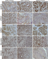
Expression of proteins encoded by `diagnostic gene set`. Pictures of SERHL, ZFP37, MIPEP, relaxin and MAGI3 in grade I, grade II and grade III canine mammary carcinomas (n = 60) obtained using Olympus BX60 microscope (x200). The examined antigen is presented as a brown precipitate. The colorimetric intensity of the IHC-stained antigen spots was counted by a computer-assisted image analyzer (Olympus Microimage™ Image Analysis, software version 4.0 for Windows, USA) and the antigen spot color intensity is expressed as mean pixel optical density on a 1–256 scale (data presented in Table 3).

Similar articles

Cited by

References

    1. Pawłowski KM, Maciejewski H, Dolka I, Mol JA, Motyl T, Król M. Gene expression profiles in canine mammary carcinoma of various grades of malignancy. BMC Vet Res. 2013;9:78. doi: 10.1186/1746-6148-9-78. - DOI - PMC - PubMed
    1. Polton G. Mammary tumors in dogs. Irish Vet J. 2009;62(1):50–56.
    1. Goldschmidt M, Pena L, Rasotto R, Zappulli V. Classification and grading of canine mammary tumors. Vet Pathol. 2011;48:117. doi: 10.1177/0300985810393258. - DOI - PubMed
    1. Karayannopoulou M, Kaldrymidou E, Constantiinidis TC, Dessiris A. Histological grading and prognosis in dogs with mammary carcinomas: application of a human grading method. J Comp Pathol. 2005;133:246–252. doi: 10.1016/j.jcpa.2005.05.003. - DOI - PubMed
    1. Król M, Pawłowski KM, Otrębska D, Motyl T. DNA microarrays – future in oncology research and therapy. JPCCR. 2008;2:091–096.

Publication types

MeSH terms