. 2013 Sep;17(9):1146-59.
doi: 10.1111/jcmm.12094. Epub 2013 Jul 11.

Dynamic and differential regulation in the microRNA expression in the developing and mature cataractous rat lens

Affiliations

Dynamic and differential regulation in the microRNA expression in the developing and mature cataractous rat lens

Eri Kubo et al. J Cell Mol Med. 2013 Sep.

Abstract

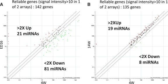
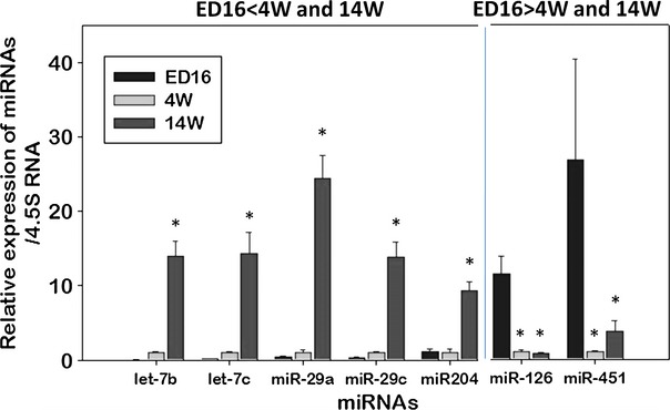
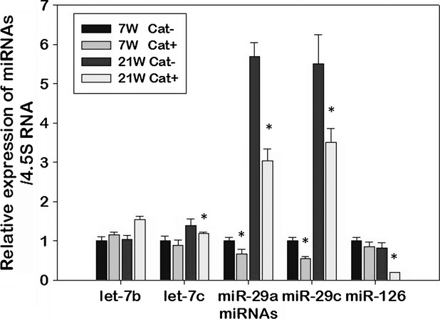
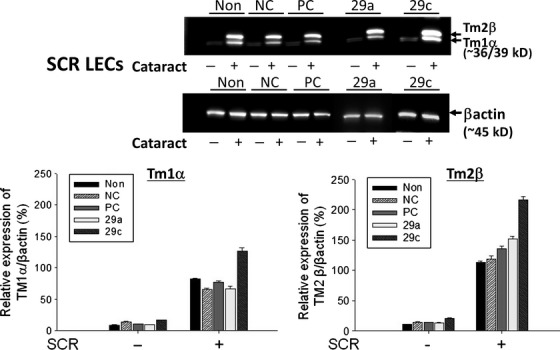
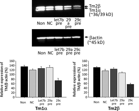
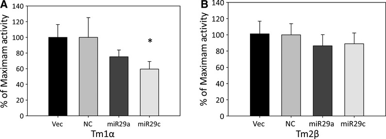
Recent evidence supports a role for microRNAs (miRNAs) in regulating gene expression, and alterations in gene expression are known to affect cells involved in the development of ageing disorders. Using developing rat lens epithelial cells (LECs), we profiled the expression of miRNAs by a microarray-based approach. Few gene expression changes known to be involved in pathogenesis or cytoprotection were uniquely influenced by miRNA expression. Most miRNAs increased or decreased in abundance (let 7b, let 7c, miR29a, miR29c, miR126 and miR551b) in LECs/lenses during late embryonic and post-natal development and in cataract. Among them, miR29a, miR29c and miR126 were dramatically decreased in cataractous LECs from Shumiya Cataract Rats (SCRs). Specifically, the cytoskeleton remodelling genes tropomyosin (Tm) 1α and 2β, which have been implicated in the initiation of pathophysiology, were targets of miR29c and were over-stimulated as demonstrated by inhibitor experiments. In transfection experiments, increasing the level of miR29c caused a corresponding decrease in the expression of Tm1α and 2β, suggesting that miR29c may regulate the translation of Tm1α and 2β. 3'UTR luciferase activity of Tm1α, not 2β, was significantly decreased in miR29c-transfected mouse LECs. These findings demonstrate changes in miRNAs expression, and target molecules have potential as diagnostic indicators of ageing and as a foundation of miR-based therapeutics for age-related diseases.

Keywords: ageing; cataract; lens development; microRNA; tropomyosin.


Figures

Fig. 1
Fig. 1
Scatter plot analysis. (A) Scatter plot matrix of each miRNA array for the ED16 (y-axis) and 4W (x-axis) rat lenses showing mean intensities in the reference channel. (B) Scatter plot of miRNA expression in 14W (y-axis) and 4W (x-axis) lenses. The scatter plots show representative data, using mRNA from rat lens epithelial cells. Each dot represents one gene. Reliable genes (signal intensity >10 in array); 142 genes (A) and 135 genes (B). A twofold change was considered to provide reliability of gene expression.
Fig. 2
Fig. 2
RT-PCR validation of relative changes in miRNA expression in lenses from ED16, 4W and 14W rats. Expression of selected and functionally significant miRNAs was further validated using RT-PCR as described in Materials and methods. Relative expression levels of miRNAs, let-7b, let-7c, miR-29a, miR-29c, miR-204, miR-126, miR-451 in ED16, 4W and 14W lenses are represented as histograms with normalized averages ± SD. *P < 0.01 or 0.0005 by Student's t-test.
Fig. 3
Fig. 3
Analysis of changes in miRNA expression in lenses from Shumiya Cataract Rats (SCR) with cataract or without cataract. Relative expression of miRNAs let7b, let7c, miR-29a, miR29c and miR126, which are significantly up- or down-regulated during the progression of lens development, were measured following RNA extraction using RT-PCR. Histograms represent normalized mean results ± SD. *P < 0.05 or P < 0.001 values are considered statistically significant compared with control. Internal control was taken to evaluate integrity of RNA. Data revealed that let7c, miR29a and miR29c were significantly up-regulated in 21W lenses compared with 7W SCR without cataract (Cat−) or SCR with cataract (Cat+). However, let7c, miR-29a, miR29c and miR126 were significantly down-regulated in SCR with cataract lenses compared with SCR without cataract lenses at both ages 7 and 21 weeks.
Fig. 4
Fig. 4
Elevated expression of Tm1α/2β proteins in lens epithelial cells (LECs) transfected with miR29a and 29c inhibitors. Lens epithelial cells isolated from Shumiya Cataract Rats (SCR) with cataract (+) or SCR without cataract (−) were cultured and processed for transfection with miRNA inhibitors as described in Materials and methods. At a pre-defined time, cellular extracts were prepared and subjected to protein expression analysis using antibody specific to Tm1α/2β. Immunoblotting experiments revealed greater abundance of Tm1α/2β protein in LECs transfected with miR29a and 29c inhibitor. Representative immunoblots show modulated expression of specific miRNA targeted gene Tm1α/2β. Blotted membranes were probed with antibody specific to Tm1α/2β and reprobed with β-actin antibody as internal and protein loading control. Histograms represent normalized mean results ± SD. *P < 0.05 is considered to be statistically significant. Non: No transfection; NC: Negative control; PC: Positive control.
Fig. 5
Fig. 5
Lens epithelial cells (LECs) from Shumiya Cataract Rats (SCR) with cataract transfected with let7b, miR29a and 29c precursors display reduced expression of Tm1α/2β proteins. We used SCR with cataract LECs because their Tm1α/2β expressions were higher than those of SCR without cataract LECs. Cells were overexpressed with let7b, miR29a and miR29c by transfecting them with miRNA precursors. Immunoblotting experiments were conducted after processing of cellular extracts isolated from transfected cells. Blotted membranes were probed with antibody specific to Tm1α/2β and reprobed with β-actin antibody as internal and protein loading control. Immunoblots are representative of experiments and show reduced expression of Tm1α/2β protein in SCR with cataract LECs overexpressing miRNA 29c. Non: No transfection; NC: Negative control. Data are means ± SD from three independent experiments.
Fig. 6
Fig. 6
Luciferase reporter assay. Mouse lens epithelial cells were cotransfected with rat TM1α (A) or rat TM2β (B) with rno-miR29a mimic, rno-miR29c mimics in pEZX-MR04 vector with eGFP reporter gene, precursor miRNA scrambled control for pEZX-MR04 or empty pEZX-MR04 vector. The luciferase activity of firefly-luc and renilla-luc were measured at 48 hrs after transfection. The firefly-luc activity was normalized to Renilla-luc expression. Data are means ± SD from three independent experiments, each containing four replicates. *P < 0.02 values are considered statistically significant compared with vector control.

Similar articles

Cited by

References

    1. Fatma N, Kubo E, Sharma P, et al. Impaired homeostasis and phenotypic abnormalities in Prdx6-/-mice lens epithelial cells by reactive oxygen species: increased expression and activation of TGFbeta. Cell Death Differ. 2005;12:734–50. - PubMed
    1. Kubo E, Singh DP, Akagi Y. Gene expression profiling of diabetic and galactosaemic cataractous rat lens by microarray analysis. Diabetologia. 2005;48:790–8. - PubMed
    1. Kubo E, Singh DP, Fatma N, et al. Cellular distribution of lens epithelium-derived growth factor (LEDGF) in the rat eye: loss of LEDGF from nuclei of differentiating cells. Histochem Cell Biol. 2003;119:289–99. - PubMed
    1. Reddy VN, Lin LR, Ho YS, et al. Peroxide-induced damage in lenses of transgenic mice with deficient and elevated levels of glutathione peroxidase. Ophthalmologica. 1997;211:192–200. - PubMed
    1. Spector A, Kuszak JR, Ma W, et al. The effect of aging on glutathione peroxidase-i knockout mice-resistance of the lens to oxidative stress. Exp Eye Res. 2001;72:533–45. - PubMed

Publication types

MeSH terms