. 2014 Jan 10;20(2):247-66.
doi: 10.1089/ars.2012.4953. Epub 2013 Aug 17.

Molecular mechanisms of the crosstalk between mitochondria and NADPH oxidase through reactive oxygen species-studies in white blood cells and in animal models

Affiliations

Molecular mechanisms of the crosstalk between mitochondria and NADPH oxidase through reactive oxygen species-studies in white blood cells and in animal models

Swenja Kröller-Schön et al. Antioxid Redox Signal. .

Abstract

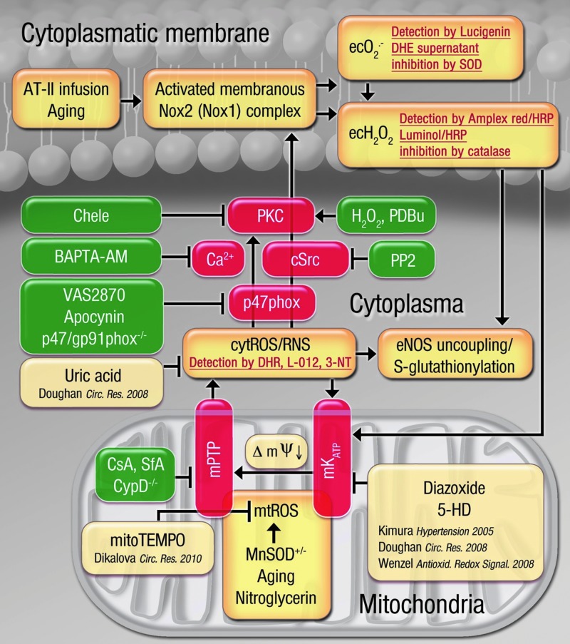
Aims: Oxidative stress is involved in the development of cardiovascular disease. There is a growing body of evidence for a crosstalk between different enzymatic sources of oxidative stress. With the present study, we sought to determine the underlying crosstalk mechanisms, the role of the mitochondrial permeability transition pore (mPTP), and its link to endothelial dysfunction.

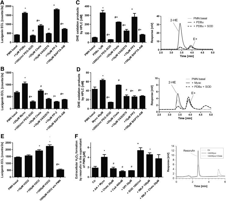
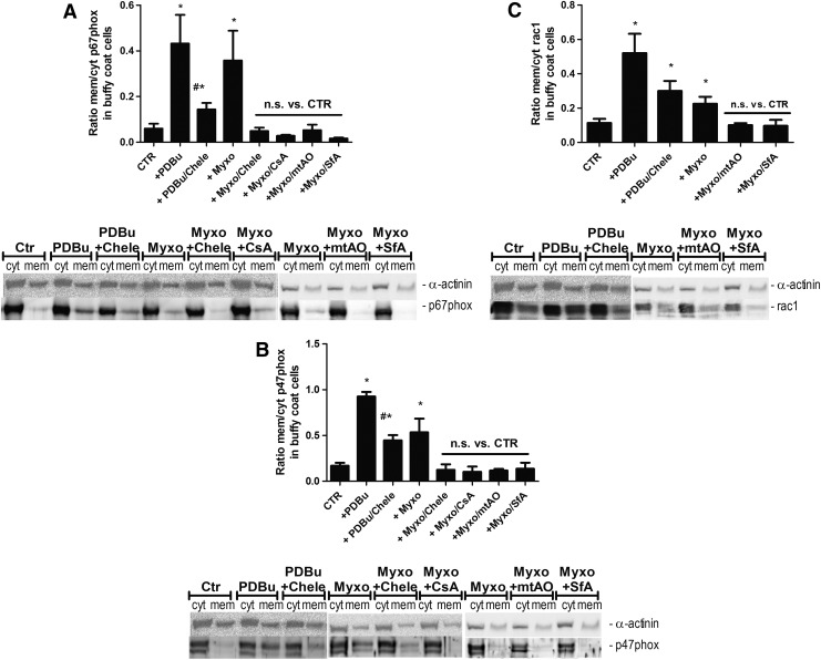
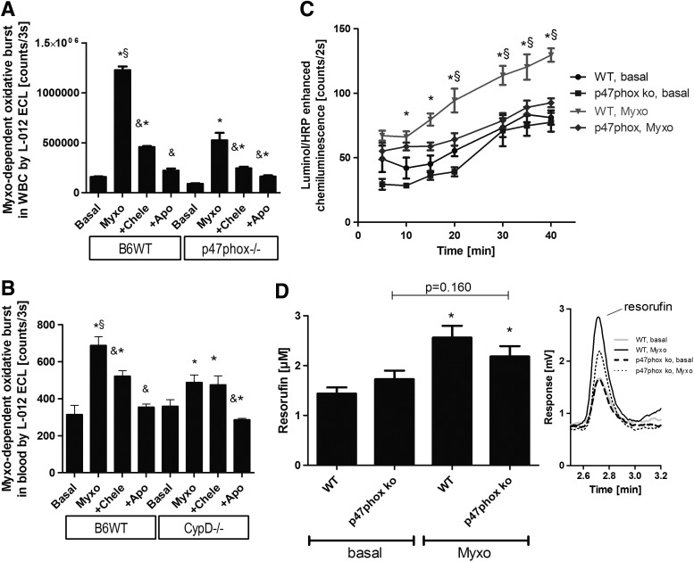
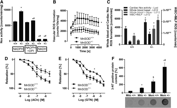
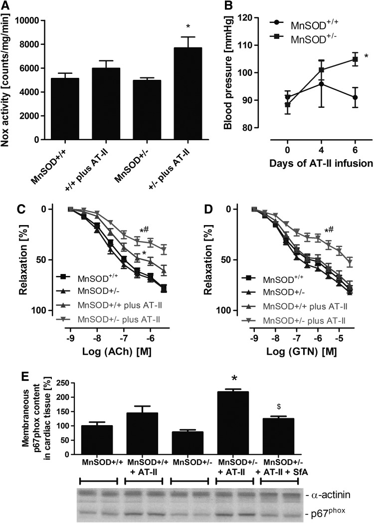
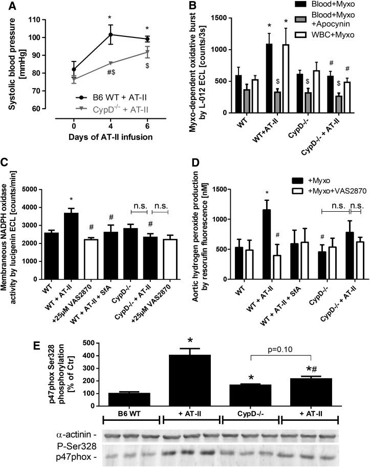
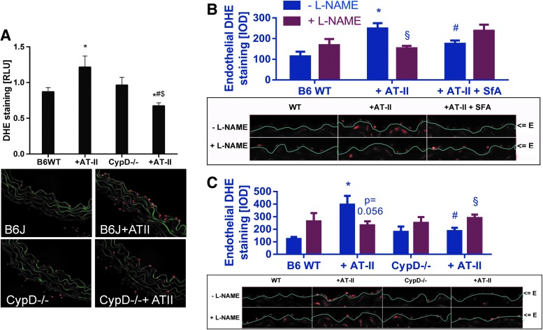
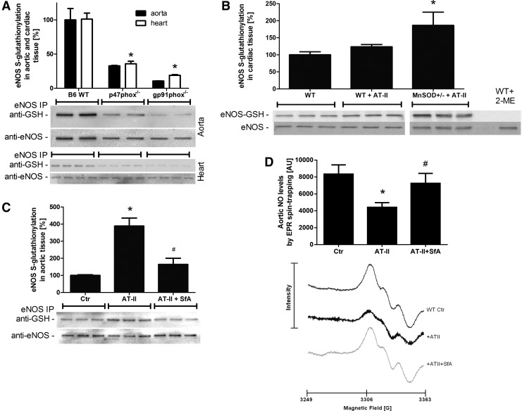
Results: NADPH oxidase (Nox) activation (oxidative burst and translocation of cytosolic Nox subunits) was observed in response to mitochondrial reactive oxygen species (mtROS) formation in human leukocytes. In vitro, mtROS-induced Nox activation was prevented by inhibitors of the mPTP, protein kinase C, tyrosine kinase cSrc, Nox itself, or an intracellular calcium chelator and was absent in leukocytes with p47phox deficiency (regulates Nox2) or with cyclophilin D deficiency (regulates mPTP). In contrast, the crosstalk in leukocytes was amplified by mitochondrial superoxide dismutase (type 2) (MnSOD(+/-)) deficiency. In vivo, increases in blood pressure, degree of endothelial dysfunction, endothelial nitric oxide synthase (eNOS) dysregulation/uncoupling (e.g., eNOS S-glutathionylation) or Nox activity, p47phox phosphorylation in response to angiotensin-II (AT-II) in vivo treatment, or the aging process were more pronounced in MnSOD(+/-) mice as compared with untreated controls and improved by mPTP inhibition by cyclophilin D deficiency or sanglifehrin A therapy.

Innovation: These results provide new mechanistic insights into what extent mtROS trigger Nox activation in phagocytes and cardiovascular tissue, leading to endothelial dysfunction.

Conclusions: Our data show that mtROS trigger the activation of phagocytic and cardiovascular NADPH oxidases, which may have fundamental implications for immune cell activation and development of AT-II-induced hypertension.


Figures

<b>FIG. 1.</b>
FIG. 1.
Determination of mitochondrial superoxide/hydrogen peroxide triggered NADPH oxidase activation in isolated human neutrophils by oxidative burst measurement. (A) PDBu or (B) myxothiazol stimulated oxidative burst in isolated PMN (5×10⁵/ml) pon 20 min (A) or 15 min (B) of incubation in PBS containing Ca2+/Mg2+ (1 mM) was determined by lucigenin ECL (250 μM) in the presence of a scavenger of extracellular superoxide (SOD), inhibitors of PKC (Chele), Nox2 (VAS2870), tyrosine kinase cSrc (PP2), or an intracellular calcium chelator (BAPTA-AM). Lucigenin ECL detects extracellular superoxide. The signal (counts/2s) was measured with a chemiluminescence plate reader (Centro 960). (C) PDBu or (D) myxothiazol-stimulated oxidative burst in isolated PMN (1×10⁶/ml) on 20 min (C) or 15 min (D) of incubation was determined by HPLC-based quantification of 2-hydroxyethidium (2-HE) in the presence of the same scavengers/inhibitors as given earlier. 2-HE in the supernatant is a specific marker of extracellular superoxide formation. Inhibitors were preincubated for 20 min. Representative chromatograms are shown for each HPLC data set. (E) Hydrogen peroxide was used as a mimic of mtROS, and subsequent Nox2-dependent superoxide formation (oxidative burst) in isolated PMN (5×10⁵/ml) was determined by lucigenin (250 μM) ECL on 20 min of incubation. (F) Antimycin A (20 μg/ml) and myxothiazol (20 μM)-stimulated oxidative burst in isolated PMN (3×10⁶/ml) on 20 min of incubation was determined by amplex red (100 μM)/peroxidase (HRP, 0.1 μM)) by HPLC-based quantification of the fluorescent oxidation product resorufin with or without PKC (Chele), Nox (DPI), or mPTP (CsA) inhibitors, a superoxide scavenger (SOD), or a chemotactic peptide (fMLP). Resorufin formation in the presence of HRP is a specific marker of extracellular hydrogen peroxide formation. Inhibitors were preincubated for 5 min. Representative chromatograms are shown for each HPLC data set. The data are mean±SEM of 8 (A, B), 3 (C, D), 8 (E) and 3–12 (F) independent experiments. *p<0.05 versus unstimulated control; #p<0.05 versus stimulated group (antimycin A, myxothiazole or phorbol ester [PDBu]). BAPTA-AM, 1,2-Bis(2-aminophenoxy)ethane-N,N,N′,N′-tetraacetic acid tetrakis(acetoxymethyl ester); CsA, cyclosporine A; DPI, diphenylene iodonium; ECL, enhanced chemiluminescence; fMLP, formyl-methionyl-leucyl-phenylalanine; HPLC, high performance liquid chromatography; HRP, horseradish peroxidase; mPTP, mitochondrial permeability transition pore; mtROS, mitochondrial ROS; PDBu, phorbol ester dibutyrate; PKC, protein kinase C; PMN, polymorphonuclear leukocyte; PP2, 4-Amino-3-(4-chlorophenyl)-1-(t-butyl)-1H-pyrazolo[3,4-d]pyrimidine; ROS, reactive oxygen species; VAS2870, 1,3-Benzoxazol-2-yl-3-benzyl-3H-[1,2,3]triazolo[4,5-d]pyrimidin-7-yl sulfide.
<b>FIG. 2.</b>
FIG. 2.
Determination of mitochondrial superoxide/hydrogen peroxide triggered NADPH oxidase activation in isolated human neutrophils by determination of the translocation of cytosolic subunits. (A) Phorbol ester (PDBu, 1 μM) or myxothiazol (Myxo, 20 μM)-stimulated translocation of cytosolic subunits in isolated leukocytes (10×106/ml) was determined by membranous and cytosolic content of the NADPH oxidase subunits p67phox (A), p47phox (B), and Rac1 (C). The effect of different inhibitors and antioxidants was assessed (see list of abbreviations, mtAO means mitoTEMPO). For applied concentrations, refer to Supplementary Figure S1E. Western blotting was applied with specific antibodies, and all signals were normalized to α-actinin. Representative blots are shown at the bottom of each densitometric quantification graph. The data are mean±SEM of 4–10 (A), 3–7 (B) and 4–9 (C) independent experiments. *p<0.05 versus unstimulated control; #p<0.05 versus stimulated group (antimycin A, myxothiazole, or phorbol ester [PDBu]). CTR, control; mitoTEMPO, (2-(2,2,6,6-Tetramethylpiperidin-1-oxyl-4-ylamino)-2-oxoethyl) triphenylphosphonium chloride; n.s., not significant.
<b>FIG. 3.</b>
FIG. 3.
Determination of mitochondrial superoxide/hydrogen peroxide triggered NADPH oxidase activation in whole blood and isolated leukocytes from mice by oxidative burst measurement. (A) Myxothiazol-stimulated oxidative burst in isolated WBC (2×10⁵/ml) from wild-type and p47phox knockout mice was determined by L-012 ECL with or without chelerythrine (Chele) or apocynin (Apo). L-012 ECL detects intra- and extracellular ROS and RNS (sensitivity: peroxynitrite>superoxide>hydrogen peroxide). The signal (counts/3 s) was measured after an incubation time of 20 min with a chemiluminescence plate reader (Centro 960). (B) Myxothiazol-stimulated oxidative burst in whole blood (1:50) from wild-type and CypD knockout mice was determined by L-012 ECL with or without inhibitors. (C) Myxothiazol (20 μM)-stimulated oxidative burst in isolated WBC (5×10⁴/ml) from wild-type and p47phox knockout mice was determined by luminol (100 μM)/HRP (0.1 μM) ECL. Luminol oxidation in the presence of HRP is specific for extracellular hydrogen peroxide (theoretically also peroxynitrite). The signal (counts/2 s) was measured with a chemiluminescence plate reader (Centro 960). (D) Myxothiazol (20 μM)-stimulated oxidative burst in isolated WBC (5×10⁵/ml) from wild-type and p47phox knockout mice on incubation for 30 min was also determined by amplex red (100 μM)/peroxidase (HRP, 0.1 μM)) by HPLC-based quantification of the fluorescent oxidation product resorufin. Resorufin formation in the presence of HRP is a specific marker of extracellular hydrogen peroxide formation. Representative chromatograms are shown for each HPLC data set. The data are mean±SEM of three (A, B), eight (C) and three (D) independent experiments. *p<0.05 versus control (untreated); &p<0.05 versus respective Myxo-stimulated group; §p<0.05 versus p47phox or CypD knockout+Myxo group. CypD, cyclophilin D; L-012, 8-amino-5-chloro-7-phenylpyrido[3,4-d]pyridazine-1,4-(2H,3H)dione sodium salt; RNS, reactive nitrogen species; WBC, white blood cells; WT, wild type.
<b>FIG. 4.</b>
FIG. 4.
Effects of partial mitochondrial superoxide dismutase (type 2) (MnSOD) deficiency on cardiovascular and blood cell oxidative stress as well as endothelial function in old (age: 12 months) MnSOD+/+ versus MnSOD+/ mice. (A) Cardiac NADPH oxidase activity was assessed by lucigenin (5 μM) ECL in membranous fractions from murine hearts in the presence of NADPH (200 μM), DPI (10 μM) or NADH (200 μM). In this assay, lucigenin ECL detects NADPH oxidase-derived superoxide. The signal (counts/min) was measured after an incubation time of 5 min with a chemiluminometer (Lumat 9507). (B) Vascular oxidative stress was assessed by Diogenes ECL in phorbol ester (200 nM)-stimulated murine aortic ring segments. This chemiluminescence assay detects extracellular hydrogen peroxide. (C) Whole blood or isolated WBC (2×10⁵/ml) oxidative burst was assessed by L-012 (100 μM) ECL in unstimulated, myxothiazol (Myxo, 20 μM) or fMLP (20 μM)-treated samples. Again, cardiac Nox activity was measured in the presence of NADPH (200 μM) by lucigenin (5 μM) ECL. (D, E) Endothelial and vascular function was determined by isometric tension recording and relaxation in aortic ring segments in response to an endothelium-dependent (acetylcholine [Ach]) and endothelium-independent (nitroglycerin, GTN) vasodilator. (F) Cardiac oxidative stress was also assessed by dot blot quantification of 3-nitrotyrosine-positive proteins, a surrogate parameter for peroxynitrite formation in biological samples. The data are mean±SEM of 4 (A), 12 (B), 3–4 for blood cells and 12 for cardiac Nox activity (C), 4 (D and E) and 3 (F) independent experiments. *p<0.05 versus respective control group (+/+); #p<0.05 versus MnSOD-deficient mice (+/−) w/o treatment; $p<0.05 versus respective mitochondrial sample.
<b>FIG. 5.</b>
FIG. 5.
Effects of partial MnSOD deficiency and chronic AT-II treatment on oxidative stress, endothelial function, and blood pressure in young (age: 3 months) mice. (A) Cardiac oxidative stress was assessed by lucigenin (5 μM) ECL in membranous fractions from murine hearts in the presence of NADPH (200 μM). This assay is specific for NADPH oxidase-derived superoxide formation. The signal (counts/min) was measured after an incubation time of 5 min with a chemiluminometer (Lumat 9507). (B) Blood pressure was assessed by the tail cuff method in AT-II (0.2 mg/kg/day for 7 days)-treated MnSOD+/+ and MnSOD+/− mice. (C, D) Endothelial and vascular function was determined by isometric tension recording and relaxation in aortic ring segments in response to an endothelium-dependent (ACh, C) and endothelium-independent (GTN, D) vasodilator. (E) Cardiac Nox activation was determined by quantification of the translocation of the cytosolic NADPH oxidase subunit p67phox (its membranous content) by Western blotting. Effect of in vivo treatment with the mPTP blocker SfA (10 mg/kg/day) is also shown. The data are mean±SEM of 22 (A), 5–8 (B), 16–21 (C) and 3–5 (E) independent experiments. *p<0.05 versus control mice (+/+); #p<0.05 versus control mice (+/+) with AT-II treatment; $p<0.05 versus MnSOD-deficient mice (+/−) with AT-II treatment. AT-II, angiotensin-II; SfA, sanglifehrin A.
<b>FIG. 6.</b>
FIG. 6.
Effects of cyclophilin D deficiency and AT-II treatment on whole blood and cardiovascular oxidative stress, NADPH oxidase activation as well as blood pressure in mice. (A) Blood pressure was assessed by the tail cuff method in AT-II (1 mg/kg/day for 7 days)-treated wild-type and CypD−/− mice. *p<0.05 versus wild-type group at day 0; #p<0.05 versus wild-type group at day 4; $p<0.05 versus CypD−/− group at day 0. (B) Myxothiazol-stimulated oxidative burst in whole blood (1:50) or isolated WBC (1×10⁴/ml) from wild-type and CypD knockout mice was determined by L-012 ECL with or without in vivo AT-II (AT-II) treatment. The effect of apocynin in vitro was tested in the whole blood assay. L-012 ECL detects intra- and extracellular reactive species (sensitivity: peroxynitrite>superoxide>hydrogen peroxide). The signal (counts/3 s) was measured after an incubation time of 20 min with a chemiluminescence plate reader (Centro 960). *p<0.05 versus wild-type control (untreated); #p<0.05 versus w/o AT-II group; $p<0.05 versus w/o apocynin group. (C) Cardiac oxidative stress was assessed by lucigenin (5 μM) ECL in membranous fractions from murine hearts in the presence of NADPH (200 μM). The mPTP blocker SfA was administrated in vivo, and the Nox2 inhibitor VAS2870 (25 μM, white bars) was used in vitro (preincubation with heart tissue for 30 min on ice before homogenization). This assay is specific for NADPH oxidase-derived superoxide formation. The signal (counts/min) was measured after an incubation time of 5 min with a chemiluminometer (Lumat 9507). (D) Aortic hydrogen peroxide was measured by amplex red (100 μM) oxidation in the presence of HRP (0.2 μM) and subsequent HPLC-based quantification of resorufin fluorescence. One aortic ring segment (4 mm) was used for one data point. The Nox2 inhibitor VAS2870 (25 μM, white bars) was used in vitro (preincubation with aortic ring segments for 20 min at 37°C). This assay is specific for extracellular hydrogen peroxide formation. Samples were measured after an incubation time of 60 min at 37°C. (E) Activation of p47phox-dependent NADPH oxidase in aortic tissue was determined by phosphorylation of p47phox at serine 328 using a specific antibody. *p<0.05 versus wild-type control; #p<0.05 versus wild type with AT-II treatment. The data are mean±SEM of 3–4 (A), 4–8 (B), 11–30 (C), and 6–8 for basal and 3–6 for VAS2870 (D) and 3 (E) independent experiments.
<b>FIG. 7.</b>
FIG. 7.
Effects of cyclophilin D deficiency and AT-II treatment on vascular oxidative stress as well as eNOS uncoupling in mice. (A) Vascular oxidative stress was assessed by dihydroethidine (DHE, 1 μM)-dependent fluorescence microtopography in aortic cryo-sections. Representative microscope images are shown below the densitometric quantifications (color, green=autofluorescence of the basal laminae, red=ROS and RNS (mainly superoxide) induced fluorescence). (B, C) eNOS uncoupling was assessed by endothelial specific quantification of DHE fluorescence in the presence of L-NAME. The eNOS inhibitor increases the signal in the endothelial cell layer with functional eNOS (by suppression of the superoxide scavenger NO) and decreases the signal in endothelial cells with uncoupled eNOS (by inhibition of eNOS-derived superoxide formation). Representative microscope images are shown below the densitometric quantifications (red=ROS and RNS (mainly superoxide) induced fluorescence). “E” means endothelial cell layer. *p<0.05 versus wild-type control; #p<0.05 versus wild type with AT-II treatment; $p<0.05 versus untreated CypD−/− mice; §p<0.05 versus respective L-NAME group. The data are mean±SEM of 12 (A) and 5–6 (B and C) independent experiments. Specificity of the eNOS uncoupling assay is shown by the use of D-NAME (see Supplementary Fig. S7). eNOS, endothelial nitric oxide synthase; L-NAME, L-NG-nitroarginine methyl ester. To see this illustration in color, the reader is referred to the web version of this article at www.liebertpub.com/ars
<b>FIG. 8.</b>
FIG. 8.
Effects of genetic deficiencies and pharmacological treatments on S-glutathionylation of eNOS. eNOS functional state was determined in aortic and heart tissue by quantification of its S-glutathionylation, an oxidative redox modification causing dysfunction or even uncoupling of eNOS. eNOS S-glutathionylation of aortic and/or cardiac tissue from wild-type mice versus p47phox−/− or gp91phox−/− (A), wild type versus WT plus AT-II treatment (0.2 mg/kg/day for 7 days) or MnSOD+/− plus AT-II treatment (B), wild type versus WT plus AT-II or WT plus AT-II plus SfA (10 mg/kg/day) (C). Treatment with 2-mercaptoethanol (2-ME) served as a negative control. Western blotting was applied with specific antibodies, and all signals were normalized to α-actinin. Representative blots are shown at the bottom of each densitometric quantification graph. (D) Aortic NO formation was measured by EPR spin trapping using Fe(DETC)2. Each spectrum was measured from one murine aorta. The representative spectra below the bar graph are the mean of all measurements. The data are mean±SEM of two for aorta (each pooled from two mice) and 4 for heart (A), 3 (B), 6 (C), and 7 (D) independent experiments. *p<0.05 versus control mice (B6 WT or+/+); #p<0.05 versus control mice with AT-II treatment. EPR, electron paramagnetic resonance.
<b>FIG. 9.</b>
FIG. 9.
Postulated molecular mechanisms of the crosstalk between mitochondria and NADPH oxidase through superoxide, hydrogen peroxide, and peroxynitrite based on studies in WBCs and in genetic/pharmacological animal models. The brown boxes represent fundamental processes (e.g., aging, MnSOD deficiency, and AT-II infusion) involved in the process of the crosstalk between mitochondria and NADPH oxidase through ROS and the genetic/pharmacological stress factors that trigger this crosstalk. The red boxes contain important enzymatic constituents of the mitochondrial-Nox redox signaling axis and highlight the important role of cytosolic calcium levels. The green boxes represent genetic and pharmacological inhibitors and activators of this crosstalk. The boxes with the red script show the detection assays used for the involved reactive species (superoxide, hydrogen peroxide, and peroxynitrite). The pale brown boxes represent previous findings providing the basis for our understanding of the crosstalk concept (23, 24, 31, 61). To see this illustration in color, the reader is referred to the web version of this article at www.liebertpub.com/ars

Similar articles

Cited by

References

    1. Ago T, Kuroda J, Pain J, Fu C, Li H, and Sadoshima J. Upregulation of Nox4 by hypertrophic stimuli promotes apoptosis and mitochondrial dysfunction in cardiac myocytes. Circ Res 106: 1253–1264, 2010 - PMC - PubMed
    1. Ahmad M, Kelly MR, Zhao X, Kandhi S, and Wolin MS. Roles for Nox4 in the contractile response of bovine pulmonary arteries to hypoxia. Am J Physiol Heart Circ Physiol 298: H1879–H1888, 2010 - PMC - PubMed
    1. Balsari A, Marolda R, Gambacorti-Passerini C, Sciorelli G, Tona G, Cosulich E, Taramelli D, Fossati G, Parmiani G, and Cascinelli N. Systemic administration of autologous, alloactivated helper-enriched lymphocytes to patients with metastatic melanoma of the lung. A phase I study. Cancer Immunol Immunother 21: 148–155, 1986 - PMC - PubMed
    1. Basso E, Fante L, Fowlkes J, Petronilli V, Forte MA, and Bernardi P. Properties of the permeability transition pore in mitochondria devoid of Cyclophilin D. J Biol Chem 280: 18558–18561, 2005 - PubMed
    1. Block K, Gorin Y, and Abboud HE. Subcellular localization of Nox4 and regulation in diabetes. Proc Natl Acad Sci U S A 106: 14385–14390, 2009 - PMC - PubMed

Publication types

MeSH terms

LinkOut - more resources