. 2013 Jun;24(2):86-98.
doi: 10.1089/humc.2013.019.

From bench to bedside: preclinical evaluation of a self-inactivating gammaretroviral vector for the gene therapy of X-linked chronic granulomatous disease

Affiliations

From bench to bedside: preclinical evaluation of a self-inactivating gammaretroviral vector for the gene therapy of X-linked chronic granulomatous disease

Stefan Stein et al. Hum Gene Ther Clin Dev. 2013 Jun.

Abstract

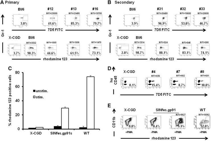
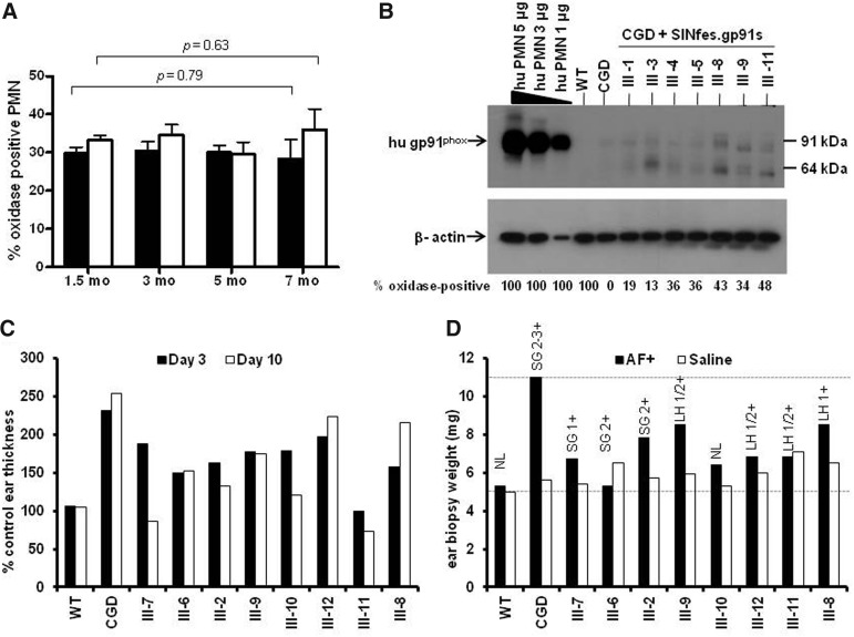
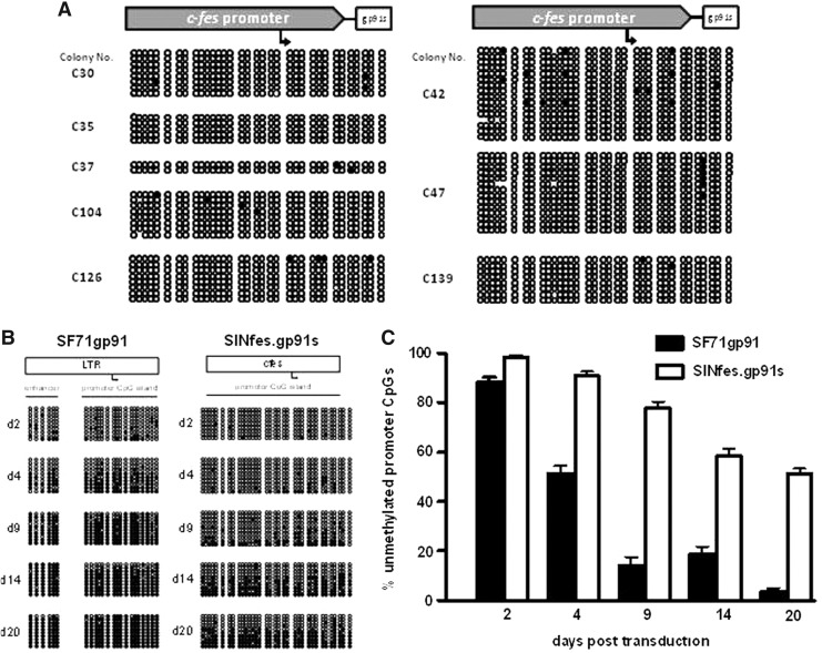
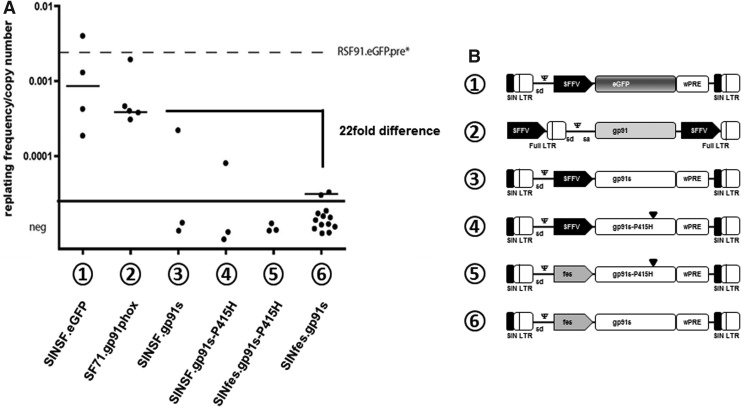
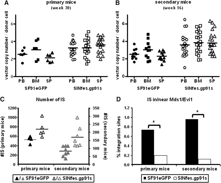
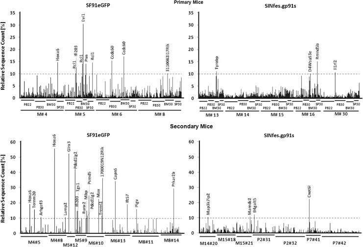
Chronic granulomatous disease (CGD) is a primary immunodeficiency characterized by impaired antimicrobial activity in phagocytic cells. As a monogenic disease affecting the hematopoietic system, CGD is amenable to gene therapy. Indeed in a phase I/II clinical trial, we demonstrated a transient resolution of bacterial and fungal infections. However, the therapeutic benefit was compromised by the occurrence of clonal dominance and malignant transformation demanding alternative vectors with equal efficacy but safety-improved features. In this work we have developed and tested a self-inactivating (SIN) gammaretroviral vector (SINfes.gp91s) containing a codon-optimized transgene (gp91(phox)) under the transcriptional control of a myeloid promoter for the gene therapy of the X-linked form of CGD (X-CGD). Gene-corrected cells protected X-CGD mice from Aspergillus fumigatus challenge at low vector copy numbers. Moreover, the SINfes.gp91s vector generates substantial amounts of superoxide in human cells transplanted into immunodeficient mice. In vitro genotoxicity assays and longitudinal high-throughput integration site analysis in transplanted mice comprising primary and secondary animals for 11 months revealed a safe integration site profile with no signs of clonal dominance.


Figures

FIG. 1.
FIG. 1.
Gp91phox expression and functional reconstitution of superoxide production by SINfes.gp91s-transduced cells in vivo. Representative examples of gp91phox expression (A and B, upper panels) and superoxide production (A and B, lower panels) in granulocytes of primary (A) and secondary (B) animals transplanted with SINfes.gp91s-transduced lin− cells. Transgene expression analysis was performed 18 (primary) and 16 (secondary) weeks after transplantation. Intracellular staining using a monoclonal antibody (7D5) was used to detect gp91phox expression. NADPH oxidase activity (A and B, lower panels) was assessed in the same samples as above by the oxidative burst assay (DHR). The percentage of gp91phox expressing Gr-1+ cells and superoxide production in CD11b+ Gr-1+ donor cells as well as the MFI of rhodamine 123 is shown. (C) Reconstitution of oxidase activity in the progeny of primary human CD34+ cells from X-CGD patients transduced with SINfes.gp91s after in vitro differentiation. NADPH oxidase activity was measured in CD11b+ cells by the DHR assay after in vitro differentiation of nontransduced (X-CGD) or transduced cells. In vitro differentiated CD34+ cells from a healthy donor served as positive control (WT). (D) Reconstitution of gp91phox expression in primary human hematopoietic cells in vivo. X-CGD CD34+ cells were transduced with SINfes.gp91s or left untreated (X-CGD). About 107 cells were transplanted into sublethally irradiated (2.5 Gy) immunodeficient mice. Three weeks after transplantation, blood was obtained from the tail vein of the animals and gp91phox expression was analyzed in human cells (huCD45) by fluorescence-activated cell sorting. (E) Reconstitution of oxidase activity in vivo. Bone marrow cells obtained from NOD/scid/IL-2Rγ null mice transplanted either with nontransduced (X-CGD) or SINfes.gp91s-transduced X-CGD CD34+ cells or cells from a healthy donor (WT) were differentiated ex vivo for 2 weeks in the presence of G-CSF. NADPH oxidase activity was measured after phorbol-12-myristate-13-acetate stimulation in the huCD45+ cell fraction by the DHR assay. Cells were costained for CD11b expression. The percentage of superoxide production in huCD45+ CD11b+ cells and the MFI of rhodamine-123-positive cells are indicated. DHR, dihydrorhodamine-123; MFI, mean fluorescence intensity; NADPH, nicotinamide dinucleotide phosphate; WT, wild type; X-CGD, X-linked form of chronic granulomatous disease.
FIG. 2.
FIG. 2.
Suppression of hyperinflammatory response in X-CGD mice transplanted with SINfes.gp91s-transduced cells. (A) Oxidase activity in peripheral blood PMNs over time after transplantation of either 1×10⁶ (solid bars) or 2×10⁶ (open bars) SINfes.gp91s-transduced lin− cells into X-CGD animals. Mean values±SEM are given. p-Values according to two-tailed t-test. (B) Human gp91phox expression in peripheral blood PMNs of animals transplanted with SINfes.gp91s-transduced cells. PMNs were obtained from the indicated animals 7 months after transplantation. Gp91phox was detected by Western blotting using the CL5 antibody (Sadat et al., 2009). Cell extracts from human PMN were used at different concentrations as control. No cross-reaction with murine gp91phox was observed as indicated by the lack of hybridization in cell extracts obtained from wild-type mice (WT). The percentage of oxidase-positive cells is indicated below. (C and D) X-CGD animals transplanted with SINfes.gp91s-transduced cells are protected from Aspergillus fumigatus (AF) challenge. Seven months after transplantation, sterile AF hyphae were injected intradermally into one of the ears of the animals. As a control, saline was injected into the contra lateral ear. The degree of inflammation at the AF injection site was estimated at 3 and 10 days after injection from the ear thickness at the injection site (C) or the weight of a 5 mm ear punch biopsy taken 30 days after injection (D). Black bars, AF injection; white bars, saline injection. Values between 1 and 3 denote the severity of the inflammation, with 1 being low and 3 high. LH, lymphocytic/histiocytic (macrophages) infiltration; NL, normal; PMNs, polymorphonuclear leukocytes; SEM, standard error of the mean; SG, suppurative/granulomatous.
FIG. 3.
FIG. 3.
The c-FES promoter is less prone to CpG methylation in hematopoietic cells. (A) Assessment of CpG methylation at the c-FES promoter in CFC. DNA was extracted from individual vector-positive but oxidase-negative colonies derived from the BM of transplanted X-CGD mice 30 weeks after transplantation and subjected to bisulfite conversion and sequencing. (B) The c-FES promoter is less sensitive than the SFFV promoter to CpG methylation. P19 cells were transduced with SF71gp91phox or SINfes.gp91s. DNA was extracted at different time points and analyzed for CpG methylation by bisulfite sequencing. (C) Schematic diagram summarizing the data presented in (B). Mean values±SEM are shown. BM, bone marrow; CFC, colony-forming cells; SFFV, spleen focus forming virus.
FIG. 4.
FIG. 4.
Frequency of immortalization of lin− cells by SINfes.gp91s in vitro. (A) lin− cells from X-CGD mice were transduced on two consecutive days at an MOI of 10 per day with different gp91phox or eGFP coding vectors, expanded in bulk for 14 days as described in the Supplementary Materials and Methods and plated at a density of 100 cells per well in 96-well plates. After 14 days, plates were screened for colony growth, and the frequency of replating was calculated based on Poisson statistics using L-Calc software. The graphic shows the frequency of replating cells corrected for the average vector copy number as measured by quantitative PCR on day 4 after transduction. The limit of detection of the assay is indicated as a thick black line. The dotted line reflects the immortalization frequency of RSF91.eGFPpre*, an LTR-driven gammaretroviral vector expressing eGFP. (B) Schematic diagram of the vectors used in this study. lin−, lineage negative; LTR, long terminal repeat; MOI, multiplicity of infection; PCR, polymerase chain reaction.
FIG. 5.
FIG. 5.
Vector copy numbers and total number of ISs per mouse in SINfes.gp91s- and SF91eGFP-transplanted animals. Vector copy numbers in DNA samples obtained from PB, BM, and SP of transplanted primary (A) and secondary (B) animals. (C) Total number of ISs per mouse. ISs were obtained from serially transplanted SF91eGFP and SINfes.gp91s mice by ligation-mediated/linear-amplification-mediated PCR and subsequent 454 sequencing. (D) Percentage of ISs found in or close to the Mds1/Evi1 locus for primary and secondary transplanted mice. With SINfes.gp91s, significantly less ISs were detected in primary and in secondary animals, respectively. *p=0.077 and 0.026 for primary and secondary mice, respectively, as determined by χ2-test. BM, bone marrow; ISs, integration sites; PB, peripheral blood; SP, spleen.
FIG. 6.
FIG. 6.
Retrieval frequency of individual ISs in SINfes.gp91s- and SF91eGFP-transplanted animals. Retrieval frequency of individual sequences in PB, BM, and SP of SF91eGFP- and SINfes.gp91s-transplanted primary (upper diagram) and secondary animals (lower diagram) at different time points after transplantation. DNA was extracted from the indicated samples, and retroviral vector integrations sites were retrieved by ligation-mediated/linear-amplification-mediated PCR and subsequent 454 high-throughput sequencing. The relative sequence count gives a semiquantitative estimation for the contribution of clones carrying a unique IS to total gene-marked hematopoiesis. A threshold of 10% was arbitrarily set to define a significant contribution of a clone to hematopoiesis. All ISs with a sequence retrieval frequency above 10% are marked.

Similar articles

Cited by

References

    1. Aiuti A. Bacchetta R. Seger R., et al. Gene therapy for primary immunodeficiencies: part 2. Curr. Opin. Immunol. 2012;24:585–591. - PubMed
    1. Avedillo Diez I. Zychlinski D. Coci E.G., et al. Development of novel efficient SIN vectors with improved safety features for Wiskott-Aldrich Syndrome stem cell based gene therapy. Mol. Pharmaceutics. 2011;8:1525–1537. - PubMed
    1. Baum C. Von Kalle C. Staal F.J., et al. Chance or necessity? Insertional mutagenesis in gene therapy and its consequences. Mol. Ther. 2004;9:5–13. - PubMed
    1. Baum C. Modlich U. Gohring G., et al. Concise review: managing genotoxicity in the therapeutic modification of stem cells. Stem Cells. 2011;29:1479–1484. - PubMed
    1. Biffi A. Bartolomae C.C. Cesana D., et al. Lentiviral vector common integration sites in preclinical models and a clinical trial reflect a benign integration bias and not oncogenic selection. Blood. 2011;117:5332–5339. - PubMed

Publication types

MeSH terms