

Porous silicon advances in drug delivery and immunotherapy
- PMID: 23845260
- PMCID: PMC3795885
- DOI: 10.1016/j.coph.2013.06.006
Porous silicon advances in drug delivery and immunotherapy
Abstract
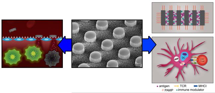
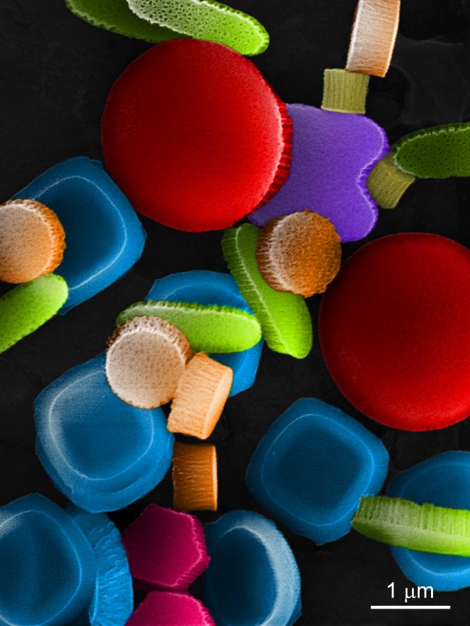
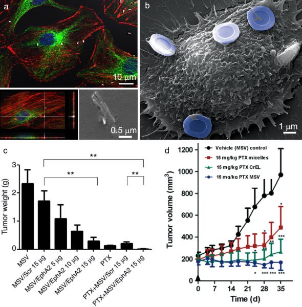
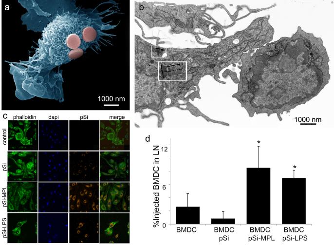
Biomedical applications of porous silicon include drug delivery, imaging, diagnostics and immunotherapy. This review summarizes new silicon particle fabrication techniques, dynamics of cellular transport, advances in the multistage vector approach to drug delivery, and the use of porous silicon as immune adjuvants. Recent findings support superior therapeutic efficacy of the multistage vector approach over single particle drug delivery systems in mouse models of ovarian and breast cancer. With respect to vaccine development, multivalent presentation of pathogen-associated molecular patterns on the particle surface creates powerful platforms for immunotherapy, with the porous matrix able to carry both antigens and immune modulators.
Copyright © 2013 Elsevier Ltd. All rights reserved.
Figures
Similar articles
-
Tailoring Porous Silicon for Biomedical Applications: From Drug Delivery to Cancer Immunotherapy.Adv Mater. 2018 Jun;30(24):e1703740. doi: 10.1002/adma.201703740. Epub 2018 Mar 13. Adv Mater. 2018. PMID: 29534311 Review.
-
Recent advances in porous silicon technology for drug delivery.Ther Deliv. 2013 Jul;4(7):811-23. doi: 10.4155/tde.13.52. Ther Deliv. 2013. PMID: 23883125 Review.
-
Porous silicon nanomaterials: recent advances in surface engineering for controlled drug-delivery applications.Nanomedicine (Lond). 2019 Dec;14(24):3213-3230. doi: 10.2217/nnm-2019-0167. Nanomedicine (Lond). 2019. PMID: 31855121
-
Particle platforms for cancer immunotherapy.Int J Nanomedicine. 2013;8:1683-96. doi: 10.2147/IJN.S31756. Epub 2013 Apr 29. Int J Nanomedicine. 2013. PMID: 23761969 Free PMC article. Review.
-
Drug delivery via porous silicon: a focused patent review.Pharm Pat Anal. 2017 Mar;6(2):77-85. doi: 10.4155/ppa-2016-0042. Epub 2017 Mar 1. Pharm Pat Anal. 2017. PMID: 28248125 Review.
Cited by
-
Particle-Based therapies for antigen specific treatment of type 1 diabetes.Int J Pharm. 2023 Jan 25;631:122500. doi: 10.1016/j.ijpharm.2022.122500. Epub 2022 Dec 15. Int J Pharm. 2023. PMID: 36529362 Free PMC article. Review.
-
Recent Developments in Porous Silicon Nanovectors with Various Imaging Modalities in the Framework of Theranostics.ChemMedChem. 2022 May 18;17(10):e202200004. doi: 10.1002/cmdc.202200004. Epub 2022 Mar 18. ChemMedChem. 2022. PMID: 35212460 Free PMC article. Review.
-
Cancer vaccines from cryogenically silicified tumour cells functionalized with pathogen-associated molecular patterns.Nat Biomed Eng. 2022 Jan;6(1):19-31. doi: 10.1038/s41551-021-00795-w. Epub 2021 Nov 1. Nat Biomed Eng. 2022. PMID: 34725505 Free PMC article.
-
Mesoporous Silicon Particles Favor the Induction of Long-Lived Humoral Responses in Mice to a Peptide-Based Vaccine.Materials (Basel). 2018 Jun 26;11(7):1083. doi: 10.3390/ma11071083. Materials (Basel). 2018. PMID: 29949862 Free PMC article.
-
Porous Si Microparticles Infiltrated with Magnetic Nanospheres.Nanomaterials (Basel). 2020 Mar 4;10(3):463. doi: 10.3390/nano10030463. Nanomaterials (Basel). 2020. PMID: 32143523 Free PMC article.
References
-
- Tsang CK, Kelly TL, Sailor MJ, Li YY. Highly stable porous silicon-carbon composites as label-free optical biosensors. Acs Nano. 2012;6(12):10546–10554. - PubMed
-
- Uhlir A JBSTJ, 35: 2. Electrolytic Shaping of Germanium and Silicon. Mar, 1956. pp. 333–347.
-
- Canham LT. Silicon quantum wire array fabrication by electrochemcal and chemical dissolutions of wafers. appl Phys Lett. 1990;57(1046-1048)
-
- Lehmann V, Gosele U. Porous silicon formation - a quantum wire effect. Applied Physics Letters. 1991;58(8):856–858.
-
- Sailor MFopspPsipp, characterization, and applications. First Ed Wiley-VCH Verlag GmbH & Co. KGaA; 2012. pp. 1–42.
Publication types
MeSH terms
Substances
Grants and funding
LinkOut - more resources
Full Text Sources
Other Literature Sources
