. 2013 Aug 23;288(34):24465-79.
doi: 10.1074/jbc.M113.465633. Epub 2013 Jul 11.

Individual interactions of the b subunits within the stator of the Escherichia coli ATP synthase

Affiliations

Individual interactions of the b subunits within the stator of the Escherichia coli ATP synthase

Karsten Brandt et al. J Biol Chem. .

Abstract

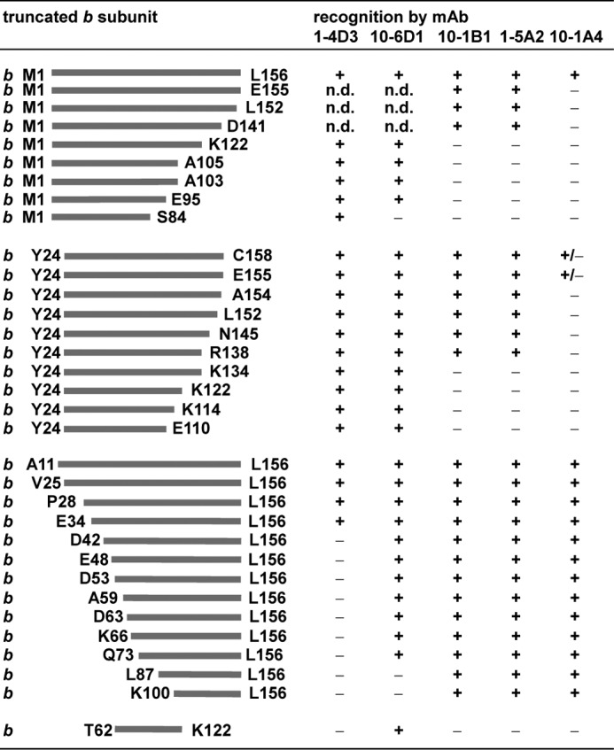
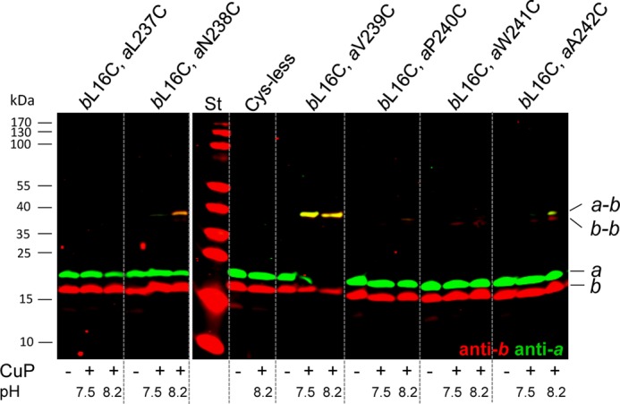
FOF1 ATP synthases are rotary nanomotors that couple proton translocation across biological membranes to the synthesis/hydrolysis of ATP. During catalysis, the peripheral stalk, composed of two b subunits and subunit δ in Escherichia coli, counteracts the torque generated by the rotation of the central stalk. Here we characterize individual interactions of the b subunits within the stator by use of monoclonal antibodies and nearest neighbor analyses via intersubunit disulfide bond formation. Antibody binding studies revealed that the C-terminal region of one of the two b subunits is principally involved in the binding of subunit δ, whereas the other one is accessible to antibody binding without impact on the function of FOF1. Individually substituted cysteine pairs suitable for disulfide cross-linking between the b subunits and the other stator subunits (b-α, b-β, b-δ, and b-a) were screened and combined with each other to discriminate between the two b subunits (i.e. bI and bII). The results show the b dimer to be located at a non-catalytic α/β cleft, with bI close to subunit α, whereas bII is proximal to subunit β. Furthermore, bI can be linked to subunit δ as well as to subunit a. Among the subcomplexes formed were a-bI-α, bII-β, α-bI-bII-β, and a-bI-δ. Taken together, the data obtained define the different positions of the two b subunits at a non-catalytic interface and imply that each b subunit has a different role in generating stability within the stator. We suggest that bI is functionally related to the single b subunit present in mitochondrial ATP synthase.

Keywords: ATP Synthase; Disulfide; Escherichia coli; F-ATPase; Membrane Proteins; Peripheral Stator Stalk; Protein Cross-linking.


Figures

FIGURE 1.
FIGURE 1.
Epitope mapping by binding of mAbs to N- or C-terminally truncated b subunits. Cells synthesizing plasmid-encoded N- or C-terminally truncated b subunits (1100ΔuncFH/pRPG derivatives (45), KF92rA/pBMF derivatives (46), and DK8/pSD or pDM derivatives (23, 29, 30, 32)) were separated on SDS-PAGE (20 μg of cell protein/lane) and analyzed by immunoblotting with different mAbs using the ECL detection system for immunodecoration. The amount of protein was determined on the basis of the calculation that 1 ml of cells with an optical density (578 nm) of 1.0 contains ∼160 μg of protein (58). The differences observed for b and bsyn truncated for the C-terminal amino acid bLeu-156 obviously depend on the varying expression level of the corresponding proteins because of different plasmid backgrounds. +, mAb binding; ±, weak mAb binding; -, no binding; n.d., not determined.
FIGURE 2.
FIGURE 2.
Epitope mapping by binding of mAbs to mutated subunit b or bsyn carrying single amino acid substitutions. Right panel, sections of immunoblot analyses comparing the binding of mAbs to cells synthesizing plasmid-encoded mutated b or bsyn subunits. The position of the amino acid substitution in each mutant is indicated. In most cases, the addressed amino acid residue has been substituted with cysteine. For exceptions, see supplemental Table S1 summarizing the complete analysis. The immunoblot analyses were performed as described in legend to Fig. 1. To verify that residue bAla-68 is not a part of the epitope of mAb GDH 10-6D1, GDH 1-5A2 has been shown as a control, revealing that, in general, the expression of the bsynA68C variant is lower. The differences observed for b and bsyn depend on the varying expression level of the corresponding proteins because of different plasmid backgrounds. Left panel, structural model of FOF1 with the b dimer in blue and the other subunits in light gray as drawn by RasMol 2.7.2.1.1 to mark the positions of the epitopes recognized by mAbs in subunit b. The structural homology model of E. coli FOF1 is a composite of several partial structures combined with biochemical data, as described in detail by Junge et al. (1).
FIGURE 3.
FIGURE 3.
Biochemical characteristics of anti-b mAbs. A, binding of mAb GDH 10-1A4 to purified ATP synthase. Equimolar amounts of mAb GDH 10-1A4 and purified FOF1 complexes were incubated for 1 h at 37 °C prior to separation of the protein complexes by size exclusion chromatography on a Superdex SPX-200, as described under “Experimental Procedures.” Peak fraction A was separated in comparison to purified FOF1 under non-reducing conditions on SDS-PAGE, silver-stained (61) (lanes 1 and 2), and analyzed by immunoblotting (lanes 3 and 4) using exclusively goat anti-mouse IgG antibodies conjugated with horseradish peroxidase for ECL detection. Lanes 1 and 3, purified FOF1; lanes 2 and 4, peak fraction A. mAU, milliabsorption unit. B, competition between bound subunit b and free membrane vesicles. Microtiter plates were coated with purified subunit b (0.1 μg/ml) and subsequently incubated with a mixture of different amounts of free membrane vesicles and culture supernatant of mAb GDH 10-1A4. The competitive inhibition ELISA was performed as described by Jäger et al. (57). ■ (dark gray), F1-containing inverted membrane vesicles of the atp wild-type strain ML308-225; ♦ (black), F1-stripped inverted membrane vesicles of strain ML308-225; ▴ (light gray), inverted membrane vesicles of the atp mutant strain CM1470. The data represent average values of three independent measurements. C, influence of subunit b-specific mAb GDH 10-1A4 on the function of FOF1. Membrane vesicles (MV; 2 mg/ml) were incubated in the absence or presence of mAb GDH 10-1A4 (50 μg/ml) as described under “Experimental Procedures.” NADH-driven proton translocation by respiration and ATP-driven proton translocation by FOF1 were measured via ACMA fluorescence quenching. DCCD-sensitive ATPase activities were determined as described. The presence of mAb GDH 10-1A4 on the membrane vesicles after the washing steps was determined by competitive inhibition ELISA before measuring ATPase and proton translocation activities. a, NADH-dependent ACMA fluorescence quenching after incubation of F1-stripped membrane vesicles with DCCD (40 μm, 20 min at room temperature) for inhibition of FO. b, rebinding of F1 to F1-stripped inverted membrane vesicles was performed after incubation with mAb, as described by Deckers-Hebestreit et al. (53).
FIGURE 4.
FIGURE 4.
Cu2+-catalyzed cross-linking between cysteine substitutions of subunit b at position 92 and subunit α or β. Inverted membrane vesicles of different mutant strains (termed by the cysteine substitutions present within FOF1 subunits) were incubated in the absence (−) or presence (+) of CuP, separated by SDS-PAGE (20 μg/lane) under non-reducing conditions, and analyzed by immunoblotting using two antibodies simultaneously, as described under “Experimental Procedures.” A, cross-linking between bA92C and residues of subunit α of region α464–483 exposed at the surface of the α3β3 hexamer. Cross-linking yields obtained after incubation with CuP at pH 7.5 and 8.2, respectively, were compared. For immunolabeling, mouse anti-b antibodies (GDH 10-1B1, green) and rabbit anti-α antibodies (red) were applied. In each mutant, endogenous cysteine residues in subunits b (bCys-21) and α (αCys-47, αCys-90, αCys-193, and αCys-243) were substituted by alanine. St, molecular mass standard. B, cross-linking of bA92C with residues of subunit β located at a non-catalytic cleft of F1. Rabbit anti-b (red) and mouse anti-β antibodies (green) were applied for detection. In each mutant, endogenous cysteine residues in subunits b (bCys-21) and β (βCys-137) were changed to alanine. St, molecular mass standard; unspec., unspecific immunolabeling. C, structure of the E. coli α3β3 hexamer (63) drawn by RasMol 2.7.2.1.1 with marked amino acid residues of subunits α and β, which were cross-linked to subunit b at position bA92C. Red, high cross-linking yield; orange, intermediate cross-linking yield; yellow, low cross-linking yield; white, no cross-linking.
FIGURE 5.
FIGURE 5.
Cu2+-catalyzed cross-linking between cysteine substitutions of subunit b at position 16 in the N-terminal transmembrane helix and subunit a. Inverted membrane vesicles of different mutant strains (termed by the cysteine substitutions present within FOF1 subunits) were incubated in the absence (-) or presence (+) of CuP, separated by SDS-PAGE (20 μg/lane) under non-reducing conditions, and analyzed by immunoblotting using two antibodies simultaneously as described under “Experimental Procedures.” St, molecular mass standard. Cross-linking yields obtained after incubation with CuP at pH 7.5 and 8.2, respectively, were compared. For immunolabeling rabbit anti-b antibodies (red) and mouse anti-a antibodies (green) were applied. In each mutant, the endogenous cysteine residue in subunit b (bCys-21) was substituted by alanine.
FIGURE 6.
FIGURE 6.
Cu2+-catalyzed cross-linking between cysteine substitutions of subunits b, α, and β combined with a b-a (bL16C/aV239C) cross-linking pair to distinguish both b subunits. A, immunoblot analysis of the a-bI-α and bII-β complexes, respectively, generated by combination of the following cysteine pairs: b-a, bL16C/aV239C; b-α, bE118C/αK118C; b-β, bE118C/βE117C; and b-α,β, bE118C/αK118C/βE117C. Inverted membrane vesicles of the corresponding mutant strains were incubated with CuP as described in the legend to Fig. 4. Immunodecoration: left panel, rabbit anti-b (red) and mouse anti-a (green) antibodies. Right panel, mouse anti-α (green) and rabbit anti-β antibodies (red). In each mutant, the corresponding endogenous cysteine residues in subunits b, α, and β (compare legend to Fig. 4) were substituted by alanine. Because of the use of 10% N-[2-hydroxy-1,1-bis(hydroxymethyl)ethyl]glycine-SDS-polyacrylamide gels (56), a high resolution of protein bands is observed within the range up to 50 kDa, whereas bands with higher molecular weights are observed as dense, highly focused bands (compare also the molecular mass standard). The yellow intensities observed in the blot analysis at the right side arise from a partial overlay because of the close proximity of the bands representing α and β as well as to a slight degradation of subunit α. St, molecular mass standard. B, sections of immunoblot analyses comparable with A showing the cross-linking products obtained by several combinations of different cysteine-substituted subunits b, α, and β as announced in the corresponding boxes at the left side. a-b always represents the presence of cross-linking pair bL16C-aV239C. For clarity, during the simultaneous presence of cysteine substitutions in subunits α and β for interaction with subunit b (last lane of the blot membranes), the complexes formed are marked in green for complexes involving subunit α and in red for those containing subunit β.
FIGURE 7.
FIGURE 7.
Schematic showing zero-length cross-links of b-α and b-β with single cysteine residues present simultaneously in each subunit and with one of the b subunits tagged with subunit a. The schematic summarizes the data obtained for lanes b-a/b-α and b-a/b-β (A) and for lanes b-a/b-α,β (B) in Fig. 6. The numbering indicates the amino acid residues substituted by cysteine within the corresponding subunit. Solid bars, high cross-linking yields; dashed bars, low cross-linking yields.
FIGURE 8.
FIGURE 8.
Cu2+-catalyzed cross-linking between cysteine substitutions of subunits b, α, and β generating an α-b-b-β complex. A, immunoblot analysis of an α-b-b-β complex generated by combination of the following cysteine pairs: b-b, bA68C; b-α, bE118C/αK118C; and b-β, bQ106C/βT287C. Inverted membrane vesicles of the corresponding mutant strains were incubated with CuP as described in the legend to Fig. 4. For immunolabeling, rabbit anti-b antibodies (red) and mouse anti-α (green, left panel) or mouse anti-β antibodies (green, right panel) were applied. In each mutant, the corresponding endogenous cysteine residues in subunits b, α, and β (compare legend to Fig. 4) were substituted by alanine. St, molecular mass standard. B, schematic to illustrate the disulfide bonds formed between the different stator subunits allowing a clear discrimination between both b subunits present in FOF1. Black bars, zero-length cross-links present to obtain the quaternary α-bI-bII-β complex; gray bar, zero-length cross-link used to mark subunit bI with subunit a.; unspec., unspecific reaction of the secondary antibody IRDyeTM 700DX-labeled goat-anti rabbit IgG (H+L).
FIGURE 9.
FIGURE 9.
Cu2+-catalyzed cross-linking between cysteine substitutions of subunits b, a, and δ generating an a-b-δ complex. A, immunoblot analysis of an a-b-δ complex generated by combination of the following cysteine pairs: a-b, bL16C/aV239C and b-δ, bE155C/δI160C. Inverted membrane vesicles of the corresponding mutant strains were incubated with CuP as described in the legend to Fig. 4. For immunolabeling, mouse anti-δ (top panel), anti-b (center panel; GDH 1-4D3), and anti-a antibodies (bottom panel) were applied. In each mutant, the corresponding endogenous cysteine residues in subunits b (bCys-21) and δ (δCys-64 and δCys-140) were substituted by alanine. unspec., unspecific immunolabeling. B, schematic to illustrate the disulfide bonds formed between the different stator subunits. Black bars, zero-length cross-links to obtain the a-bI-δ complex; gray bar, zero-length cross-links used for the α-bI-bII-β complex. C, immunoblot analysis corresponding to the top panel in A using two antibodies simultaneously: mouse anti-δ (green) and rabbit anti-b (red) antibodies. The prominent yellow band observed in each lane is due to an unspecific binding of antibodies (compare with A), which has not been marked for clarity. St, molecular mass standard. D, detection of the b-δ cross-linking product (bE155C/δI160C or bE155C/δG162C) by different anti-b mAbs. bdegrad, degradation product of subunit b detected by mAb.

Similar articles

Cited by

References

    1. Junge W., Sielaff H., Engelbrecht S. (2009) Torque generation and elastic power transmission in the rotary FOF1-ATPase. Nature 459, 364–370 - PubMed
    1. von Ballmoos C., Wiedenmann A., Dimroth P. (2009) Essentials for ATP synthesis by F1FO ATP synthases. Annu. Rev. Biochem. 78, 649–672 - PubMed
    1. von Ballmoos C., Cook G. M., Dimroth P. (2008) Unique rotor ATP synthase and its biological diversity. Annu. Rev. Biophys. 37, 43–64 - PubMed
    1. Cross R. L., Müller V. (2004) The evolution of A-, F-, and V-type ATP synthases and ATPases. Reversals in function and changes in the H+/ATP coupling ratio. FEBS Lett. 576, 1–4 - PubMed
    1. Muench S. P., Trinick J., Harrison M. A. (2011) Structural divergence of the rotary ATPases. Q. Rev. Biophys. 44, 311–356 - PubMed

Publication types

MeSH terms

LinkOut - more resources