. 2013 Aug 23;288(34):24956-71.
doi: 10.1074/jbc.M113.461004. Epub 2013 Jul 11.

Endocytosis of Mycobacterium tuberculosis heat shock protein 60 is required to induce interleukin-10 production in macrophages

Affiliations

Endocytosis of Mycobacterium tuberculosis heat shock protein 60 is required to induce interleukin-10 production in macrophages

Nazia Parveen et al. J Biol Chem. .

Abstract

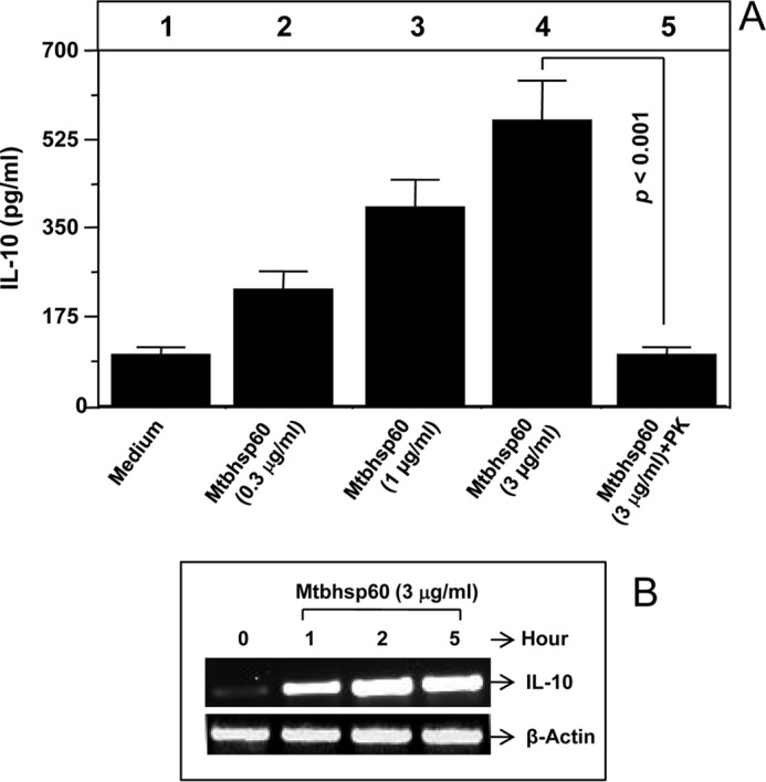
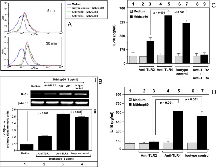
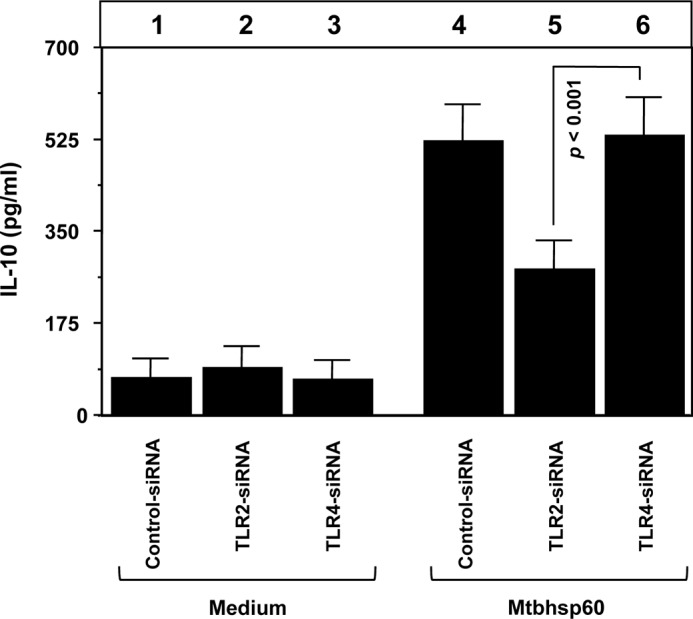
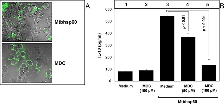
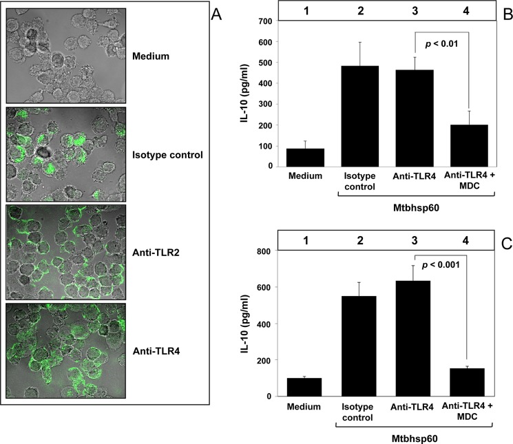
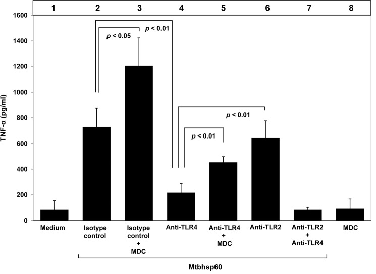
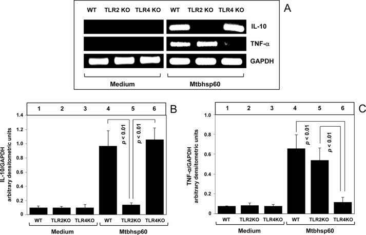
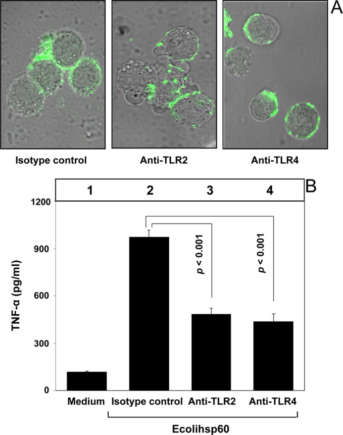
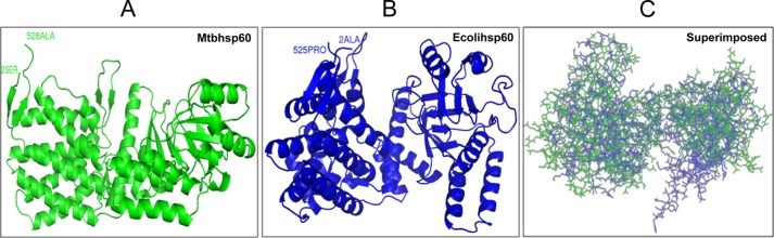
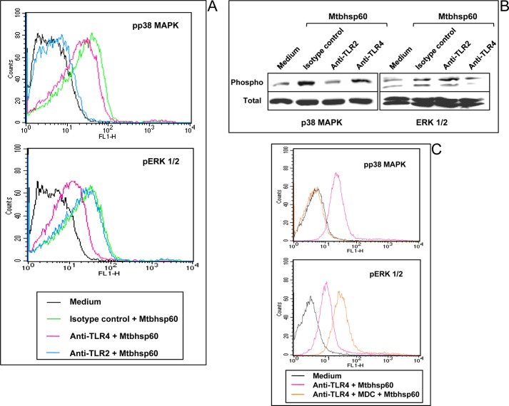
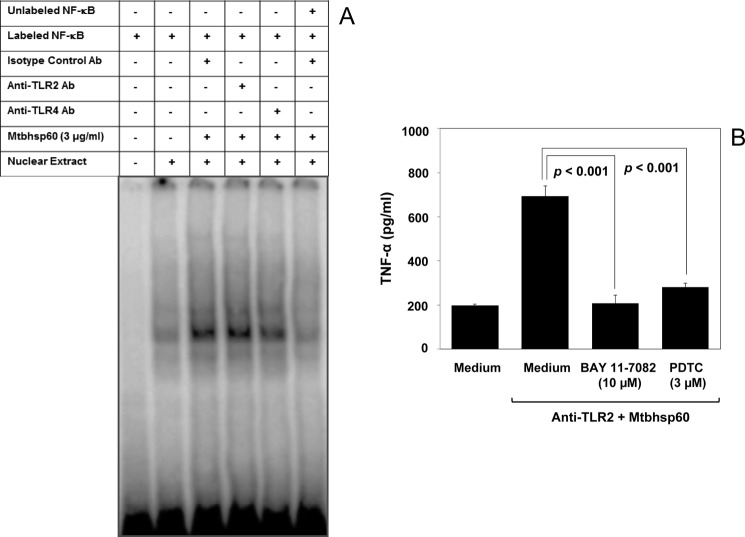
Understanding the signaling pathways involved in the regulation of anti-inflammatory and pro-inflammatory responses in tuberculosis is extremely important in tailoring a macrophage innate response to promote anti-tuberculosis immunity in the host. Although the role of toll-like receptors (TLRs) in the regulation of anti-inflammatory and pro-inflammatory responses is known, the detailed molecular mechanisms by which the Mycobacterium tuberculosis bacteria modulate these innate responses are not clearly understood. In this study, we demonstrate that M. tuberculosis heat shock protein 60 (Mtbhsp60, Cpn60.1, and Rv3417c) interacts with both TLR2 and TLR4 receptors, but its interaction with TLR2 leads to clathrin-dependent endocytosis resulting in an increased production of interleukin (IL)-10 and activated p38 MAPK. Blockage of TLR2-mediated endocytosis inhibited IL-10 production but induced production of tumor necrosis factor (TNF)-α and activated ERK1/2. In contrast, upon interaction with TLR4, Mtbhsp60 remained predominantly localized on the cell surface due to poorer endocytosis of the protein that led to decreased IL-10 production and p38 MAPK activation. The Escherichia coli homologue of hsp60 was found to be retained mainly on the macrophage surface upon interaction with either TLR2 or TLR4 that triggered predominantly a pro-inflammatory-type immune response. Our data suggest that cellular localization of Mtbhsp60 upon interaction with TLRs dictates the type of polarization in the innate immune responses in macrophages. This information is likely to help us in tailoring the host protective immune responses against M. tuberculosis.

Keywords: Cytokine; Endocytosis; MAP Kinases (MAPKs); Mycobacterium tuberculosis; Toll-like Receptors (TLR).


Figures

FIGURE 1.
FIGURE 1.
Mtbhsp60 induces IL-10 production in THP-1 macrophages. PMA-differentiated THP-1 macrophages were treated with either titrating concentrations of the purified Mtbhsp60 or a fixed concentration (3 μg/ml) of proteinase K-treated Mtbhsp60. A, IL-10 cytokine level was estimated by EIA in various culture supernatants harvested after 48 h of incubation. B, total RNA was extracted from various groups as described under “Experimental Procedures,” and IL-10 mRNA expression was measured at various time points by semi-quantitative RT-PCR. Results shown are representative of three to four different experiments.
FIGURE 2.
FIGURE 2.
IL-10 activation by Mtbhsp60 is TLR2-dependent. A, PMA-differentiated THP-1 macrophages were pretreated with either 10 μg/ml of anti-TLR2 mAb or anti-TLR4 mAb or isotype-matched control antibody for 1 h and then incubated with 10 μg/ml of biotin-labeled Mtbhsp60 at 4 °C for 5 and 30 min followed by incubation with streptavidin-FITC. The fluorescence was measured by flow cytometry. B, PMA-differentiated THP-1 macrophages were pretreated with 10 μg/ml of anti-TLR2 mAb or anti-TLR4 mAb or isotype-matched control antibody for 1 h and cultured for 2 h in the presence of 3 μg/ml of Mtbhsp60. Total RNA was extracted, and IL-10 levels were measured by semi-quantitative RT-PCR, and quantification of the IL-10 mRNA was performed by densitometric analysis using AlphaEaseFC software and the Spot Denso tool (version 7.0.1; Alpha Innotech, San Leandro, CA). Data are expressed as mean ± S.D. of three independent experiments. C, PMA-differentiated THP-1 macrophages were pretreated with neutralizing mAb to either TLR2 or TLR4, isotype-matched control antibody, or with both anti-TLR2 mAb and anti-TLR4 mAb in the absence or presence of Mtbhsp60 (3 μg/ml). After 48 h, IL-10 cytokine levels in culture supernatants from various groups were measured by EIA. D, experiments were also set to measure IL-10 levels by EIA in C57Bl/6 peritoneal macrophages treated with 10 μg/ml of anti-TLR2 Ab, anti-TLR4 Ab, or isotype control antibody in the absence or presence of Mtbhsp60 (3 μg/ml). Results shown are representative of three different experiments.
FIGURE 3.
FIGURE 3.
Silencing of TLR2 expression by siRNA down-regulates Mtbhsp60-induced IL-10 in THP-1 macrophages. PMA-differentiated THP-1 macrophages were transfected with negative control siRNA or TLR2-specific siRNA or TLR4-specific siRNA. After 24 h post-transfection, cells were incubated with Mtbhsp60 (3 μg/ml) for 48 h, and levels of IL-10 were measured by EIA in the culture supernatants. Results shown are representative of three independent experiments.
FIGURE 4.
FIGURE 4.
MDC inhibits Mtbhsp60-mediated induction of IL-10 in THP-1 macrophages. A, PMA-differentiated THP-1 macrophages in a chamber slide were preincubated for 30 min without or with MDC (100 μm) followed by incubation with 10 μg/ml of Mtbhsp60-FITC at 37 °C for 15 min. Cells were fixed and washed, and the cell bound fluorescence was analyzed by confocal laser scanning microscopy. B, PMA-differentiated THP-1 macrophages were either left untreated or pretreated with 50 or 100 μm MDC and subsequently incubated with 3 μg/ml of Mtbhsp60. IL-10 cytokine level was estimated by EIA in various culture supernatants harvested after 48 h of incubation. Results shown are representative of three different experiments.
FIGURE 5.
FIGURE 5.
IL-10 activation by Mtbhsp60 is dependent on TLR2-mediated endocytosis of Mtbhsp60. A, PMA-differentiated THP-1 macrophages were pretreated with 10 μg/ml of anti-TLR2 mAb or anti-TLR4 mAb or isotype-matched control antibody for 1 h and further incubated for 15 min in the presence of Mtbhsp60-FITC (10 μg/ml) at 37 °C. Cells were fixed and washed, and the endocytosis was examined by confocal laser scanning microscopy. PMA-differentiated THP-1 macrophages (B) or peritoneal macrophages (C) from C57Bl/6 mice were pretreated with 10 μg/ml of anti-TLR4 Ab or isotype control Ab for 1 h and then incubated with 3 μg/ml of Mtbhsp60 in the absence or presence of 100 μm MDC. After 48 h of incubation, IL-10 cytokine level was measured by EIA in various culture supernatants. Results shown are representative of at least three different experiments.
FIGURE 6.
FIGURE 6.
Interaction of Mtbhsp60 with TLR4 or TLR2 in the presence of MDC triggers TNF-α production. PMA-differentiated THP-1 macrophages were pretreated with 10 μg/ml of neutralizing mAb to either TLR2 or TLR4, or isotype-matched control antibody, or with both anti-TLR2 mAb and anti-TLR4 mAb for 1 h and subsequently incubated with Mtbhsp60 (3 μg/ml) in the absence or presence of MDC (100 μm), and TNF-α cytokine levels were quantified after 48 h in different culture supernatants by EIA. Results shown are representative of three different experiments.
FIGURE 7.
FIGURE 7.
Mtbhsp60 targets the TLR2 to induce IL-10 and the TLR4 to activate production of TNF-α in macrophages. Thioglycolate-elicited peritoneal macrophages from C57Bl/6 WT, TLR2 KO, and TLR4 KO mice were either left untreated or treated with Mtbhsp60 (3 μg/ml) for 1 h. A, total RNA was extracted, and IL-10 and TNF-α levels were measured by semi-quantitative RT-PCR. Densitometric analyses were performed for IL-10 (B) and TNF-α (C) using the AlphaEaseFC software and the Spot Denso tool, normalized for GAPDH, and reported as arbitrary densitometric units. Values are means ± S.D. of the densitometric analysis of three independent experiments.
FIGURE 8.
FIGURE 8.
Interaction of Ecolihsp60 either with TLR2 or TLR4 results in induction of TNF-α. A, PMA-differentiated THP-1 macrophages were pretreated with 10 μg/ml of anti-TLR2 mAb or anti-TLR4 mAb or isotype-matched control antibody for 1 h, followed by incubation with Ecolihsp60-FITC (10 μg/ml) at 37 °C for 15 min. Cells were fixed and washed, and endocytosis of the protein was assessed by confocal laser scanning microscopy. B, PMA-differentiated THP-1 macrophages were pretreated with 10 μg/ml of anti-TLR2 mAb or anti-TLR4 mAb or isotype-matched control antibody for 1 h and then incubated with 3 μg/ml Ecolihsp60. TNF-α levels were quantified after 48 h in different culture supernatants by EIA. Data are representative of mean ± S.D. of three different experiments.
FIGURE 9.
FIGURE 9.
Secondary structure comparison of Mtbhsp60 with Ecolihsp60. A, secondary structure of the Mtbhsp60 protein model predicted based on homology modeling with Modeler software is shown (green) after its energy minimization. B, solved crystal structure of Ecolihsp60 obtained from Protein Data bank (chain A of PDB code 2EU1) is displayed (blue). C, superimposed structures of Mtbhsp60 and Ecolihsp60.
FIGURE 10.
FIGURE 10.
Comparison of phosphorylation status of p38 MAPK and ERK1/2 mediated by Mtbhsp60 between the TLR2 receptor that undergoes endocytosis against TLR2 or TLR4 that does not undergo endocytosis. A, PMA-differentiated THP-1 macrophages were pretreated with 10 μg/ml of anti-TLR2 mAb or anti-TLR4 mAb or isotype-matched control Ab for 1 h and then treated with Mtbhsp60 (3 μg/ml) for 15 min. After permeabilization, macrophages were incubated with antibody to either phospho-p38 or phospho-ERK1/2 followed by incubation with anti-rabbit IgG-FITC or anti-mouse IgG-FITC, and fluorescence was analyzed by flow cytometry. B, in another experiment, THP-1 macrophages were pretreated with 10 μg/ml of either anti-TLR2 mAb or anti-TLR4 mAb or isotype-matched control Ab followed by incubation with Mtbhsp60 (3 μg/ml). Cells were lysed, and the levels of phosphorylated and total p38 MAPK as well as phosphorylated and total ERK1/2 levels were measured by Western blotting. C, PMA-differentiated THP-1 macrophages were pretreated with neutralizing mAb to TLR4 (10 μg/ml) for 1 h and then treated with Mtbhsp60 for 15 min in the absence or presence of MDC (100 μm). The phosphorylation status of p38 MAPK and ERK1/2 was analyzed by flow cytometry. Results are representative of three experiments.
FIGURE 11.
FIGURE 11.
Mtbhsp60 targets TLR4 to activate NF-κB transcription factors. A, PMA-differentiated THP-1 macrophages were pretreated with 10 μg/ml of either anti-TLR2 mAb or anti-TLR4 mAb or isotype-matched control Ab for 60 min and then incubated with 3 μg/ml of Mtbhsp60 for another 60 min. Cells were harvested, and nuclear extracts were prepared. The DNA-binding activity of NF-κB complex was measured by EMSA. B, in another experiment, PMA-differentiated THP-1 macrophages were pretreated with 10 μg/ml of either isotype-control Ab or anti-TLR2 Ab and further incubated with 3 μg/ml of Mtbhsp60 in the absence or presence of BAY 11-7082 (10 μm) or pyrrolidine dithiocarbamate (3 μm). After 48 h, culture supernatants were harvested, and TNF-α levels were quantified by EIA. Results shown are representative of three different experiments.

Similar articles

Cited by

References

    1. Schluger N. W., Rom W. N. (1998) The host immune response to tuberculosis. Am. J. Respir. Crit. Care Med. 157, 679–691 - PubMed
    1. Rook G. A., Dheda K., Zumla A. (2005) Immune responses to tuberculosis in developing countries: implications for new vaccines. Nat. Rev. Immunol. 5, 661–667 - PubMed
    1. Flynn J. L., Goldstein M. M., Chan J., Triebold K. J., Pfeffer K., Lowenstein C. J., Schreiber R., Mak T. W., Bloom B. R. (1995) Tumor necrosis factor-α is required in the protective immune response against Mycobacterium tuberculosis in mice. Immunity 2, 561–572 - PubMed
    1. Flynn J. L. (2004) Immunology of tuberculosis and implications in vaccine development. Tuberculosis 84, 93–101 - PubMed
    1. Quesniaux V. F., Jacobs M., Allie N., Grivennikov S., Nedospasov S. A., Garcia I., Olleros M. L., Shebzukhov Y., Kuprash D., Vasseur V., Rose S., Court N., Vacher R., Ryffel B. (2010) TNF in host resistance to tuberculosis infection. Curr. Dir. Autoimmun. 11, 157–179 - PubMed

Publication types

MeSH terms

LinkOut - more resources