Positional scanning mutagenesis of α-conotoxin PeIA identifies critical residues that confer potency and selectivity for α6/α3β2β3 and α3β2 nicotinic acetylcholine receptors
- PMID: 23846688
- PMCID: PMC3757205
- DOI: 10.1074/jbc.M113.482059
Positional scanning mutagenesis of α-conotoxin PeIA identifies critical residues that confer potency and selectivity for α6/α3β2β3 and α3β2 nicotinic acetylcholine receptors
Abstract
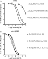
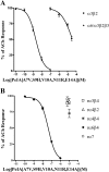
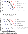
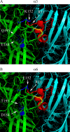
The nicotinic acetylcholine receptor (nAChR) subtype α6β2* (the asterisk denotes the possible presence of additional subunits) has been identified as an important molecular target for the pharmacotherapy of Parkinson disease and nicotine dependence. The α6 subunit is closely related to the α3 subunit, and this presents a problem in designing ligands that discriminate between α6β2* and α3β2* nAChRs. We used positional scanning mutagenesis of α-conotoxin PeIA, which targets both α6β2* and α3β2*, in combination with mutagenesis of the α6 and α3 subunits, to gain molecular insights into the interaction of PeIA with heterologously expressed α6/α3β2β3 and α3β2 receptors. Mutagenesis of PeIA revealed that Asn(11) was located in an important position that interacts with the α6 and α3 subunits. Substitution of Asn(11) with a positively charged amino acid essentially abolished the activity of PeIA for α3β2 but not for α6/α3β2β3 receptors. These results were used to synthesize a PeIA analog that was >15,000-fold more potent on α6/α3β2β3 than α3β2 receptors. Analogs with an N11R substitution were then used to show a critical interaction between the 11th position of PeIA and Glu(152) of the α6 subunit and Lys(152) of the α3 subunit. The results of these studies provide molecular insights into designing ligands that selectively target α6β2* nAChRs.
Keywords: Electrophysiology; Neurotoxin; Neurotransmitter Receptors; Nicotinic Acetylcholine Receptors; Oocyte; alpha-Conotoxin; alpha3beta2; alpha6beta2.
Figures



Similar articles
-
α-Conotoxin PeIA[S9H,V10A,E14N] potently and selectively blocks α6β2β3 versus α6β4 nicotinic acetylcholine receptors.Mol Pharmacol. 2012 Nov;82(5):972-82. doi: 10.1124/mol.112.080853. Epub 2012 Aug 22. Mol Pharmacol. 2012. PMID: 22914547 Free PMC article.
-
A novel α4/7-conotoxin LvIA from Conus lividus that selectively blocks α3β2 vs. α6/α3β2β3 nicotinic acetylcholine receptors.FASEB J. 2014 Apr;28(4):1842-53. doi: 10.1096/fj.13-244103. Epub 2014 Jan 7. FASEB J. 2014. PMID: 24398291 Free PMC article.
-
PeIA-5466: A Novel Peptide Antagonist Containing Non-natural Amino Acids That Selectively Targets α3β2 Nicotinic Acetylcholine Receptors.J Med Chem. 2019 Jul 11;62(13):6262-6275. doi: 10.1021/acs.jmedchem.9b00566. Epub 2019 Jun 27. J Med Chem. 2019. PMID: 31194549 Free PMC article.
-
Mutagenesis of α-Conotoxins for Enhancing Activity and Selectivity for Nicotinic Acetylcholine Receptors.Toxins (Basel). 2019 Feb 13;11(2):113. doi: 10.3390/toxins11020113. Toxins (Basel). 2019. PMID: 30781866 Free PMC article. Review.
-
Progress and challenges in the study of α6-containing nicotinic acetylcholine receptors.Biochem Pharmacol. 2011 Oct 15;82(8):862-72. doi: 10.1016/j.bcp.2011.06.022. Epub 2011 Jun 28. Biochem Pharmacol. 2011. PMID: 21736871 Free PMC article. Review.
Cited by
-
Rigidity of loop 1 contributes to equipotency of globular and ribbon isomers of α-conotoxin AusIA.Sci Rep. 2021 Nov 9;11(1):21928. doi: 10.1038/s41598-021-01277-4. Sci Rep. 2021. PMID: 34753970 Free PMC article.
-
The crystal structure of Ac-AChBP in complex with α-conotoxin LvIA reveals the mechanism of its selectivity towards different nAChR subtypes.Protein Cell. 2017 Sep;8(9):675-685. doi: 10.1007/s13238-017-0426-2. Epub 2017 Jun 5. Protein Cell. 2017. PMID: 28585176 Free PMC article.
-
Computational and Functional Mapping of Human and Rat α6β4 Nicotinic Acetylcholine Receptors Reveals Species-Specific Ligand-Binding Motifs.J Med Chem. 2021 Feb 11;64(3):1685-1700. doi: 10.1021/acs.jmedchem.0c01973. Epub 2021 Feb 1. J Med Chem. 2021. PMID: 33523678 Free PMC article.
-
Marine Origin Ligands of Nicotinic Receptors: Low Molecular Compounds, Peptides and Proteins for Fundamental Research and Practical Applications.Biomolecules. 2022 Jan 23;12(2):189. doi: 10.3390/biom12020189. Biomolecules. 2022. PMID: 35204690 Free PMC article. Review.
-
Monkey adrenal chromaffin cells express α6β4* nicotinic acetylcholine receptors.PLoS One. 2014 Apr 11;9(4):e94142. doi: 10.1371/journal.pone.0094142. eCollection 2014. PLoS One. 2014. PMID: 24727685 Free PMC article.
References
-
- Han Z. Y., Le Novère N., Zoli M., Hill J. A., Jr., Champtiaux N., Changeux J. P. (2000) Localization of nAChR subunit mRNAs in the brain of Macaca mulatta. Eur. J. Neurosci. 12, 3664–3674 - PubMed
-
- Endo T., Yanagawa Y., Obata K., Isa T. (2005) Nicotinic acetylcholine receptor subtypes involved in facilitation of GABAergic inhibition in mouse superficial superior colliculus. J. Neurophysiol. 94, 3893–3902 - PubMed
-
- Cox B. C., Marritt A. M., Perry D. C., Kellar K. J. (2008) Transport of multiple nicotinic acetylcholine receptors in the rat optic nerve. High densities of receptors containing α6 and β3 subunits. J. Neurochem. 105, 1924–1938 - PubMed
Publication types
MeSH terms
Substances
Grants and funding
LinkOut - more resources
Full Text Sources
Other Literature Sources

