. 2013 Aug 23;288(34):24286-92.
doi: 10.1074/jbc.M113.461996. Epub 2013 Jul 11.

Disruption of multivesicular body vesicles does not affect major histocompatibility complex (MHC) class II-peptide complex formation and antigen presentation by dendritic cells

Affiliations

Disruption of multivesicular body vesicles does not affect major histocompatibility complex (MHC) class II-peptide complex formation and antigen presentation by dendritic cells

Berta Bosch et al. J Biol Chem. .

Abstract

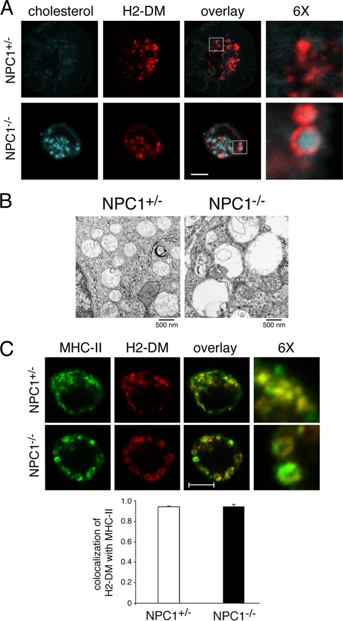
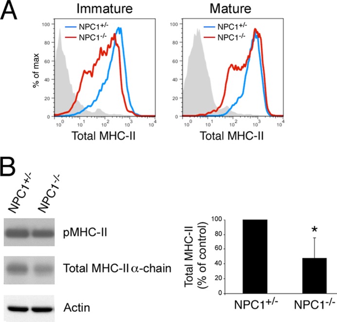
The antigen processing compartments in antigen-presenting cells (APCs) have well known characteristics of multivesicular bodies (MVBs). However, the importance of MVB integrity to APC function remains unknown. In this study, we have altered the ultrastructure of the MVB by perturbing cholesterol content genetically through the use of a deletion of the lipid transporter Niemann-Pick type C1 (NPC1). Immunofluorescence and electron microscopic analyses reveal that the antigen processing compartments in NPC1(-/-) dendritic cells (DCs) have an abnormal ultrastructure in that the organelles are enlarged and the intraluminal vesicles are almost completely absent and those remaining are completely disorganized. MHC-II is restricted to the limiting membrane of these enlarged MVBs where it colocalizes with the peptide editor H2-DM. Curiously, proteolytic removal of the chaperone protein Invariant chain from MHC-II, degradation of internalized foreign antigens, and antigenic-peptide binding to nascent MHC-II are normal in NPC1(-/-) DCs. Antigen-pulsed NPC1(-/-) DCs are able to effectively activate antigen-specific CD4 T cells in vitro, and immunization of NPC1(-/-) mice reveals surprisingly normal CD4 T cell activation in vivo. Our data thus reveal that the localization of MHC-II on the intraluminal vesicles of multivesicular antigen processing compartments is not required for efficient antigen presentation by DCs.

Keywords: Antigen Presentation; Antigen Processing; Endosomes; Major Histocompatibility Complex (MHC); Niemann-Pick Disease Type C; T Cell.


Figures

FIGURE 1.
FIGURE 1.
MVB ultrastructure and MHC-II localization are disturbed in lipid-overloaded MVBs. A, control or NPC1−/− DCs were harvested after 7 days of growth in medium and attached to polylysine-coated coverslips prior to fixation, permeabilization, and staining with filipin to detect unesterified cholesterol (blue) and a mAb recognizing H2-DM (red). 6× magnified images of the indicated regions of the merged image are shown. Scale bar represents 5 μm. B, DCs were fixed and sectioned for electron microscopy. Scale bar represents 500 nm. C, immature DCs were fixed, permeabilized, and stained with mAbs recognizing MHC-II (M5/114, green) or H2-DM (red). 6× magnified images of the indicated regions of the merged image are shown. Scale bar represents 5 μm. The extent of colocalization of H2-DM+ red pixels with MHC-II+ green pixels was determined using software provided with the LSM510 confocal microscope.
FIGURE 2.
FIGURE 2.
NPC1−/− DCs are activated normally by LPS and generate MHC-II-peptide complexes. A, immature or LPS-matured control (blue lines) or NPC1−/− (red lines) DCs were monitored by flow cytometry for surface expression of total surface MHC-II using mAb M5/114. B, LPS-matured control or NPC1−/− DCs were harvested and lysed in Triton X-100. Aliquots of the lysates were incubated with SDS-PAGE sample buffer and were either left at room temperature for 30 min or boiled for 3 min prior to SDS-PAGE. SDS stable pMHC-II complexes (53 kDa), total MHC-II α-chain (35 kDa), and actin (41 kDa) were detected by immunoblotting. The total amount of MHC-II present in each sample was quantitated by densitometry, normalized to the amount of actin present in the sample, and expressed as a percentage of total MHC-II present in control DCs.
FIGURE 3.
FIGURE 3.
Antigen uptake and proteolysis as well as Ii association and CLIP removal from MHC-II are normal in NPC1−/− DCs. A, immature NPC1+/− (control, blue) and NPC1−/− (red) DCs were incubated with the indicated amount of DQ-OVA for 1.5 h at 37 °C. Uptake and degradation were monitored by flow cytometry. The mean fluorescence intensity of DQ-OVA taken up and processed in NPC1−/− DCs was expressed relative to control DCs. B, mature NPC1+/− (control) and NPC1−/− DCs were lysed in Triton X-100, and immunoprecipitations were performed using isotype control (IgG2b) and anti-MHC-II mAb. Portions of each immunoprecipitate were analyzed by immunoblotting for the presence of intact Ii or MHC-II β-chain. The amount of Ii present in the MHC-II immunoprecipitate from NPC1−/− DCs was expressed as a percentage of Ii present in control DCs. Error bars, S.D. C, mature NPC1+/− (solid lines) and NPC1−/− DCs (dashed lines) were incubated with the MHC-II-CLIP mAb 15G4, washed, and counterstained with the pan-MHC-II mAb M5/114. The stained cells were analyzed by FACS, and cells in the MHC-II low and MHC-II high gates were analyzed separately for expression of total MHC-II and MHC-II-CLIP complexes.
FIGURE 4.
FIGURE 4.
NPC1−/− DCs effectively activate antigen-specific naïve CD4 T cells in vitro. A and B, immature NPC1+/− (control) and NPC1−/− DCs were incubated with ovalbumin protein, washed, and activated with LPS overnight. The cells were then stained with pan-MHC-II mAb, and MHC-II high cells were sorted by FACS. Aliquots of the unsorted cells (A) or isolated MHC-II high cells (B) were analyzed for total MHC-II expression by FACS and were incubated with CFSE-labeled naïve OTII CD4 T cells at a 1:10 ratio. CFSE dilution was examined 72 h later by FACS analysis. C, the percentage of CFSE-labeled OTII T cells proliferating more than once when incubated with either unsorted DCs or the isolated MHC-II high DCs was calculated. The data shown are the mean ± S.D. (error bars) from three independent experiments.
FIGURE 5.
FIGURE 5.
NPC1−/− DCs effectively activate antigen-specific naïve CD4 T cells in vivo. A, CFSE-labeled naïve OTII CD4 T cells (CD45.1) were transferred into NPC1+/− (control) and NPC1−/− mice (CD45.2) and followed by immunization 24 h later with ovalbumin protein. After 3 days CD45.1+ CD4 T cells from the spleen were isolated, and CFSE dilution was examined by FACS analysis. B, the absolute number of CD11chigh MHC-II+ DCs present in the spleens of NPC1+/− (control) and NPC1−/− mice was determined by FACS analysis. C, CD11c+ DCs were isolated from the spleens of NPC1+/− (control) and NPC1−/− mice by immunomagnetic purification. Live cells were attached to polylysine-coated coverslips prior to fixation, permeabilization, and staining with MHC-II mAb (M5/114, green) and counterstaining with filipin to detect unesterified cholesterol (red).

Similar articles

Cited by

References

    1. Berger A. C., Roche P. A. (2009) MHC class II transport at a glance. J. Cell Sci. 122, 1–4 - PMC - PubMed
    1. McCormick P. J., Martina J. A., Bonifacino J. S. (2005) Involvement of clathrin and AP-2 in the trafficking of MHC class II molecules to antigen-processing compartments. Proc. Natl. Acad. Sci. U.S.A. 102, 7910–7915 - PMC - PubMed
    1. Kleijmeer M., Ramm G., Schuurhuis D., Griffith J., Rescigno M., Ricciardi-Castagnoli P., Rudensky A. Y., Ossendorp F., Melief C. J., Stoorvogel W., Geuze H. J. (2001) Reorganization of multivesicular bodies regulates MHC class II antigen presentation by dendritic cells. J. Cell Biol. 155, 53–63 - PMC - PubMed
    1. Zwart W., Griekspoor A., Kuijl C., Marsman M., van Rheenen J., Janssen H., Calafat J., van Ham M., Janssen L., van Lith M., Jalink K., Neefjes J. (2005) Spatial separation of HLA-DM/HLA-DR interactions within MIIC and phagosome-induced immune escape. Immunity 22, 221–233 - PubMed
    1. Carstea E. D., Morris J. A., Coleman K. G., Loftus S. K., Zhang D., Cummings C., Gu J., Rosenfeld M. A., Pavan W. J., Krizman D. B., Nagle J., Polymeropoulos M. H., Sturley S. L., Ioannou Y. A., Higgins M. E., Comly M., Cooney A., Brown A., Kaneski C. R., Blanchette-Mackie E. J., Dwyer N. K., Neufeld E. B., Chang T. Y., Liscum L., Strauss J. F., 3rd, Ohno K., Zeigler M., Carmi R., Sokol J., Markie D., O'Neill R. R., van Diggelen O. P., Elleder M., Patterson M. C., Brady R. O., Vanier M. T., Pentchev P. G., Tagle D. A. (1997) Niemann-Pick C1 disease gene: homology to mediators of cholesterol homeostasis. Science 277, 228–231 - PubMed

Publication types

MeSH terms

LinkOut - more resources