. 2013 Aug 23;288(34):24569-80.
doi: 10.1074/jbc.M113.479550. Epub 2013 Jul 11.

DEAF1 is a Pellino1-interacting protein required for interferon production by Sendai virus and double-stranded RNA

Affiliations

DEAF1 is a Pellino1-interacting protein required for interferon production by Sendai virus and double-stranded RNA

Alban Ordureau et al. J Biol Chem. .

Abstract

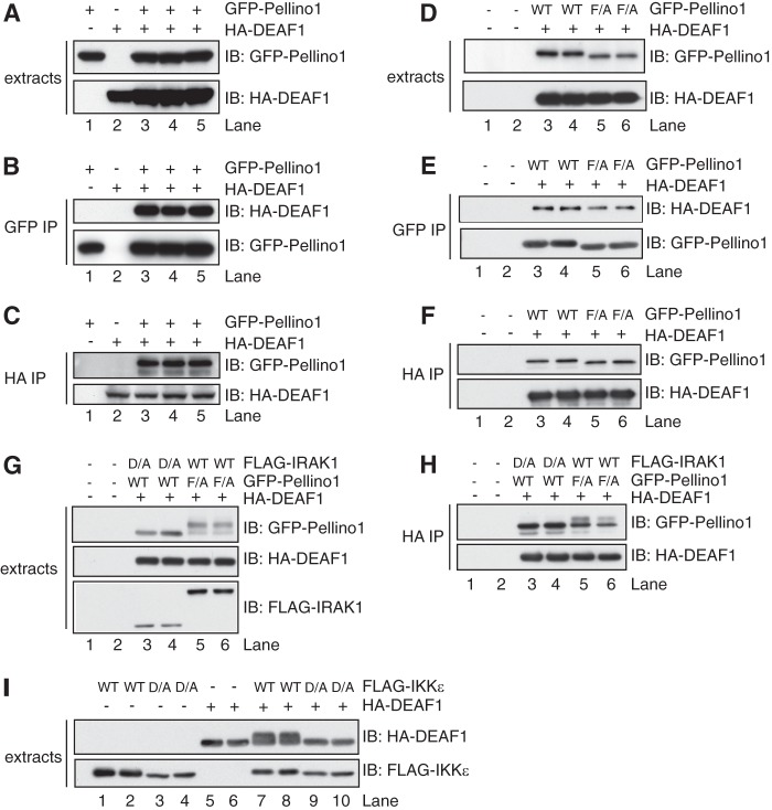
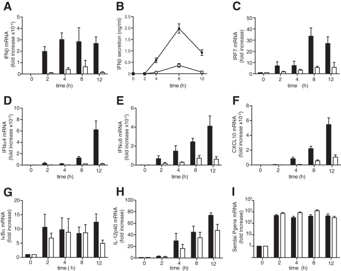
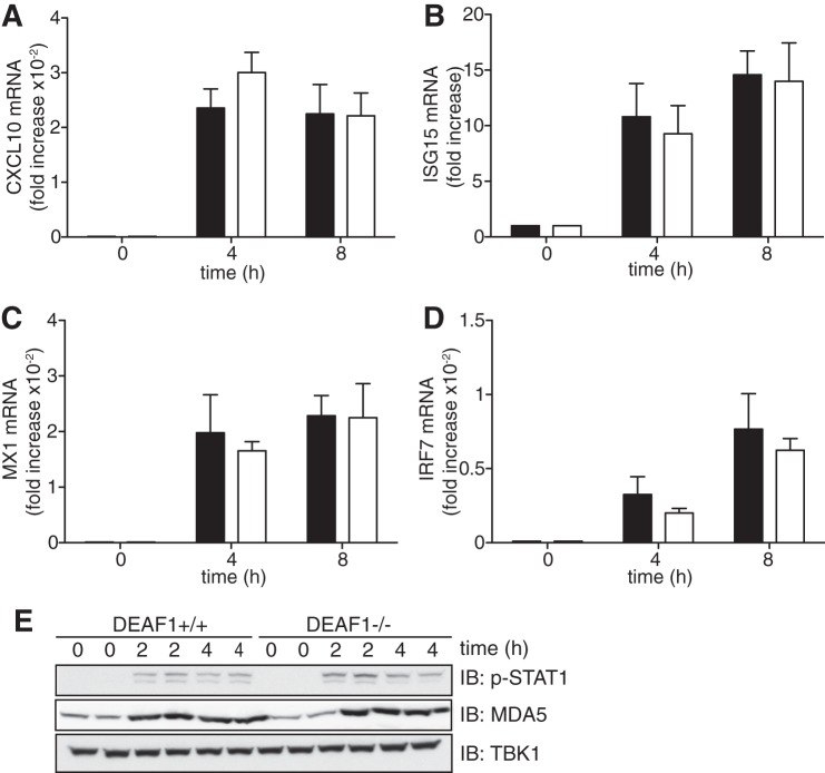
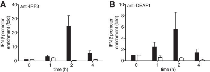
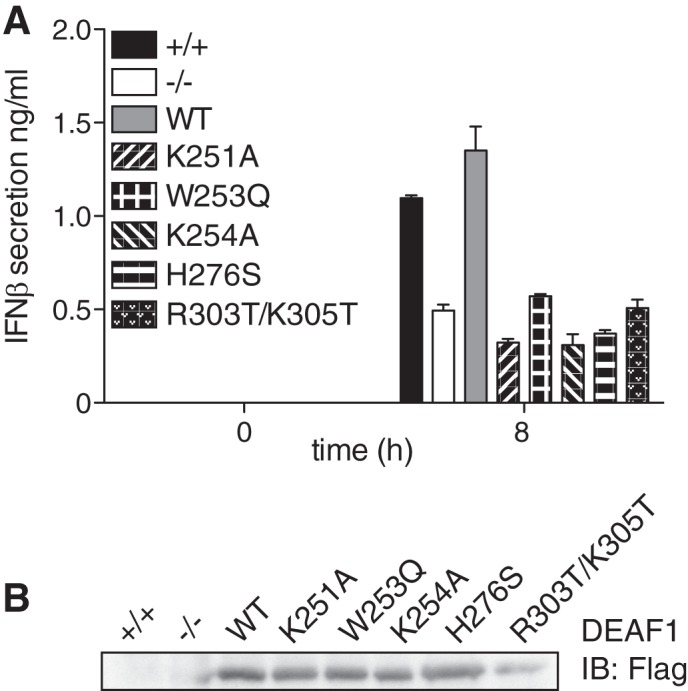
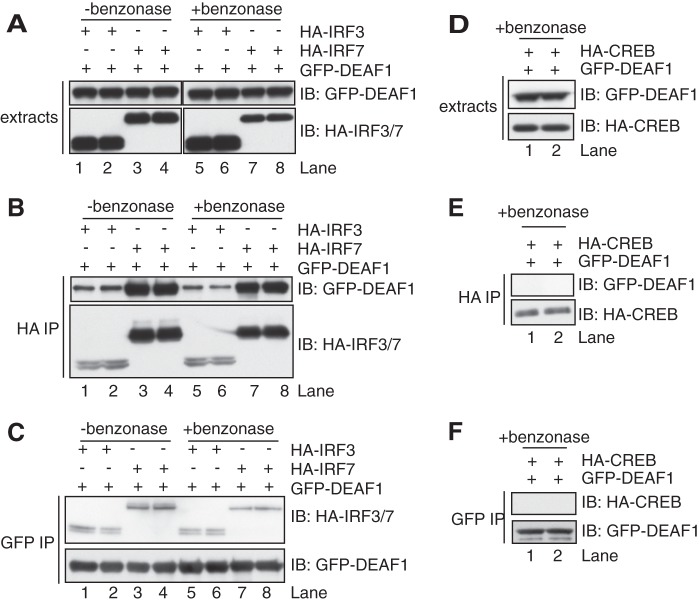
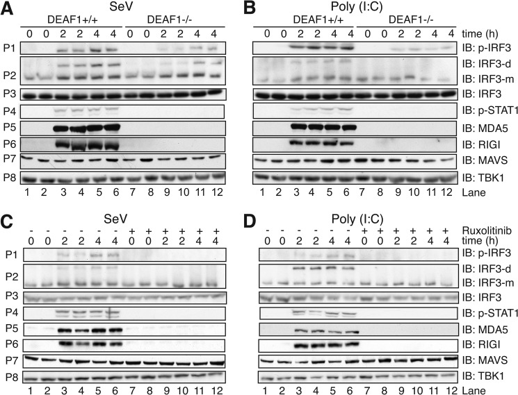
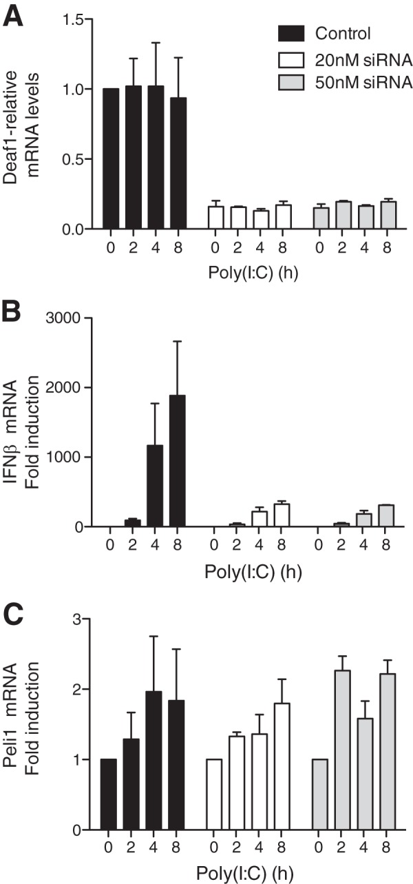
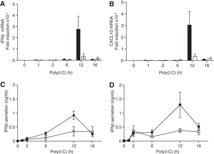
Double-stranded (ds) RNA of viral origin, a ligand for Melanoma Differentiation-associated gene 5 (MDA5) and Toll-Like Receptor 3 (TLR3), induces the TANK-Binding Kinase 1 (TBK1)-dependent phosphorylation and activation of Interferon Regulatory Factor 3 (IRF3) and the E3 ubiquitin ligase Pellino1, which are required for interferon β (IFNβ) gene transcription. Here, we report that Pellino1 interacts with the transcription factor Deformed Epidermal Autoregulatory Factor 1 (DEAF1). The interaction is independent of the E3 ligase activity of Pellino1, but weakened by the phosphorylation of Pellino1. We show that DEAF1 binds to the IFNβ promoter and to IRF3 and IRF7, that it is required for the transcription of the IFNβ gene and IFNβ secretion in MEFs infected with Sendai virus or transfected with poly(I:C). DEAF1 is also needed for TLR3-dependent IFNβ production. Taken together, our results identify DEAF1 as a novel component of the signal transduction network by which dsRNA of viral origin stimulates IFNβ production.

Keywords: Cytokines/Interferon; DEAF1; Double-stranded RNA Viruses; Fibroblast; IRF3; Interferon; Pellino1; TBK1; Transcription Factors.


Figures

FIGURE 1.
FIGURE 1.
DEAF1 and Pellino1 interact in overexpression studies. HEK293FT cells were transfected with DNA encoding HA-DEAF1 and/or GFP-Pellino1. After 36 h, the cells were lysed in buffer containing phosphatase inhibitors (see “Experimental Procedures”) plus 50 mm iodoacetamide. A, cell extracts (3 μg of protein) were denatured in SDS, subjected to SDS-PAGE, transferred on to a PVDF membrane, and immunoblotted (IB) with anti-HA and or anti-GFP to monitor the expression of the DEAF1 and Pellino1 fusion proteins. B, Pellino1 was immunoprecipitated (IP) from 0.1 mg of cell extract protein with the GFP antibody (GFP-Trap beads). The bound proteins were denatured in SDS, and immunoblotted with anti-HA and anti-GFP to detect DEAF1 and Pellino1, respectively. C, as in B except that DEAF1 was immunoprecipitated from 0.1 mg of cell lysate protein with an HA antibody. D–F, same as A–C, except that HA-DEAF1 was transfected with either wild-type (WT) or the F397A mutant (F/A) of Pellino1. G and H, same as D, E except that the cells were also transfected with FLAG-tagged wild-type (WT) IRAK1 or the catalytically inactive IRAK1[D358A] mutant (D/A). I, same as A, except that the cells were transfected with HA-DEAF1 and either wild-type FLAG-IKKϵ (WT) or the catalytically inactive FLAG-IKKϵ[D157A] mutant (D/A). The cell extracts were immunoblotted with anti-HA and anti-FLAG.
FIGURE 2.
FIGURE 2.
The production of IFNβ in Sendai virus infected MEFs is reduced in DEAF1−/− mice. A–I, immortalized MEFs from wild type mice (black bars, black circles in B) or DEAF1−/− mice (white bars, white squares in B) were infected with Sendai virus (100 HA/ml) for the times indicated. Total RNA was extracted and analyzed by qRT-PCR to measure the mRNA encoding IFNβ, IRF7, IFNα4, IFNα6, CXCL10, IκBα, IL-12p40, and the Sendai virus Pgene. The results show fold increase in mRNA relative to the values measured in unstimulated MEFs (± S.D. for triplicate determinations). In panel B, the concentration of IFNβ in the culture medium was measured by ELISA. The results are shown ± S.D. for triplicate determinations. Similar results were obtained in four independent experiments using MEFs immortalized from two different wild type and two different DEAF1−/− mice.
FIGURE 3.
FIGURE 3.
IFNβ-stimulated signal transduction does not require DEAF1. A–D, immortalized MEFs from wild type (black bars) and DEAF1−/− (white bars) mice were stimulated with 500 units/ml IFNβ for the times indicated. Total RNA was extracted and analyzed by qRT-PCR to measure the mRNA encoding CXCL10 (A), ISG15 (B), MX1 (C), and IRF7 (D). The results show fold increase in mRNA relative to the values measured in unstimulated MEFs (± S.D. for triplicate determinations). Similar results were obtained in two independent experiments. E, MEFs from wild type (DEAF1+/+) and DEAF1−/−) mice were stimulated with IFNβ as in A–D, and then lysed in the presence of phosphatase inhibitors. Aliquots of the cell extract (20 μg protein) were subjected to SDS-PAGE and, after transfer to PVDF membranes, immunoblotted with an antibody that recognizes STAT1 phosphorylated at Tyr-701 (p-STAT1) or that recognize MDA5 or TBK1.
FIGURE 4.
FIGURE 4.
IFNβ promoters associated with IRF3 are reduced in MEFs from DEAF1−/− mice. A, MEFs from wild type mice (black bars) or DEAF1−/− mice (white bars) were infected with Sendai virus for the times indicated. The cells were lysed, and ChIP assays performed after immunoprecipitating IRF3 from the extracts with a specific anti-IRF3 antibody. The ordinate shows the fold enrichment of the IFNβ promoter in the anti-IRF3 immunoprecipitates compared with that measured after immunoprecipitation from uninfected cells (time = 0). The IFNβ promoter DNA was measured by qRT-PCR, as described in “Experimental Procedures.” The results are shown ± S.D. for triplicate determinations using MEFs from two different wild type and two different DEAF1−/− mice at each time point. Similar results were obtained in two independent experiments. B, experiment was carried out as in A, except that the ChIP assay was performed after immunoprecipitating DEAF1 from the extracts of MEFs from wild type mice (black bars) or from DEAF1−/− mice (white bars) with an anti-DEAF1 antibody.
FIGURE 5.
FIGURE 5.
DEAF1 transcriptional activity and nuclear localization is needed to stimulate IFNβ production. A, immortalized MEFs from DEAF1−/− mice were stably reconstituted with either FLAG-tagged wild-type (WT) DEAF1, DEAF1[K251A], DEAF1[W253Q], DEAF1[K254A], DEAF1[H276S] and DEAF1[R303T/K305T]. These MEF cell lines, as well as the immortalized MEFs from wild type mice (+/+) and DEAF1−/− mice were infected with Sendai virus (SeV) (100 HA/ml) for the times indicated, and the concentration of IFNβ in the culture medium was measured by ELISA. The results are shown ± S.D. for triplicate determinations. B, the cell extract from A (25 μg protein) was denatured in SDS, subjected to SDS-PAGE, transferred to a nitrocellulose membrane, and immunoblotted (IB) with anti-FLAG to show the expression of the different DEAF1 mutants in the reconstituted MEFs.
FIGURE 6.
FIGURE 6.
DEAF1 interacts with IRF3 and IRF7. HEK293FT cells in 10-cm dishes were transfected with DNA (0.6 μg/ml) encoding GFP-DEAF1 and either HA-IRF3 or HA-IRF7. The cells were extracted in lysis buffer containing 50 mm iodoacetamide as in Fig. 1 and incubated for 2 h at 4 °C in the absence or presence of the DNase benzonase (50 units/ml). A, aliquots of the cell extract (3 μg protein) were subjected to SDS-PAGE and immunoblotted as in Fig. 1. B, IRF3 or IRF7 were immunoprecipitated from 0.1 mg of cell lysate protein with an HA antibody and the immunoprecipitates washed, denatured in SDS, and subjected to SDS-PAGE and immunoblotting as in A. C, as in B except that DEAF1 was immunoprecipitated using anti-GFP. D–F, as in A–C except that the cells were co-transfected with DNA encoding GFP-DEAF1 and HA-CREB.
FIGURE 7.
FIGURE 7.
The activation and synthesis of component of the interferon pathway in MEFs infected with Sendai virus or transfected with poly(I:C). A and B, immortalized MEFs from wild type mice (DEAF1+/+) or DEAF1−/− mice were infected with Sendai virus (SeV) (100 HA units/ml) (A) or transfected with poly(I:C) (10 μg/ml) (B) for the times indicated. Cell lysates (30 μg protein) were denatured in SDS, subjected to SDS/PAGE, transferred to PVDF membranes and immunoblotted (IB) with the antibodies indicated. Panel P2 shows a separate experiment in which SDS was excluded from the sample and native gel electrophoresis performed to separate the monomeric (m) and dimeric (d) forms of IRF3, which were then detected by immunoblotting. C and D, same as A and B, except that DEAF1+/+ MEFs were incubated for 1 h without (−) or with (+) 1 μm Ruxolitinib prior to infection with Sendai virus (C) or transfection with poly(I:C) (D).
FIGURE 8.
FIGURE 8.
RNA interference of DEAF1 suppresses IFNβ mRNA production in poly(I:C)-stimulated HEK293-TLR3 cells. HEK293 cells stably expressing the TLR3 receptor (termed HEK293-TLR3 cells) were transfected with “scrambled” siRNA (control) or with the indicated amounts of DEAF1-blocking siRNA. 72 h later, the cells were stimulated with poly(I:C) (50 μg/ml) for the times indicated, total RNA extracted and the mRNA encoding DEAF1 (A), IFNβ (B), and Pellino1 (C) was measured by qRT-PCR as described under “Experimental Procedures.” The results show relative mRNA levels (± S.D. for triplicate determinations) compared with the value of 1.0 measured in unstimulated control, HEK293-TLR3 cells. The experiment was performed in 12-well plates with three wells used for each condition. Similar results were obtained in two independent experiments.
FIGURE 9.
FIGURE 9.
The poly(I:C)-stimulated production of IFNβ is impaired in BMDM from DEAF1−/− mice. A and B, four wild type mice and three DEAF1−/− mice were used at each time point. BMDM from wild type (WT) (black bars) or DEAF1−/− mice (white bars) were stimulated for the times indicated with poly(I:C) (10 μg/ml) and, at each time point, the total RNA was extracted from the macrophages and the mRNA encoding IFNβ (A) and CXCL10 (B) was measured by qRT-PCR. The results show the fold increase in mRNA relative to the values measured in unstimulated, wild-type BMDMs (± S.D. for triplicate determinations). C, as in A and B except that, at each time point, the amount of IFNβ secreted into the cell culture medium from wild type BMDM (black circles) or DEAF1−/− BMDM (white circles) was measured by ELISA. D, same as C, except that BMDM from wild type (WT) mice (black squares) or Pellino1[F397A] mice (white squares) (9) were used.

Similar articles

Cited by

References

    1. Grosshans J., Schnorrer F., Nüsslein-Volhard C. (1999) Oligomerisation of Tube and Pelle leads to nuclear localisation of dorsal. Mech. Dev. 81, 127–138 - PubMed
    1. Haghayeghi A., Sarac A., Czerniecki S., Grosshans J., Schöck F. (2010) Pellino enhances innate immunity in Drosophila. Mech. Dev. 127, 301–307 - PubMed
    1. Schauvliege R., Janssens S., Beyaert R. (2006) Pellino proteins are more than scaffold proteins in TLR/IL-1R signalling: a role as novel RING E3-ubiquitin-ligases. FEBS Lett. 580, 4697–4702 - PubMed
    1. Ordureau A., Smith H., Windheim M., Peggie M., Carrick E., Morrice N., Cohen P. (2008) The IRAK-catalysed activation of the E3 ligase function of Pellino isoforms induces the Lys63-linked polyubiquitination of IRAK1. Biochem. J. 409, 43–52 - PMC - PubMed
    1. Smith H., Peggie M., Campbell D. G., Vandermoere F., Carrick E., Cohen P. (2009) Identification of the phosphorylation sites on the E3 ubiquitin ligase Pellino that are critical for activation by IRAK1 and IRAK4. Proc. Natl. Acad. Sci. U.S.A. 106, 4584–4590 - PMC - PubMed

Publication types

MeSH terms

Substances

LinkOut - more resources