. 2013 Jul 10;14(7):14301-20.
doi: 10.3390/ijms140714301.

Lipidosterolic extract of serenoa repens modulates the expression of inflammation related-genes in benign prostatic hyperplasia epithelial and stromal cells

Affiliations

Lipidosterolic extract of serenoa repens modulates the expression of inflammation related-genes in benign prostatic hyperplasia epithelial and stromal cells

Nanor Sirab et al. Int J Mol Sci. .

Abstract

Despite the high prevalence of histological Benign Prostatic Hypeplasia (BPH) in elderly men, little is known regarding the molecular mechanisms and networks underlying the development and progression of the disease. Here, we explored the effects of a phytotherapeutic agent, Lipidosterolic extract of the dwarf palm plant Serenoa repens (LSESr), on the mRNA gene expression profiles of two representative models of BPH, BPH1 cell line and primary stromal cells derived from BPH. Treatment of these cells with LSESr significantly altered gene expression patterns as assessed by comparative gene expression profiling on gene chip arrays. The expression changes were manifested three hours following in vitro administration of LSESr, suggesting a rapid action for this compound. Among the genes most consistently affected by LSESr treatment, we found numerous genes that were categorized as part of proliferative, apoptotic, and inflammatory pathways. Validation studies using quantitative real-time PCR confirmed the deregulation of genes known to exhibit key roles in these biological processes including IL1B, IL1A, CXCL6, IL1R1, PTGS2, ALOX5, GAS1, PHLDA1, IL6, IL8, NFkBIZ, NFKB1, TFRC, JUN, CDKN1B, and ERBB3. Subsequent analyses also indicated that LSESr treatment can impede the stimulatory effects of certain proinflammatory cytokines such as IL6, IL17, and IL15 in these cells. These results suggest that LSESr may be useful to treat BPH that manifest inflammation characteristics. This also supports a role for inflammation in BPH presumably by mediating the balance between apoptosis and proliferation.


Figures

Figure 1
Figure 1
(a) MTT assay showing dose ranging (10 to 200 μg/mL) effect of lipidosterolic extract of Serenoa repens (LSESr) at 24 h on the cell viability of BPH1 immortalized epithelial cell line (left) and primary stromal fibroblasts (PrSF) primary culture of stromal cells (right). The mean value of the optical density (OD) measured in the control condition (0) was defined as 100% survival with other conditions reported as a percent of the control. Error bars show standard error of the mean; (b) Microscopic observation of BPH1 epithelial cells (left) and PrSF stromal cells (right) after 1 h, 3 h, 6 h, and 24 h exposure to LSESr as compared to untreated cells (0 h). Phase contrast photomicrographs (40×) show the accumulation of vesicles in the cytoplasm of both cell types after 3 h of exposure to LSESr. In this experiment, the final concentration of LSESr is 60 μg/mL for epithelial cells and 50 μg/mL for stromal cells.
Figure 1
Figure 1
(a) MTT assay showing dose ranging (10 to 200 μg/mL) effect of lipidosterolic extract of Serenoa repens (LSESr) at 24 h on the cell viability of BPH1 immortalized epithelial cell line (left) and primary stromal fibroblasts (PrSF) primary culture of stromal cells (right). The mean value of the optical density (OD) measured in the control condition (0) was defined as 100% survival with other conditions reported as a percent of the control. Error bars show standard error of the mean; (b) Microscopic observation of BPH1 epithelial cells (left) and PrSF stromal cells (right) after 1 h, 3 h, 6 h, and 24 h exposure to LSESr as compared to untreated cells (0 h). Phase contrast photomicrographs (40×) show the accumulation of vesicles in the cytoplasm of both cell types after 3 h of exposure to LSESr. In this experiment, the final concentration of LSESr is 60 μg/mL for epithelial cells and 50 μg/mL for stromal cells.
Figure 2
Figure 2
Microarray profiling of gene expression in control (0 h) and LSESr treated BPH epithelial (left) and stromal (right) cells. BPH cells are treated in three replicates for 1, 3, and 6 h by LSESr (60 μg/mL for BPH1, 50 μg/mL for PrSF). (a) Differential analysis representing the number of up/down-regulated genes at 0 h, 1 h, 3 h, and 6 h; (b) Heat maps demonstrating the relative level of differentially expressed genes after 6 h exposure to LSESr. An FDR adjusted p-value represents the significance of the enrichment. Only annotations with a significant FDR adjusted p-value of <0.05 are shown.
Figure 3
Figure 3
Gene Ontology (GO) analysis. (a) DAVID analysis for differentially regulated genes, which belong to the categories of inflammation, apoptosis and regulation of proliferation in at least one time point. Gene Ontology (GO) based annotation was used to perform functional enrichment analysis using DAVID tools; (b) Heat maps of significantly differentially expressed genes involved in the inflammation process in BPH epithelial cells (left) and PrSF cells (right) treated by LSESr at different time-points as compared to control. Genes significantly up-regulated are in red and down-regulated are in green. Black bar is for control (0 h), red for 1 h, green for 3 h, and blue for 6 h of exposure to LSESr.
Figure 3
Figure 3
Gene Ontology (GO) analysis. (a) DAVID analysis for differentially regulated genes, which belong to the categories of inflammation, apoptosis and regulation of proliferation in at least one time point. Gene Ontology (GO) based annotation was used to perform functional enrichment analysis using DAVID tools; (b) Heat maps of significantly differentially expressed genes involved in the inflammation process in BPH epithelial cells (left) and PrSF cells (right) treated by LSESr at different time-points as compared to control. Genes significantly up-regulated are in red and down-regulated are in green. Black bar is for control (0 h), red for 1 h, green for 3 h, and blue for 6 h of exposure to LSESr.
Figure 4
Figure 4
Real Time PCR (RT-PCR) measurements confirming the results of the Microarray study. (a) qRT-PCR analysis for mRNA expression of some inflammatory markers in BPH1 cells (left): IL1B, IL1A, IL1R1, CXCL6, ALOX5, PTGS2, and ALOX5 and in PrSF cells (right) IL6, IL8, NFKBIZ, and TFRC; (b) RT-PCR showing a time-dependent regulation of mRNA expression by LSESr. The transcript expression of PTSG2 in BPH1 cells (left) was decreased significantly after 3 and 6 h of LSESr supplementation. NFKBIZ down-regulation in PrSF cells was significant only after 6 h; (c) qRT-PCR analysis for mRNA expression of Apoptosis and cell cycle related genes (GAS1, CDKN1B, PHLDA1, ERBB3, DEDD, and JUN). Bars, SD for three experiments, each performed in duplicate; *p < 0.05, **p < 0.01.
Figure 5
Figure 5
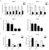
Effects of LSESr in BPH epithelial and stromal cells stimulated by inflammatory factors. (a) BrdU incorporation assay investigating cell proliferation index. Cells were treated with 10 ng/mL of recombinant human IL6, IL17, IL15, or bFGF for 24 h in presence or absence of LSESr; (b) qRT-PCR analysis for mRNA expression of some inflammatory markers in epithelial cells stimulated by IL17 (left), or in stromal cells stimulated by IL15 (right) in the presence or absence of LSESr. In BPH1 cells, PTGS2, IL1B and NFkB1 were down-regulated by LSESr.

Similar articles

Cited by

References

    1. Carter H.B., Coffey D.S. The prostate: An increasing medical problem. Prostate. 1990;16:39–48. - PubMed
    1. Lee K.L., Peehl D.M. Molecular and cellular pathogenesis of benign prostatic hyperplasia. J. Urol. 2004;172:1784–1791. - PubMed
    1. Kramer G., Mitteregger D., Marberger M. Is benign prostatic hyperplasia (BPH) an immune inflammatory disease? Eur. Urol. 2007;51:1202–1216. - PubMed
    1. Robert G., Descazeaud A., Nicolaiew N., Terry S., Sirab N., Vacherot F., Maille P., Allory Y., de la Taille A. Inflammation in benign prostatic hyperplasia: A 282 patients’ immunohistochemical analysis. Prostate. 2009;69:1774–1780. - PMC - PubMed
    1. Schenk J.M., Kristal A.R., Neuhouser M.L., Tangen C.M., White E., Lin D.W., Thompson I.M. Serum adiponectin, C-peptide and leptin and risk of symptomatic benign prostatic hyperplasia: Results from the prostate cancer prevention trial. Prostate. 2009;69:1303–1311. - PMC - PubMed

Publication types

MeSH terms