

Diacetyl increases sensory innervation and substance P production in rat trachea
- PMID: 23847039
- PMCID: PMC4637165
- DOI: 10.1177/0192623313493689
Diacetyl increases sensory innervation and substance P production in rat trachea
Abstract
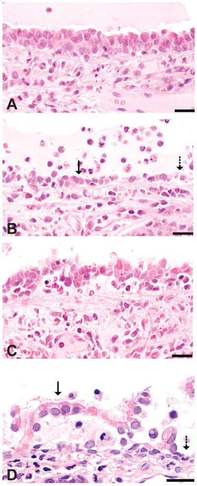
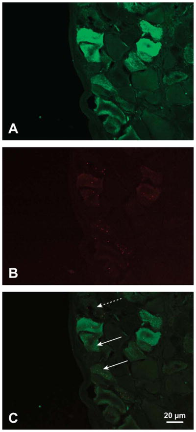
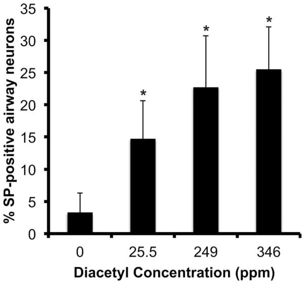
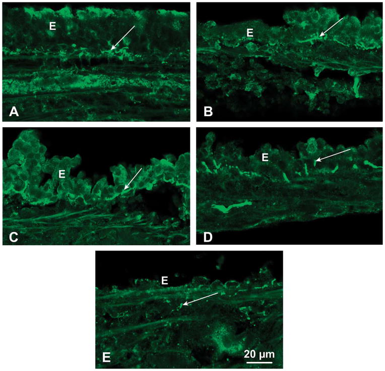
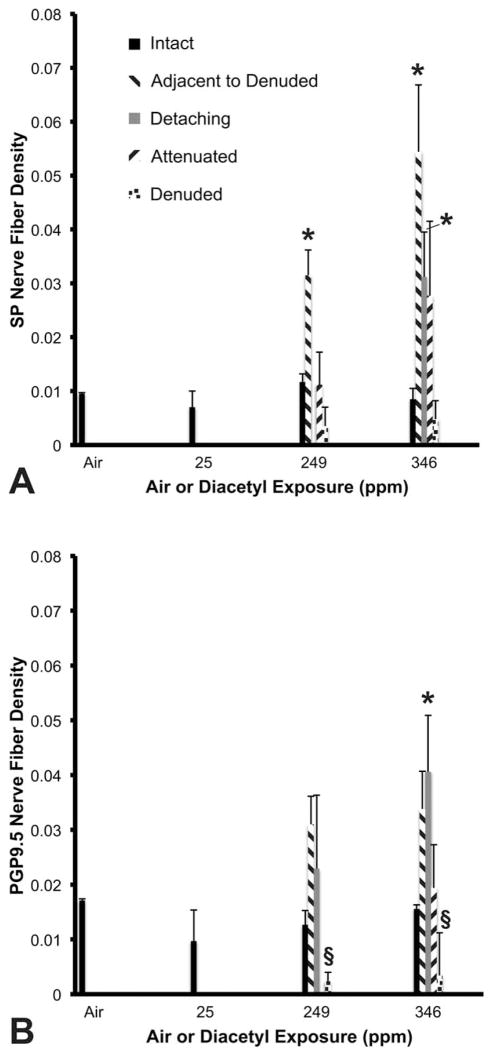
Inhalation of diacetyl, a butter flavoring, causes airway responses potentially mediated by sensory nerves. This study examines diacetyl-induced changes in sensory nerves of tracheal epithelium. Rats (n = 6/group) inhaled 0-, 25-, 249-, or 346-ppm diacetyl for 6 hr. Tracheas and vagal ganglia were removed 1-day postexposure and labeled for substance P (SP) or protein gene product 9.5 (PGP9.5). Vagal ganglia neurons projecting to airway epithelium were identified by axonal transport of fluorescent microspheres intratracheally instilled 14 days before diacetyl inhalation. End points were SP and PGP9.5 nerve fiber density (NFD) in tracheal epithelium and SP-positive neurons projecting to the trachea. PGP9.5-immunoreactive NFD decreased in foci with denuded epithelium, suggesting loss of airway sensory innervation. However, in the intact epithelium adjacent to denuded foci, SP-immunoreactive NFD increased from 0.01 ± 0.002 in controls to 0.05 ± 0.01 after exposure to 346-ppm diacetyl. In vagal ganglia, SP-positive airway neurons increased from 3.3 ± 3.0% in controls to 25.5 ± 6.6% after inhaling 346-ppm diacetyl. Thus, diacetyl inhalation increases SP levels in sensory nerves of airway epithelium. Because SP release in airways promotes inflammation and activation of sensory nerves mediates reflexes, neural changes may contribute to flavorings-related lung disease pathogenesis.
Keywords: airway epithelium; airway inflammation; airway innervation; cough; flavorings; inhalation toxicity.
Conflict of interest statement
The author(s) declared no potential conflicts of interest with respect to the research, authorship, and/or publication of this article.
Figures
References
-
- Akpinar-Elci M, Travis WD, Lynch DA, Kreiss K. Bronchiolitis obliterans syndrome in popcorn production plant workers. Eur Respir J. 2004;24:298–302. - PubMed
-
- Anderson SE, Wells J, Fedorowicz A, Butterworth L, Meade B, Munson AE. Evaluation of the contact and respiratory sensitization potential of volatile organic compounds generated by simulated indoor air chemistry. Toxicol Sci. 2007;97:355–63. - PubMed
-
- Barnes PJ. Asthma as an axon reflex. Lancet. 1986;1:242–45. - PubMed
-
- Barnes PJ. Neurogenic inflammation in airways and its modulation. Arch Int Pharmacodyn Ther. 1990;303:67–82. - PubMed
-
- Canning BJ, Mori N, Mazzone SB. Vagal afferent nerves regulating the cough reflex. Respir Physiol Neurobiol. 2006;152:223–42. - PubMed
Publication types
MeSH terms
Substances
Grants and funding
LinkOut - more resources
Full Text Sources
Other Literature Sources
