. 2013 Jul 30;110(31):12649-54.
doi: 10.1073/pnas.1307218110. Epub 2013 Jul 11.

Inactivating mutations of RNF43 confer Wnt dependency in pancreatic ductal adenocarcinoma

Affiliations

Inactivating mutations of RNF43 confer Wnt dependency in pancreatic ductal adenocarcinoma

Xiaomo Jiang et al. Proc Natl Acad Sci U S A. .

Abstract

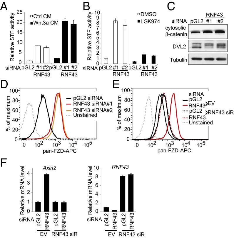
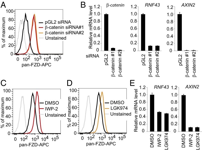
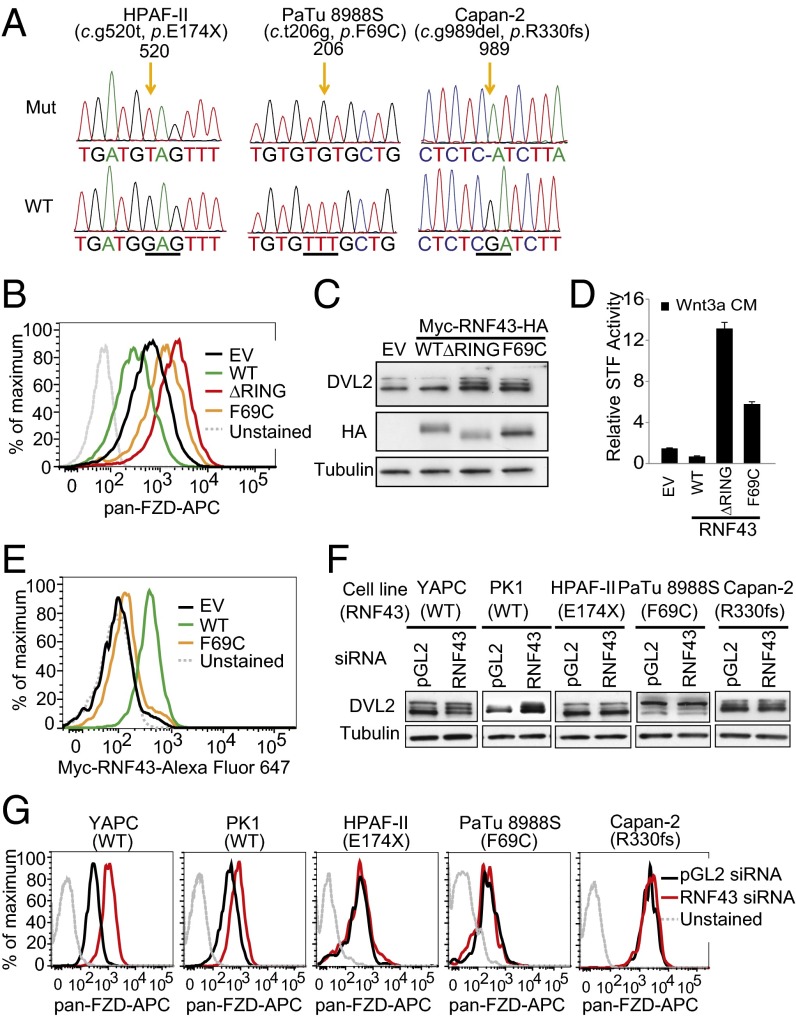
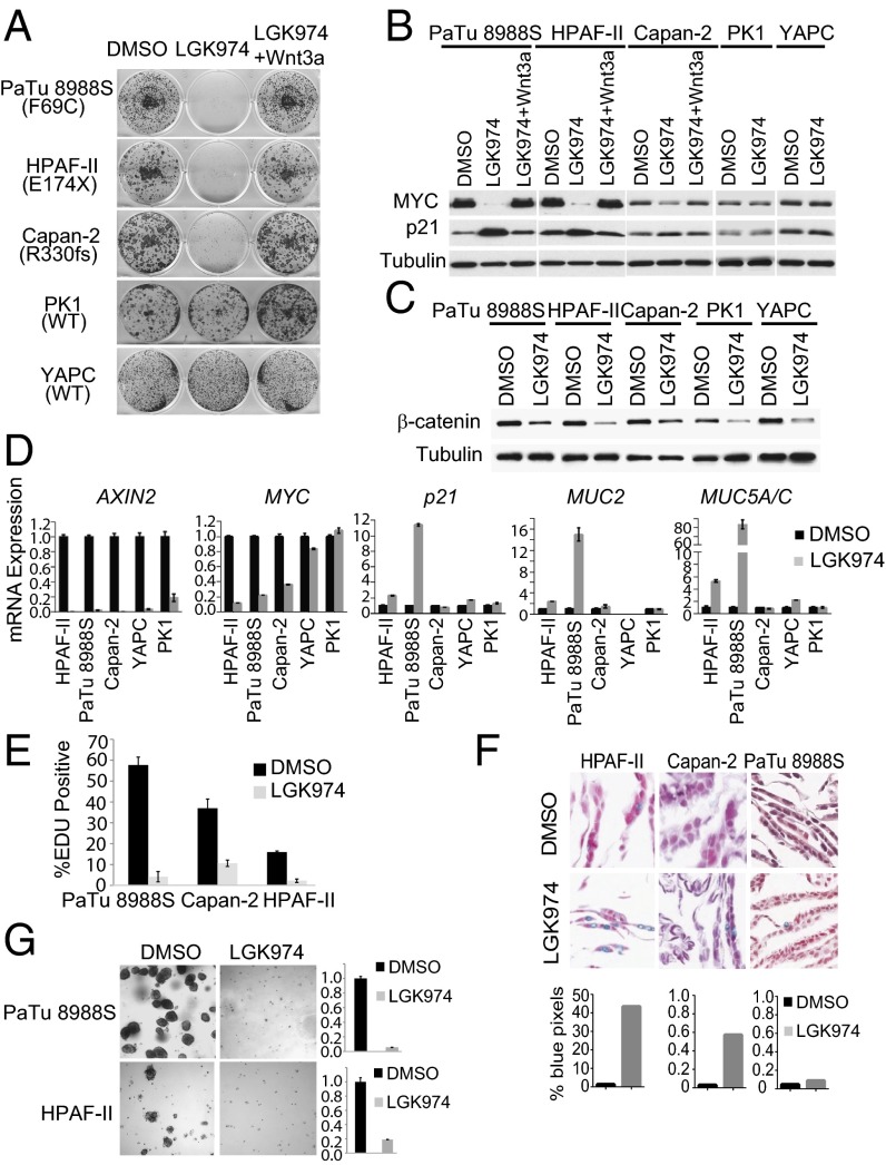
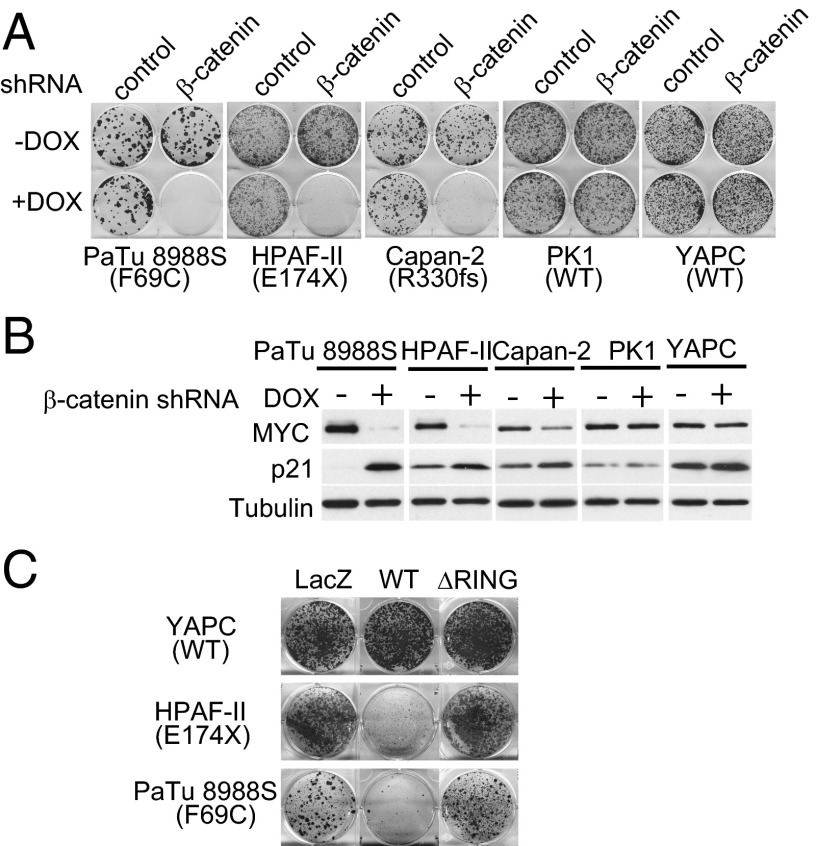
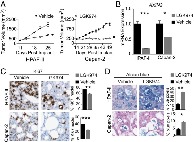
A growing number of agents targeting ligand-induced Wnt/β-catenin signaling are being developed for cancer therapy. However, clinical development of these molecules is challenging because of the lack of a genetic strategy to identify human tumors dependent on ligand-induced Wnt/β-catenin signaling. Ubiquitin E3 ligase ring finger 43 (RNF43) has been suggested as a negative regulator of Wnt signaling, and mutations of RNF43 have been identified in various tumors, including cystic pancreatic tumors. However, loss of function study of RNF43 in cell culture has not been conducted, and the functional significance of RNF43 mutations in cancer is unknown. Here, we show that RNF43 inhibits Wnt/β-catenin signaling by reducing the membrane level of Frizzled in pancreatic cancer cells, serving as a negative feedback mechanism. Inhibition of endogenous Wnt/β-catenin signaling increased the cell surface level of Frizzled. A panel of 39 pancreatic cancer cell lines was tested for Wnt dependency using LGK974, a selective Porcupine inhibitor being examined in a phase 1 clinical trial. Strikingly, all LGK974-sensitive lines carried inactivating mutations of RNF43. Inhibition of Wnt secretion, depletion of β-catenin, or expression of wild-type RNF43 blocked proliferation of RNF43 mutant but not RNF43-wild-type pancreatic cancer cells. LGK974 inhibited proliferation and induced differentiation of RNF43-mutant pancreatic adenocarcinoma xenograft models. Our data suggest that mutational inactivation of RNF43 in pancreatic adenocarcinoma confers Wnt dependency, and the presence of RNF43 mutations could be used as a predictive biomarker for patient selection supporting the clinical development of Wnt inhibitors in subtypes of cancer.


Conflict of interest statement

The authors declare no conflict of interest.

Figures

Fig. 1.
Fig. 1.
Negative regulation of Wnt signaling by RNF43 in pancreatic cancer cells. (A) Depletion of RNF43 increases STF activity in YAPC pancreatic cancer cell line. YAPC-STF cells were transfected with indicated siRNA in the absence or presence of Wnt3a conditioned medium (CM), and STF luciferase reporter activity was measured. pGL2 siRNA is a negative control. (B) RNF43 siRNA-induced activation of STF depends on endogenous Wnt. YAPC-STF cells were transfected with indicated siRNA and then treated with DMSO or 1 μM porcupine inhibitor LGK974. STF reporter activity was then measured. (C) Depletion of RNF43 increases cytosolic β-catenin and phosphorylation of DVL2. (D) Depletion of RNF43 increases the cell surface level of Frizzled (FZD). YAPC cells were transfected with indicated siRNA, and membrane levels of Frizzled were analyzed by flow cytometry using pan-Frizzled antibody 18R5. (E) Flow cytometric analysis of membrane Frizzled in YAPC cells stably expressing empty vector (EV), siRNA-resistant RNF43, and transfected with indicated siRNA. (F) Depletion of RNF43 increases expression of AXIN2.
Fig. 2.
Fig. 2.
Wnt/β-catenin signaling suppresses membrane expression of Frizzled in pancreatic cancer cells. (A) Depletion of β-catenin increases the cell surface level of Frizzled. YAPC cells were transfected by indicated siRNA, and membrane levels of Frizzled were analyzed by flow cytometry. (B) Depletion of β-catenin decreases mRNA level of AXIN2 and RNF43. Cells were treated as in A, and relative mRNA levels of indicated genes were analyzed by quantitative RT-PCR. (CE) Porcupine inhibitor increases the cell surface level of Frizzled. YAPC cells were treated with 3 μM IWP-2 or 1 μM LGK974 and subjected to flow cytometry analysis for membrane Frizzled (C and D) or gene expression analysis by quantitative RT-PCR (E).
Fig. 3.
Fig. 3.
Characterization of RNF43 mutations in pancreatic cancer. (A) Mutations of RNF43 in HPAF-II, PaTu 8988S, and Capan-2. Corresponding sequence of wild-type RNF43 is shown at the bottom. (B) Flow cytometric analysis of membrane Frizzled in YAPC cells stably expressing empty vector (EV), wild-type (WT) RNF43, or mutant (ΔRING or F69C) RNF43. (C) DVL2 phosphorylation was analyzed by immunoblotting in the same cells described in B. (D) Wnt3a-induced STF reporter activity in YAPC-STF cells expressing empty vector (EV), wild-type (WT) RNF43, or mutant (ΔRING, or F69C) RNF43. (E) Flow cytometric analysis of membrane Myc-RNF43 in YAPC cells stably expressing empty vector (EV), wild-type (WT) RNF43, or mutant (F69C) RNF43. (F) Depletion of RNF43 increases DVL2 phosphorylation in pancreatic cell lines with wild-type RNF43 (YAPC and PK1), but not those with mutant RNF43 (HPAF-II, PaTu 8988S, and Capan-2). (G) Flow cytometric analysis of membrane Frizzled in pancreatic cancer cell lines treated with RNF43 siRNA.
Fig. 4.
Fig. 4.
LGK974 specifically inhibits the growth of pancreatic cancer cell lines harboring RNF43 mutation. (A) LGK974 specifically inhibits the growth of pancreatic cancer cell lines with RNF43 mutation. Pancreatic cancer cell lines were treated with DMSO, 1 μM LGK974, or LGK974 together with recombinant Wnt3a in foci formation assay. (B) LGK974 decreases the protein expression of MYC and increases the protein expression of p21 in RNF43 mutant pancreatic cancer lines. (C) LGK974 decreases cytosolic β-catenin in all pancreatic cancer cell lines. (D) LGK974 decreases mRNA expression of MYC and induces mRNA expression of p21, MUC2, and MUC5A/C in RNF43-mutant pancreatic cancer lines. (E) LGK974 inhibits EDU incorporation in pancreatic cancer lines with RNF43 mutation. (F) Representative images of Alcian blue staining for mucin of cells treated with DMSO or LGK974 (Upper). (F, Lower) Percentage of blue positive pixels. (G) LGK974 inhibits the growth of pancreatic cancer cell lines with RNF43 mutation in soft agar assay. Representative images of cells treated with DMSO or LGK974 were shown (Left). The number of surviving cells was quantified by Alama blue staining (Right).
Fig. 5.
Fig. 5.
Depletion of β-catenin or reexpression of RNF43 inhibits the proliferation of pancreatic cancer cells with RNF43 mutation. (A) Depletion of β-catenin specifically inhibits the growth of pancreatic cancer cells with RNF43 mutation in foci formation assay. The expression of β-catenin shRNA was induced by doxycycline (DOX). (B) Depletion of β-catenin decreases the protein expression of MYC and increases the protein expression of p21 in pancreatic cancer cells with RNF43 mutation. (C) Reexpression of RNF43 inhibits the proliferation of pancreatic cancer cells with RNF43 mutation. Cells were infected with retrovirus encoding LacZ, wild-type (WT), or mutant (ΔRING) RNF43 and subjected to foci formation assay.
Fig. 6.
Fig. 6.
LGK974 inhibits tumor growth of RNF43-mutant pancreatic tumors in vivo. (A) HPAF-II (Left) or Capan-2 (Right) xenograft-tumor-bearing mice were treated with 10 mL/kg vehicle, p.o., BID (filled circles) or 5 mg/kg LGK974, p.o., BID (open circles) for 14 d (HPAF-II) or 35 d (Capan-2). The tumor volume of vehicle and LGK974-treated mice is plotted as the mean ± SEM (n = 8 per treatment group). (B) Quantitative RT-PCR of tumor AXIN2 levels after 3 d of treatment, 7 h after final dose. Graphs represent mean ± SEM (n = 3 per treatment group). (C) Representative images of Ki67 staining by immunohistochemistry (Left) after 14 d of treatment. (Right) Percentage of nuclei positive for Ki67 after 14 d of treatment. Graphs represent mean ± SEM (n = 3 per treatment group). (D) Representative images of Alcian blue staining for mucin (Left) after 14 d of treatment. (Right) Percentage of blue pixels after 14 d of treatment. Graphs represent mean ± SEM (n = 3 per treatment group). *P < 0.05; **P < 0.01; ***P < 0.001.

References

    1. Logan CY, Nusse R (2004) The Wnt signaling pathway in development and disease. Ann Rev Cell Dev Biol 20:781–810. - PubMed
    1. Clevers H. Wnt/beta-catenin signaling in development and disease. Cell. 2006;127(3):469–480. - PubMed
    1. MacDonald BT, Tamai K, He X. Wnt/beta-catenin signaling: Components, mechanisms, and diseases. Dev Cell. 2009;17(1):9–26. - PMC - PubMed
    1. Herr P, Hausmann G, Basler K. WNT secretion and signalling in human disease. Trends Mol Med. 2012;18(8):483–493. - PubMed
    1. Takada R, et al. Monounsaturated fatty acid modification of Wnt protein: Its role in Wnt secretion. Dev Cell. 2006;11(6):791–801. - PubMed

MeSH terms