. 2013:8:2333-50.
doi: 10.2147/IJN.S31678. Epub 2013 Jun 28.

Systematic dielectrophoretic analysis of the Ara-C-induced NB4 cell apoptosis combined with gene expression profiling

Affiliations

Systematic dielectrophoretic analysis of the Ara-C-induced NB4 cell apoptosis combined with gene expression profiling

Yi Lv et al. Int J Nanomedicine. 2013.

Abstract

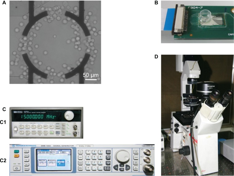
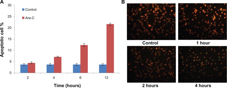
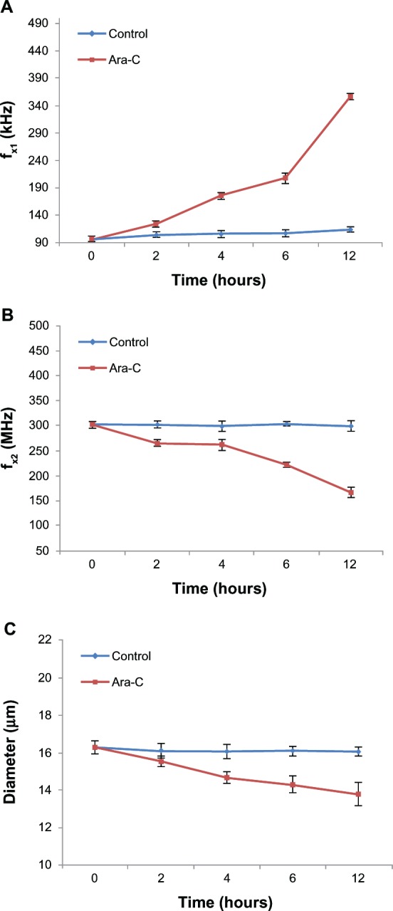
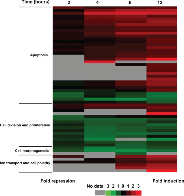
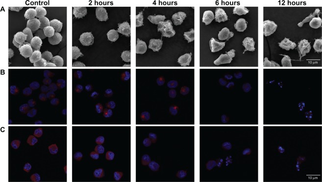
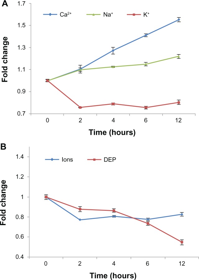
Dielectrophoresis (DEP) can be used to noninvasively measure the dielectric state of the cell, and this data can be used to monitor cell health or apoptosis. In this study, we followed events associated with cytosine arabinoside (Ara-C)-induced apoptosis in NB4 cells using DEP analysis. Our data showed that the membrane capacitance of NB4 cells decreases from 9.42 to 7.63 mF/m(2) in the first 2 hours following treatment with Ara-C, and that this decreased capacitance persists for >12 hours. Additionally, cytoplasmic conductivity decreases from 0.217 to 0.190 S/m within 2 hours of Ara-C treatment; this level is maintained for a short period of time before decreasing. We also investigated these events molecularly at the level of gene expression using microarray analysis and showed that the expression of genes related to membrane capacitance and cytoplasmic conductivity change dramatically as early as 2 hours post-Ara-C treatment, and further demonstrated a temporal relationship between the dielectric properties and key events in apoptosis. This study, integrating physical electrical properties of the cell membrane and cytoplasm with those of conductivity-related gene networks, provides new insights into the molecular mechanisms underlying the initiation of apoptosis, establishing a systematic foundation for DEP application in follow-up drug screening and development of medicines for treating leukemia.

Keywords: apoptosis; dielectrophoresis; gene expression profiling; leukemia.


Figures

Figure 1
Figure 1
Principal components of the DEP assay system. (A) Nonclosed ring gold electrodes, with width of 20 μm and inner diameter of 200 μm. (B) The assembled chip with a reservoir modified from a 200-μL Eppendorf tube. (C) Signal generator. C1: HP33120A, C2: SMB100A. (D) CCD camera coupled to a fluorescence microscope. Abbreviation: DEP, dielectrophoresis.
Figure 2
Figure 2
Detection of apoptosis by Annexin V and JC-1 assays. (A) Percentage of apoptotic cells after different incubation periods with Ara-C as measured using FCA and Annexin V/PI assays. (B) Viable cells display greenish orange fluorescence and apoptotic cells display green fluorescence. Note: Our results suggest that NB4 cell apoptosis initiated by Ara-C can be detected at 2 h post-treatment using the JC-1 assay. Abbreviations: Ara-C, cytosine arabinoside; FCA, flow cytometric analysis.
Figure 3
Figure 3
Monitoring of Ara-C induced apoptosis by DEP analysis. (A and B) Distinct differences in crossover frequency occur between control and the apoptotic NB4 cells as early as 2 h post-exposure to Ara-C. ƒx1 increased from 96 ± 4.73 to 354 ± 6.11 KHz, while ƒx2 decreased from 301 ± 7.09 to 165 ± 7.78 MHz over the 12-h time course. (C) Cell diameter decreased from 16.30 ± 0.35 to 13.81 ± 0.62 μm in apoptotic cells. Note: No significant change was observed for control cells. Abbreviations: Ara-C, cytosine arabinoside; DEP, dielectrophoresis.
Figure 4
Figure 4
Relationship of molecular events to the time course of Ara-C-induced apoptosis. Note: Cluster image of gene expression data of the four main categories: apoptosis, cell division and proliferation, cell morphogenesis, and ion transport and cell polarity. Abbreviation: Ara-C, cytosine arabinoside.
Figure 5
Figure 5
Changes in morphological features of cells over the 12-h time course of Ara-C induced apoptosis. (A) SEM images of NB4 cells after treatment for 0 (control), 2, 4, 6, and 12 h with Ara-C. The cells displayed progressive changes in membrane morphology as apoptosis developed. (B and C) Merged confocal scanning laser microscopy images represent the changes in microtubule-related proteins over the 12-h time course: (B) γ-tubulin and (C) KIF20A. Note: Cell nuclei are stained with DAPI (blue). Abbreviations: Ara-C, cytosine arabinoside, SEM, scanning electron microscopy; DAPI, 4′,6-diamidino-2-phenylindole dihydrochloride.
Figure 6
Figure 6
Relative dynamic and temporal changes in intracellular ion levels and cytoplasmic conductivity over the 12-h time course. (A) Fold changes in intracellular calcium, sodium, and potassium from the 0- to 12-hour time points during Ara-C-induced apoptosis. The data represent averages of three independent experiments. (B) Estimated changes in average cytoplasmic conductivity based on changes in intracellular ions levels (blue) or DEP analysis (red) over the 12-h time course. Abbreviations: Ara-C, cytosine arabinoside; DEP, dielectrophoresis.

References

    1. Grant S. Ara-C: Cellular and molecular pharmacology. Adv Cancer Res. 1998;72:197–233. - PubMed
    1. Schlenk RF, Germing U, Hartmann F, et al. High-dose cytarabine and mitoxantrone in consolidation therapy for acute promyelocytic leukemia. Leukemia. 2005;19(6):978–983. - PubMed
    1. Thompson CB. Apoptosis in the pathogenesis and treatment of disease. Science. 1995;267(5203):1456–1462. - PubMed
    1. Ly JD, Grubb DR, Lawen A. The mitochondrial membrane potential (ΔΨm) in apoptosis; an update. Apoptosis. 2003;8(2):115–128. - PubMed
    1. Fadok VA, Voelker DR, Campbell PA, Cohen JJ, Bratton DL, Henson PM. Exposure of phosphatidylserine on the surface of apoptotic lymphocytes triggers specific recognition and removal by macrophages. J Immunol. 1992;148(7):2207–2216. - PubMed

Publication types