. 2013 Jul 9:5:29.
doi: 10.3389/fnagi.2013.00029. eCollection 2013.

Molecular mechanisms of cognitive dysfunction following traumatic brain injury

Affiliations

Molecular mechanisms of cognitive dysfunction following traumatic brain injury

Kendall R Walker et al. Front Aging Neurosci. .

Abstract

Traumatic brain injury (TBI) results in significant disability due to cognitive deficits particularly in attention, learning and memory, and higher-order executive functions. The role of TBI in chronic neurodegeneration and the development of neurodegenerative diseases including Alzheimer's disease (AD), Parkinson's disease (PD), Amyotrophic Lateral Sclerosis (ALS) and most recently chronic traumatic encephalopathy (CTE) is of particular importance. However, despite significant effort very few therapeutic options exist to prevent or reverse cognitive impairment following TBI. In this review, we present experimental evidence of the known secondary injury mechanisms which contribute to neuronal cell loss, axonal injury, and synaptic dysfunction and hence cognitive impairment both acutely and chronically following TBI. In particular we focus on the mechanisms linking TBI to the development of two forms of dementia: AD and CTE. We provide evidence of potential molecular mechanisms involved in modulating Aβ and Tau following TBI and provide evidence of the role of these mechanisms in AD pathology. Additionally we propose a mechanism by which Aβ generated as a direct result of TBI is capable of exacerbating secondary injury mechanisms thereby establishing a neurotoxic cascade that leads to chronic neurodegeneration.

Keywords: Alzheimer's; Aβ; CTE; TBI; cognition; tau.


Figures

Figure 1
Figure 1
Schematic representation of primary and secondary phases of injury leading to cognitive dysfunction following TBI. The primary injury force (direct physical impact or rotational accelaration/decelaration) gives rise to either a focal or diffuse injury which initiates secondary systemic complications and cellular injury mechanisms leading to cell death, axonal injury, and impaired synaptic plasticity contributing to the cognitive dysfunction observed following a TBI. The extent of cell death and axonal injury correlate strongly with neurological outcome following brain injury. TBI induced impairments in synaptic plasticity characterized by impaired long-term potentiation (LTP) and enhanced long-term depression (LTD), two well-known molecular mechanisms controlling memory formation, also contribute to cognitive dysfunction particularly in mild TBI where no overt cell loss is detected despite chronic cognitive deficits being observed.
Figure 2
Figure 2
Pathophysiological mechanisms of cell death following TBI. In moderate to severe cases of TBI the extent of cell death strongly correlates with cognitive outcome. Following injury cell death can occur by two main pathways, necrosis and programmed cell death. Necrotic cell death is a rapid uncontrolled form of cell loss mediated primarily by calpains and spilled lysosomal cathepsins and contributes to the inflammatory response following TBI. In comparison programmed cell death mechanisms including apoptosis, necroptosis or autophagy occur in a delayed fashion following TBI. A key initiating event in the cell death pathways is TBI induced glutamate excitotoxicity due to damaged cell membranes, enhanced vesicular release of glutamate and impaired glutamate reuptake by glia resulting in increased glutamate levels at the synapse. Increased extracellular glutamate levels activate ionotrophic and metabotropic glutamate receptors leading to intracellular calcium overload and activation of secondary cellular injury mechanisms including calcium sensitive phosphatase and protease activation, mitochondrial oxidative stress, inflammation and proapoptotic gene activation initiating cell death pathways.
Figure 3
Figure 3
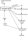
Schematic representation of the proposed mechanism by which TBI induced increases in Aβ create a vicious neurotoxic cascade leading to chronic neurodegeneration. Soluble Aβ levels are increased both clinically and experimentally following TBI and are known to impair synaptic plasticity and hence cognitive function by inhibiting LTP and enhancing LTD mechanisms of memory formation. The increased Aβ observed following TBI presumably occurs through a combination of increased generation and impaired clearance mechanisms. BACE1 levels are elevated following experimental TBI and directly contribute to the observed increases in Aβ. Caspase-3 mediated cleavage of BACE1 interacting adaptor proteins GGA1 and GGA3 has been demonstrated to modulate BACE1 levels and Aβ production following experimental TBI. The increased Aβ levels observed due to mechanisms including caspase mediated depletion of GGA1 and GGA3 could theoretically create a vicious feed forward mechanism leading to further propagation of Aβ. In vitro evidence suggests that Aβ is capable of initiating and mediating many of the cellular injury mechanisms that lead to programmed cell death following TBI.
Figure 4
Figure 4
Schematic representation of potential molecular mechanisms modulating Aβ and Tau pathology following injury. Loss of neurons, synapses, and dendritic spines as well as elevated soluble Aβ levels, amyloid plaque deposition along with elevated phosphorylated tau levels and the formation of neurofibrillary tangles (NFT's) are pathological features common to both traumatic brain injury (TBI) and Alzheimer's disease (AD). Not surprisingly the pathophysiological mechanisms leading to cell death, synaptic dysfunction and altered Aβ and tau pathology also overlap. Glutamate mediated excitotoxicity is a key initiator of cell death following traumatic brain injury (TBI). Excessive glutamate induced activation of extrasynaptic NMDA receptors leads to dramatically elevated intracellular calcium levels. Impaired calcium homeostasis in turn initiates a number of crucial downstream cellular injury processes including mitochondrial oxidative stress as well as the activation of the calcium sensitive proteases (caspases and calpains) which have been demonstrated to induce cell death in vitro (and hence loss of neurons and synapses) via caspase-3 activation and the cleavage of tau into a 17 kDa toxic tau fragment. TBI induced inflammatory events including activation of microglia and astrocytes and hence increased cytokine and chemokine production plays an important role in cell death following injury. Inflammation may amplify excitotoxic cell death by reducing glial glutamate transporters leading increased glutamate at the synapse exacerbating calcium dysgregulation. Experimental evidence suggests calcium-induced activation of calpain and calcineurin induces dendritic spine remodeling and loss and hence impairs synaptic transmission. Additionally, calpain and calcinuerin have both been implicated in NFT formation via cleavage induced activation of GSK-3β leading to hyperphosphorylation of tau. TBI induces elevated levels of soluble Aβ which is capable of inducing synaptic dysfunction. β-secretase (BACE1) is a key rate limiting enzyme in the production of Aβ and we have demonstrated a molecular mechanism by caspase-3 mediated depletion of the adaptor proteins GGA1 and GGA3 modulates BACE1 levels and Aβ production following experimental TBI. Experimental evidence suggests that soluble Aβ is capable of directly mediating many of the cellular injury processes characteristic of the secondary phase of TBI and hence elevated Aβ levels may be key initiating events in the development of a “feed forward” toxic cascade that links TBI to chronic neurodegeneration.
Figure 5
Figure 5
Cognitive reserve as a risk modifier for cognitive dysfunction and dementia following TBI. A number of environmental and genetic factors determine a persons level of cognitive reserve, those individuals with higher cognitive reserve are predicted to have a better cognitive outcome and a reduced risk of developing dementia following TBI. Individuals with lower cognitive reserve are predicted to have a poorer cognitive outcome and an increased risk of developing dementia following TBI. Cognitive reserve can be viewed as either an active (inherent compensatory mechanisms) or a passive (level of damage that can be tolerated by brain) process.

References

    1. Abdul H. M., Sama M. A., Furman J. L., Mathis D. M., Beckett T. L., Weidner A. M., et al. (2009). Cognitive decline in Alzheimer's disease is associated with selective changes in calcineurin/NFAT signaling. J. Neurosci. 29, 12957–12969 10.1523/JNEUROSCI.1064-09.2009 - DOI - PMC - PubMed
    1. Abrahamson E. E., Ikonomovic M. D., Ciallella J. R., Hope C. E., Paljug W. R., Isanski B. A., et al. (2006). Caspase inhibition therapy abolishes brain trauma-induced increases in Abeta peptide: implications for clinical outcome. Exp. Neurol. 197, 437–450 10.1016/j.expneurol.2005.10.011 - DOI - PubMed
    1. Acosta S. A., Tajiri N., Shinozuka K., Ishikawa H., Grimmig B., Diamond D., et al. (2013). Long-term upregulation of inflammation and suppression of cell proliferation in the brain of adult rats exposed to traumatic brain injury using the controlled cortical impact model. PLoS ONE 8:e53376 10.1371/journal.pone.0053376 - DOI - PMC - PubMed
    1. Albensi B. C., Sullivan P. G., Thompson M. B., Scheff S. W., Mattson M. P. (2000). Cyclosporin ameliorates traumatic brain-injury-induced alterations of hippocampal synaptic plasticity. Exp. Neurol. 162, 385–389 10.1006/exnr.1999.7338 - DOI - PubMed
    1. Alberdi E., Sánchez-Gómez M. V., Cavaliere F., Pérez-Samartín A., Zugaza J. L., Trullas R., et al. (2010). Amyloid β oligomers induce Ca2+ dysregulation and neuronal death through activation of ionotropic glutamate receptors. Cell Calcium 47, 264–272 10.1016/j.ceca.2009.12.010 - DOI - PubMed