

Hippocampal protein expression is differentially affected by chronic paroxetine treatment in adolescent and adult rats: a possible mechanism of "paradoxical" antidepressant responses in young persons
- PMID: 23847536
- PMCID: PMC3703543
- DOI: 10.3389/fphar.2013.00086
Hippocampal protein expression is differentially affected by chronic paroxetine treatment in adolescent and adult rats: a possible mechanism of "paradoxical" antidepressant responses in young persons
Abstract
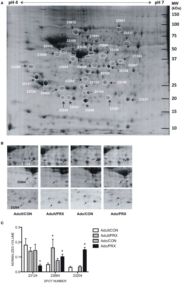
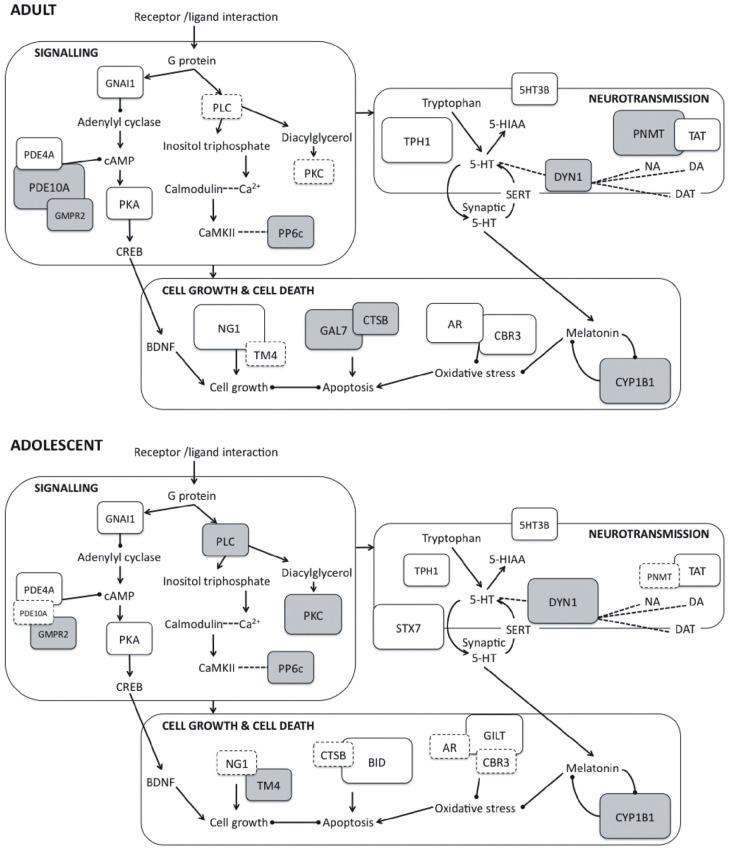
Selective serotonin reuptake inhibitors (SSRIs) are commonly recognized as the pharmacological treatment of choice for patients with depressive disorders, yet their use in adolescent populations has come under scrutiny following reports of minimal efficacy and an increased risk of suicidal ideation and behavior in this age group. The biological mechanisms underlying these effects are largely unknown. Accordingly, the current study examined changes in hippocampal protein expression following chronic administration of paroxetine in drinking water (target dose = 10 mg/kg for 22 days) to adult and adolescent rats. Results indicated age-specific changes in protein expression, with paroxetine significantly altering expression of 8 proteins in adolescents only and 10 proteins solely in adults. A further 12 proteins were significantly altered in both adolescents and adults. In adults, protein changes were generally suggestive of a neurotrophic and neuroprotective effect of paroxetine, with significant downregulation of apoptotic proteins Galectin 7 and Cathepsin B, and upregulation of the neurotrophic factor Neurogenin 1 and the antioxidant proteins Aldose reductase and Carbonyl reductase 3. Phosphodiesterase 10A, a signaling protein associated with major depressive disorder, was also downregulated (-6.5-fold) in adult rats. Adolescent rats failed to show the neurotrophic and neuroprotective effects observed in adults, instead displaying upregulation of the proapoptotic protein BH3-interacting domain death agonist (4.3-fold). Adolescent protein expression profiles also suggested impaired phosphoinositide signaling (Protein kinase C: -3.1-fold) and altered neurotransmitter transport and release (Syntaxin 7: 5.7-fold; Dynamin 1: -6.9-fold). The results of the present study provide clues as to possible mechanisms underlying the atypical response of human adolescents to paroxetine treatment.
Keywords: adolescent; antidepressant; hippocampus; paroxetine; proteomics; rat.
Figures
Similar articles
-
Contrasting regional Fos expression in adolescent and young adult rats following acute administration of the antidepressant paroxetine.Brain Res Bull. 2016 Mar;121:246-54. doi: 10.1016/j.brainresbull.2016.02.008. Epub 2016 Feb 11. Brain Res Bull. 2016. PMID: 26876759
-
Behavioral and serotonergic consequences of decreasing or increasing hippocampus brain-derived neurotrophic factor protein levels in mice.Neuropharmacology. 2008 Nov;55(6):1006-14. doi: 10.1016/j.neuropharm.2008.08.001. Epub 2008 Aug 12. Neuropharmacology. 2008. PMID: 18761360 Review.
-
Downregulation of Ccnd1 and Hes6 in rat hippocampus after chronic exposure to the antidepressant paroxetine.Acta Neuropsychiatr. 2008 Dec;20(6):307-13. doi: 10.1111/j.1601-5215.2008.00334.x. Acta Neuropsychiatr. 2008. PMID: 25384412
-
[Central serotonin receptors and chronic treatment with selective serotonin reuptake inhibitors in the rat: comparative effects of fluoxetine and paroxetine].Encephale. 1995 Mar-Apr;21(2):123-32. Encephale. 1995. PMID: 7781583 French.
-
Paroxetine: an update of its use in psychiatric disorders in adults.Drugs. 2002;62(4):655-703. doi: 10.2165/00003495-200262040-00010. Drugs. 2002. PMID: 11893234 Review.
Cited by
-
Nicotine and fluoxetine alter adolescent dopamine-mediated behaviors via 5-HT1A receptor activation.Front Psychiatry. 2024 Jun 11;15:1380123. doi: 10.3389/fpsyt.2024.1380123. eCollection 2024. Front Psychiatry. 2024. PMID: 38919632 Free PMC article.
-
What do DNA methylation studies tell us about depression? A systematic review.Transl Psychiatry. 2019 Feb 4;9(1):68. doi: 10.1038/s41398-019-0412-y. Transl Psychiatry. 2019. PMID: 30718449 Free PMC article.
-
The Role of Cathepsins in Memory Functions and the Pathophysiology of Psychiatric Disorders.Front Psychiatry. 2020 Jul 24;11:718. doi: 10.3389/fpsyt.2020.00718. eCollection 2020. Front Psychiatry. 2020. PMID: 32793006 Free PMC article. Review.
-
Disturbance of neurotransmitter metabolism in drug-naïve, first-episode major depressive disorder: a comparative study on adult and adolescent cohorts.Eur Arch Psychiatry Clin Neurosci. 2022 Oct;272(7):1283-1296. doi: 10.1007/s00406-022-01406-8. Epub 2022 Apr 11. Eur Arch Psychiatry Clin Neurosci. 2022. PMID: 35410391
-
Impact of escitalopram on vagally mediated cardiovascular function in healthy participants: implications for understanding differential age-related, treatment emergent effects.Psychopharmacology (Berl). 2014 Jun;231(11):2281-90. doi: 10.1007/s00213-013-3374-4. Epub 2013 Dec 15. Psychopharmacology (Berl). 2014. PMID: 24337078 Clinical Trial.
References
-
- Avissar S., Schreiber G. (1994). Antidepressant and antibipolar treatments' effects on receptor-coupled G proteins measures in lymphocytes of patients with mood disorders. Neuropsychopharmacology 10, 170S
LinkOut - more resources
Full Text Sources
Other Literature Sources