. 2013 Jul 8:4:83.
doi: 10.3389/fendo.2013.00083. eCollection 2013.

The Novel Actions of the Metabolite GnRH-(1-5) are Mediated by a G Protein-Coupled Receptor

Affiliations

The Novel Actions of the Metabolite GnRH-(1-5) are Mediated by a G Protein-Coupled Receptor

Darwin Omar Larco et al. Front Endocrinol (Lausanne). .

Abstract

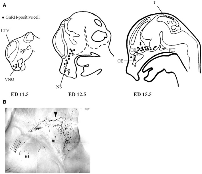
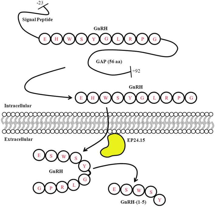
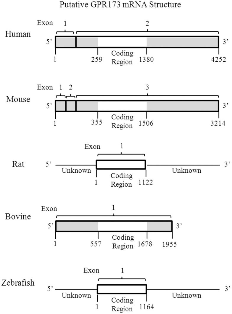
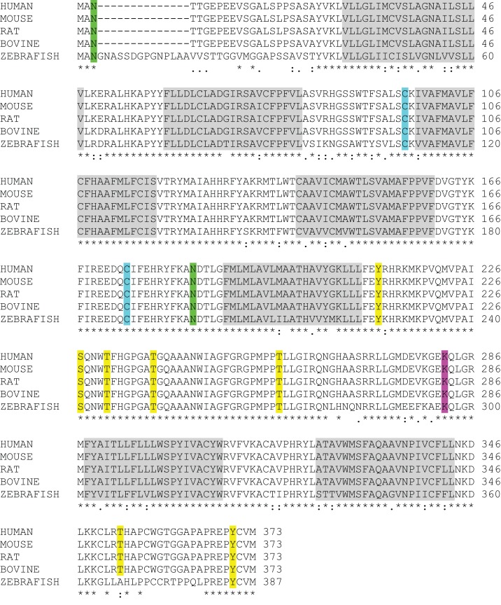
The gonadotropin-releasing hormone (GnRH) was originally isolated from the mammalian hypothalamus for its role as the primary regulator of reproductive function. Since its discovery, GnRH has also been shown to be located in non-hypothalamic tissues and is known to have diverse functions. Although the regulation of GnRH synthesis and release has been extensively studied, there is additional evidence to suggest that the processing of GnRH to the metabolite GnRH-(1-5) represents another layer of regulation. The focus of this review will be on the current evidence for the action of the pentapeptide metabolite GnRH-(1-5) in regulating cellular migration. We discuss the potential role of GnRH-(1-5) in regulating GnRH neuronal migration during development. Furthermore, we demonstrate these actions are mediated by the activation of a G protein-coupled receptor. Our findings suggest that GnRH-(1-5) may play a developmental function in addition to regulating developing cells.

Keywords: EP24.15; GPCR; GPR173; GnRH; SREB3; migration.


Figures

Figure 1
Figure 1
Gonadotropin-releasing hormone neuronal migratory route during development. (A) GnRH neurons are born in the nasal placode near the vomeronasal organ (VMO) at approximately embryonic day (ED) 11.5 in the mouse. As development progresses GnRH neurons begin to enter the CNS (dashed lines) at ED12.5 and begin to descend within the basal forebrain region at ED15.5. (B) Most GnRH neurons are within the CNS at ED15.5 however the remaining GnRH neurons in the nasal septum (NS) have a morphologically distinct migratory pattern relative to neurons found near the telencephalon (tel). Arrow indicates descending GnRH neurons of a mouse ED15.5 brain (Wu, TJ unpublished data). LTV, left telencephalic vesicle; OB, olfactory bulb; OE, olfactory epithelium; PIT, pituitary; MV, mesencephalic vesicle; T, tectum.
Figure 2
Figure 2
Gonadotropin-releasing hormone Peptide Processing. The prepro-GnRH is processed intracellularly to generate the mature GnRH peptide. In the extracellular matrix, GnRH is metabolized in a two step mechanism generating the metabolite GnRH-(1-5). Modified from Roberts et al. (11).
Figure 3
Figure 3
Putative mRNA Structure of GPR173 across Species. Analysis of the putative GPR173 mRNA structure across species reveals a significant degree of conservation within the coding region. These structures were generated from NCBI website (Bethesda, MD, USA). The mRNA accession number can be found inTable 1.
Figure 4
Figure 4
GPR173 Protein Sequence Alignment. A protein sequence analysis of GPR173 reveals a high degree of conservation across species. Additionally, many sites within the intracellular loop 3 are putative phosphorylation sites (yellow) with a conserved lysine residue (purple), which may undergo ubiquitination. A highly conserved arginine residue (green) in the N-terminal tail indicates a putative site for N-glycosylation. “*” indicate conserved residues; “.” or “:” indicate similarity between residues. Shaded regions indicate putative transmembrane spanning domains (TM). Sequence alignment analysis was conducted by using the UniProt consortium (www.uniprot.org).
Figure 5
Figure 5
Putative Model of Mouse GPR173 Disposition in the Membrane. The structure sketch map of Mouse GPR173 shows a large intracellular loop with multiple sites that may undergo post-translational modification. Yellow denotes putative phosphorylation site; purple denotes putative ubiquitination site; and green denotes putative N-glycosylation site. This model of GPR173 was generated with the aid of the UniProt consortium (www.uniprot.org).

Similar articles

Cited by

References

    1. Millar RP. GnRHs and GnRH receptors. Anim Reprod Sci (2005) 88:5–2810.1016/j.anireprosci.2005.05.032 - DOI - PubMed
    1. Moenter SM, Caraty A, Karsch FJ. The estradiol-induced surge of gonadotropin-releasing hormone in the ewe. Endocrinology (1990) 127:1375–8410.1210/endo-127-3-1375 - DOI - PubMed
    1. Chongthammakun S, Terasawa E. Negative feedback effects of estrogen on luteinizing hormone-releasing hormone release occur in pubertal, but not prepubertal, ovariectomized female rhesus monkeys. Endocrinology (1993) 132:735–4310.1210/en.132.2.735 - DOI - PubMed
    1. Ng Y, Wolfe A, Novaira HJ, Radovick S. Estrogen regulation of gene expression in GnRH neurons. Mol Cell Endocrinol (2009) 303:25–3310.1016/j.mce.2009.01.016 - DOI - PMC - PubMed
    1. Krsmanovic LZ, Hu L, Leung PK, Feng H, Catt KJ. The hypothalamic GnRH pulse generator: multiple regulatory mechanisms. Trends Endocrinol Metab (2009) 20:402–810.1016/j.tem.2009.05.002 - DOI - PMC - PubMed

LinkOut - more resources